4962
Compensating T2 blurring in 3D TSE with Cartesian acquisition based arterial spin labeled MRI1Radiology, UT Southwestern Medical Center, Dallas, TX, United States, 2Pediatrics, UT Southwestern Medical Center, Dallas, TX, United States, 3Advanced Imaging Research Center, UT Southwestern Medical Center, Dallas, TX, United States
Synopsis
3D fast/turbo spin echo (FSE/TSE) acquisitions are preferred for arterial spin labeled (ASL) MRI due to their higher SNR and compatibility with background suppression. However, 3D TSE suffers from T2 blurring caused by the T2 decay of the ASL signal along the prolonged echo train lengths, which may degrade image quality. This is often more noticeable in 3D TSE with Cartesian acquisitions. In this study, a truncated k-space filter is designed to compensate the T2 blurring of 3D TSE with Cartesian acquisitions and improve sharpness of ASL brain perfusion images.
Introduction
3D fast/turbo spin echo (FSE/TSE) acquisitions are preferred for arterial spin labeled (ASL) MRI due to their higher signal to noise ratio (SNR) and compatibility with highly efficient background suppression (BGS). Although, 3D ASL using stack-of-stars (SOS) or GRASE has been recommended by the ISMRM perfusion study group for brain perfusion imaging, these acquisitions are challenging in areas with increased B0 inhomogeneities (e.g. skull base). 3D TSE with Cartesian acquisitions have been shown to be robust in such applications [1]. However, the T2 decay due to prolonged echo train lengths (ETLs) of 3D TSE broadens local point spread functions, and leads to image blurring. Nevertheless, T2 blurring can be addressed through multiplicative k-space filtering [2], which is well-suited for ASL applications, since the majority of the signal is from a single tissue component. In this study, such a truncated k-space filter was designed to reduce image blurring while minimizing SNR loss in 3D ASL images of the brain.Methods
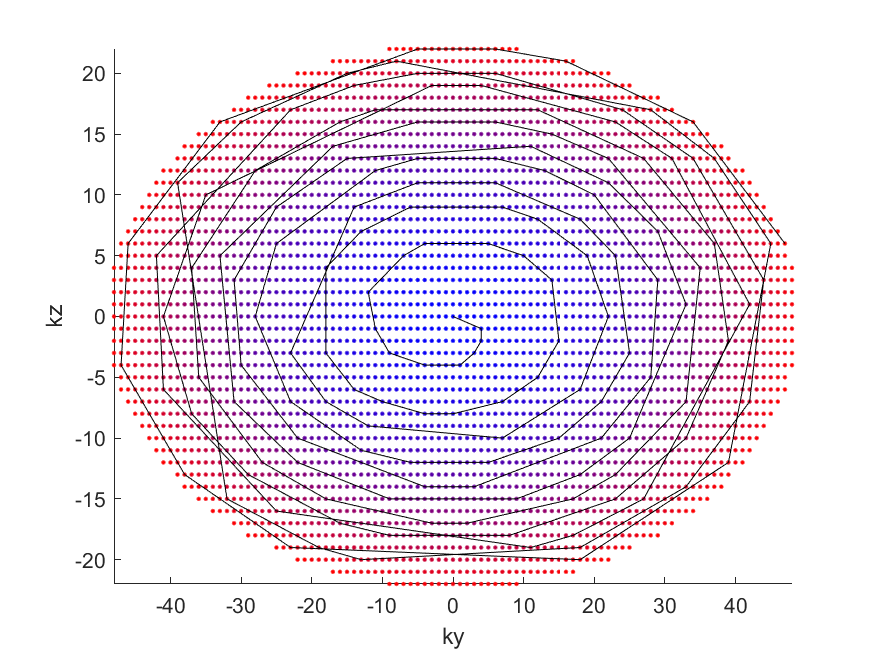
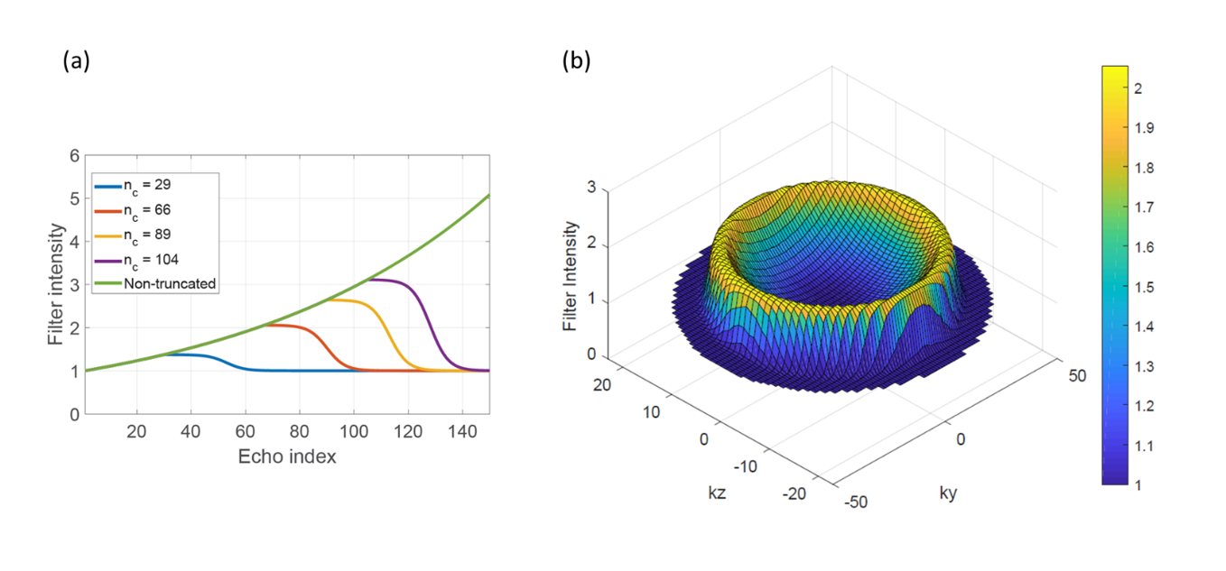
Pseudo-continuous ASL (pCASL) with optimized background suppression was used to acquire brain perfusion images [1]. With optimized background suppression, the signals of the perfusion-weighted images are mostly from the labeled blood, and thus the blood T2 of 275 ms (at 3T) was used for the filter design [3]. A 3D TSE Cartesian Acquisition using Spiral Profile Reordering (CASPR) was used for data acquisition (Fig. 1). Due to sampling of the center of k-space at the beginning of each echo train, CASPR has been shown to improve the robustness of ASL signal preparation similar to a spiral trajectory, but still maintaining acquisition on a Cartesian grid, thus increasing its robustness to B0 inhomogeneities. The truncated k-space filter was designed to compensate T2 blurring, such that, \begin{equation} I(n) =\ \left\{ \begin{array}{**lr**} e^{\left[(n-1) \cdot \frac{\Delta t }{T_2} \right] }, &n<n_c &\\ \frac{e^{ \left[(n_c-1)\cdot \frac{\Delta t }{T_2} \right] }-1}{e^{\left[k\cdot(n-n_c)-p\right]}+1}+1, & otherwise\\ \end{array} , \right. \end{equation} where Δt is the echo spacing, n is the echo index within an echo train, nc is the cut-off echo index where the truncation begins, k, p are two constants related to the truncation shape. The filter intensity I increases exponentially with n when n < nc, and progressively decreases to 1 when n exceeds nc, following the Fermi-Dirac function (Fig. 2). This filter compensates the T2 signal decay in the central k-space (up to nc), without amplifying the later echoes at the higher spatial frequencies, potentially avoiding significant SNR loss. The filter was applied to 3D ASL images of the brain in 7 volunteers, acquired using 3D pCASL with CASPR view ordering at 3T. The imaging parameters were: TR/TE = 6800/12 ms, FOV = 200x200x155 mm3, matrix = 68x67 with 77 slices, acquired resolution = 3x3x3 mm3, reconstructed resolution = 0.69x0.69x1.50 mm3, label duration = 1.8 s, post-label delay = 1.8 s, 4 BGS pulses and total acquisition time = 5.5 minutes. The Just Noticeable Blur (JNB) metric, a no-reference objective image sharpness metric, was used to evaluate the 3D pCASL image sharpness [4].Results
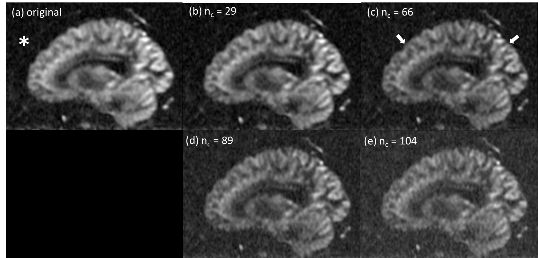
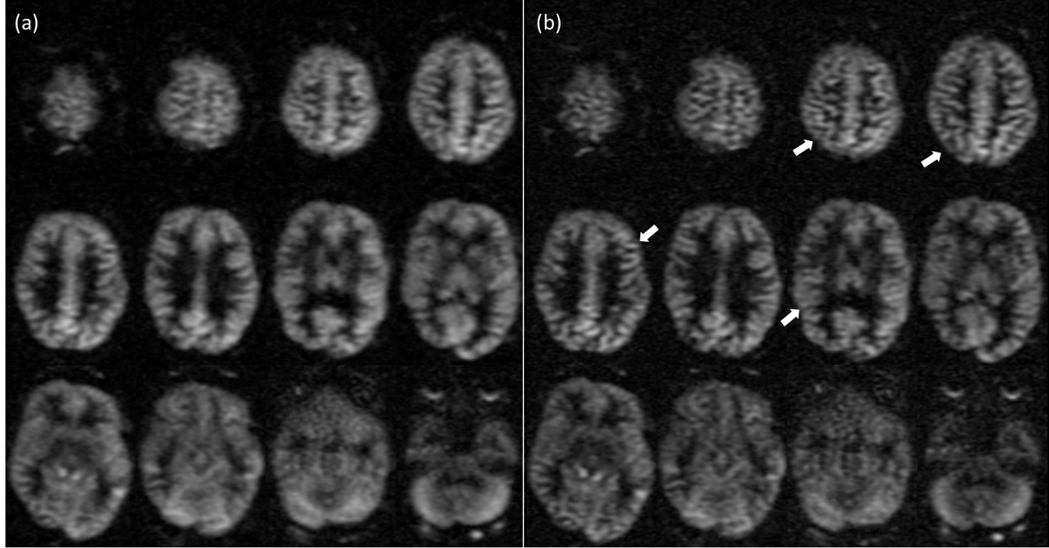
The k, p were found to be optimal at 0.3 and 7 considering the filter shape, the ETL, and the echo spacing used. The cut-off echo index, nc was found to be optimal at 66 and about 75% of the total echoes were multiplied with filter intensities greater than 1. This optimal combination improved image sharpness, without increasing the background noise (Fig. 3). The axial reformats from a sagittal acquisition showed improved image sharpness across the entire brain (Fig. 4). The JNB metric showed improvement across all slices (Fig. 5a), with an average improvement of 17% across all subjects (Fig. 5b).Discussion and Conclusion
The truncated k-space filter designed in this study can be used to retrospectively improve image sharpness of 3D pCASL images. While the improvements are shown in brain perfusion images acquired with 3D CASPR, this retrospective filter can also be applied to other 3D TSE acquisitions as well as to other anatomies such as kidneys. Future studies will consider optimizing the refocusing flip angles along with k-space filtering for prospective acquisitions [5].Acknowledgements
This work was supported by the NIH/NCI (grant U01CA207091).References
[1] Greer, JS et al. ISMRM 2017:3628.
[2] Zhou, X et al. JMRI 1993; 3(5):803-7.
[3] Stanisz, GJ et al. MRM 2005; 54:507-512.
[4] Ferzli, R et al. IEEE TIP 2009; 18(4):717-728.
[5] Zhao, L et al. MRM 2018; 80(4):1391-1401.
Figures