4957
Investigation of the effects of age and gender on normal cerebral blood flow in infants using arterial spin labeling MRI1Shaanxi University of Chinese Medicine, Xianyang, China, 2Baoji Center Hospital, Baoji, China, 3GE Healthcare China, Beijing, China
Synopsis
This study systematically revealed normal values of cerebral blood flow (CBF) in different age groups of infants using three-dimensional pseudocontinuous arterial spin labeling (3D PCASL) technique. Our results demonstrated a significantly lower CBF value in neonates than in other age groups. We also found a significant positive correlation between age and various regional mean gray matter (GM) and white matter (WM) CBF values in infants. Taken together, our findings demonstrated benefits of the application of the infants perfusion imaging technology to the clinical field by using arterial spin labeling (ASL) to provide information of metabolic status and neurodevelopmental outcomes.
Introduction
The brain is the organ with the most vigorous metabolism and the greatest demand for oxygen in the human body1. Therefore, the maintenance of normal brain function is highly dependent on the continuous and sufficient blood supply. Structural and functional imaging methods have been reported to provide new insights into normal and abnormal brain development1,2. Among these methods, ASL is a noninvasive magnetic resonance (MR) perfusion technique, based on the use of magnetically labeled blood-water protons as a nominally diffusible flow tracer3. ASL has been applied to evaluate changes in perfusion during brain maturation and aging1,4. However, the effects of age and gender on regional CBF during brain maturation have not yet been systematically studied. Here we revealed a reference set of normal values of CBF in different age groups of infants using 3D PCASL technique, and also investigated the effect of gender on CBF. The correlation between age and the CBF values of GM as well as WM was evaluated.Methods
Forty-four normal infants (age: 3.45 ± 3.42 months; age range: 4 days–12 months) joined this study with written informed parental consent after ethical approval. The subjects were divided into four age groups that included the first group (neonate, <28 days, 11 cases), the second group (1–3 months, 14 cases), the third group (4–6 months, 11 cases), and the fourth group (7–12 months, 8 cases). All data were acquired on a 3T MRI system (DISCOVERY MR 750W, GE). The 3D PCASL sequence was performed with following parameters: post-labeling delay = 1. 5 s / TR = 4599 ms / TE = 10.8 ms / NEX = 3 / FOV = 20 x 20 cm2 / slice thickness = 4 mm, and the pulse labeling plane was placed just below the volume of interest. Data post-processing was performed on AW4.6 GE workstation using Functool Brain stat Software for 3D ASL with automated generation of quantitative perfusion and CBF maps. The mean values of CBF in frontal GM, frontal WM, parietal GM, parietal WM, temporal GM, temporal WM, occipital GM, occipital WM, caudate nucleus, shell nucleus, pallium, and thalamus4 were obtained by averaging the data in manually selected regions of interest (ROIs) (7–10 mm2) on the CBF maps overlaid on T1-weighted images acquired with the same planning.Results
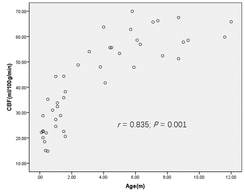
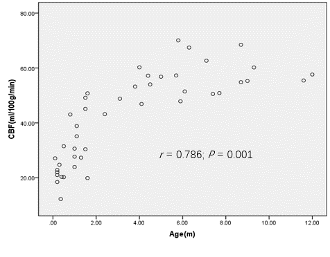
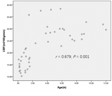
There was no significant difference in mean CBF values between all interested brain regions (P > 0.05), while mean CBF values in deep GM nuclei were slightly higher than cortical GM and WM. Comparing mean CBF values across gender, we found that except for the thalamus, the CBF values in all brain regions were slightly higher in male than in female subjects. However, there was no significant difference in the overall CBF values between male and female subjects (P > 0.05). Comparing mean CBF values between different age groups, a significantly lower CBF value in neonates than in other age groups (P < 0.05) was observed. Further correlation analysis between age and CBF values in all interested brain regions, we found that except for the temporal WM (r = 0.07, P > 0.05), there was a significant positive correlation between age and CBF values in all interested regions (Fig. 1–3).Discussion
During growth and development of the human brain, neurons and synapses are produced vastly, reaching its maximum capacity and the steady state by the 12th year5. Previous studies have shown that the metabolic level of newborns is generally low and gradually increases with age6. Our results echoed their findings by demonstrating a significantly lower CBF value in neonates than in other age groups, and a significant positive correlation between age and various regional mean GM and WM CBF values in infants. It can be seen that the application of neonates and infants perfusion imaging technology to the clinical field has many benefits, because it can help to predict metabolic status and neurodevelopmental outcomes. Determination of perfusion patterns that change with normal brain development and establishing a reference set of normal values of CBF during early life may be important preconditions for diagnosing brain disorders in children.Acknowledgements
No acknowledgement found.References
[1] Kim HG, Lee JH, Choi JW, et al. Multidelay Arterial Spin-Labeling MRI in Neonates and Infants: Cerebral Perfusion Changes during Brain Maturation. AJNR Am J Neuroradiol. 2018;39(10):1912-1918.
[2] Wang Z, Fernández-Seara M, Alsop DC, et al. Assessment of functional development in normal infant brain using arterial spin labeled perfusion MRI. Neuroimage. 2008;39(3):973-978.
[3] Detre JA, Leigh JS, Williams DS, and Koretsky AP. Perfusion imaging. Magn Reson Med. 1992;23(1):37-45.
[4] Soni N, Jain A, Kumar S, et al. Arterial spin labeling magnetic resonance perfusion study to evaluate the effects of age and gender on normal cerebral blood flow. Neurol India. 2016;64(7):32-38.
[5] Chugani HT. Biological basis of emotions: Brain systems and brain development. Pediatrics. 1998;102(5 Suppl E):1225-1229.
[6] Peterson BS. Brain imaging studies of the anatomical and functional consequences of preterm birth for human brain development. Ann N Y Acad Sci. 2003;1008:219-237.
Figures