4955
Alternative Slice Acquisition Orders for High-Resolution MB-EPI PCASL Imaging with Background Suppression1Radiology-Center for Magnetic Resonance Research, University of Minnesota, Minneapolis, MN, United States
Synopsis
Relative static tissue signal differences between neighboring slices across slice bands in MB-EPI PCASL imaging with background suppression (BS) are dramatically larger than those in MB-EPI PCASL imaging without BS, and can result in severe subtraction errors/artifacts for imaging data with large subject motion that sometimes cannot be corrected or removed by motion correction. To resolve this issue, alternative slice acquisition orders are proposed and evaluated. Our results suggest that the proposed alternative slice acquisition orders can improve the robustness of MB-EPI PCASL imaging with BS, providing comparable CBF estimates with minimized subtraction errors.
Purpose
Recent studies have suggested that 2D MB-EPI PCASL is a viable and valuable approach for high-resolution whole brain perfusion imaging (1-3). Background suppression (BS) of static tissue using combined pre-saturation and inversion RF pulses (4) has been proposed for ASL imaging to increase the temporal stability of the perfusion signal (5), and can also benefit MB-EPI PCASL imaging, although the beneficial effects will reduce with increases in the number of slices acquired within a slice band, as well as with increases in the slice acquisition time for high-resolution imaging.
However, relative static tissue signal differences between neighboring slices across slice bands in MB-EPI PCASL imaging with BS are dramatically larger than those in MB-EPI PCASL imaging without BS, and can result in severe subtraction errors/artifacts for imaging data with large subject motion that sometimes cannot be corrected or removed by motion correction. To improve the robustness of MB-EPI PCASL imaging with BS, we propose to use alternative slice acquisition orders to minimize relative static tissue signal differences between neighboring slices across slice bands.
Methods
Four alternative slice acquisition orders were proposed for MB-EPI PCASL imaging with BS (Figure 1). MRI studies using healthy volunteers were performed on a Siemens 3T Prisma MRI scanner under an IRB-approved protocol with written informed consent. The body coil was used for RF transmission and a 32-channel phased array head coil for signal reception.
After a scout localizer, Siemens Auto-Align scan was used for automatic imaging prescription followed by T1-weighted anatomic scans. High-resolution MB-EPI PCASL imaging scans with BS were performed using a single post-bolus delay and different alternative slice acquisition orders. The BS was achieved with in-plane pre-saturation and two inversion RF pulses optimized for the suppression of grey and white matter (4). To investigate whether alternative slice acquisition orders improve the robustness of high-resolution MB-EPI PCASL imaging with BS, imaging scans with significant motions were performed. To avoid potential bias in the comparisons of imaging results using different slice acquisition orders, long imaging scans were performed with different slice acquisition orders applied in an interleaved fashion. The major imaging parameters for MB-EPI PCASL imaging were as follows: TR/TE = 3570/19 ms; FOV = 215 x 215 mm2; matrix size = 86 x 86; slice thickness/gap in percentage = 2.27 mm/10; partial Fourier = 6/8; multi-band acceleration factor = 6; labeling duration = 1.5 s; post-bolus delay = 1.6 s; and number of fully relaxed M0 images acquired at the end = 2. A total of 60 label and control images were acquired for each type of slice acquisition order. To facilitate image distortion correction due to an inhomogeneous B0 field, spin echo EPI acquisitions with readouts matching those of the PCASL acquisitions were acquired along with ones using the reverse PE direction.
Post-processing, such as motion correction, was performed with SPM. CBF quantification was achieved using the single-blood compartment model (4). Statistical analyses were performed using GraphPad Prism software.
Results and Discussion
Our results indicate that the proposed alternative slice acquisition orders consistently improved the robustness of MB-EPI PCASL imaging with BS and greatly minimized motion-associated subtraction errors/artifacts. The results from imaging scans using C2POF and P2COF slice orders were very similar to those using C2PEF and P2CEF. Therefore, only results from imaging using the C2PEF and P2CEF slice orders are presented.
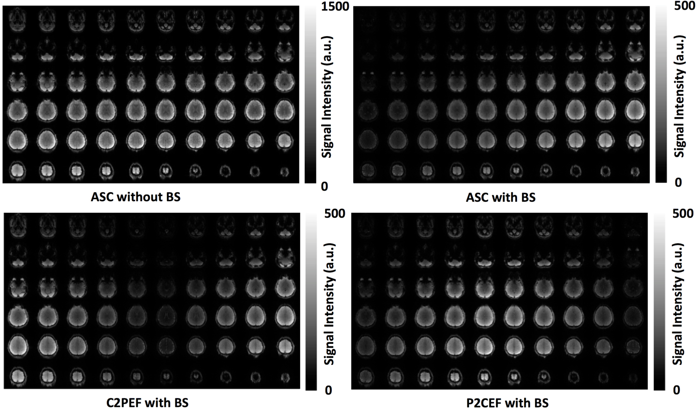
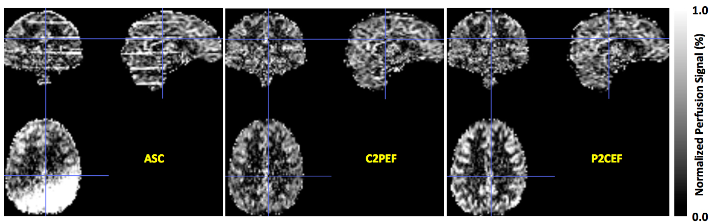
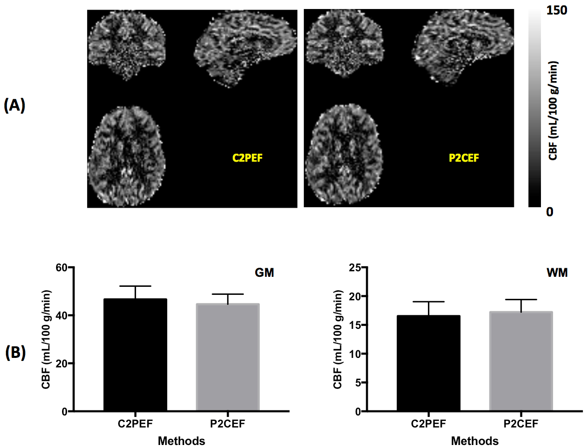
Figure 2 shows control images from perfusion scans using different slice acquisition orders. The reduction of static tissue signal with BS ranges from about 94% for the first acquired slices to about 65% for the last acquired slices of 6 slice bands. Figure 3 shows normalized perfusion-weighted images from scans using different slice acquisition orders. The perfusion-weighted images from imaging acquisition using the ASC slice order have obvious artifacts in slices across slice bands. CBF maps from one typical subject and mean CBF measurements from five subjects are presented in Figure 4. Statistical analyses suggested that perfusion imaging acquisitions using two alternative slice acquisition orders, C2PEF and P2CEF, provided comparable CBF estimates for both grey and white matter.
Because of relaxation effects with multi-slice acquisitions, the extent of static tissue suppression reduces for later acquired slices (Figure 2). This can be alleviated with interleaved C2PEF or P2CEF slice ordering.
Conclusions
The proposed alternative slice acquisition orders can improve the robustness of MB-EPI PCASL imaging with BS, providing comparable CBF estimates with minimized subtraction errors.Acknowledgements
P41 EB015894, P30 NS076408 and Human Connectome Projects (U01 MH109589 and U01 AG052564), and UL1TR000114. This research work is also supported by the University of Minnesota Foundation. The content is solely the responsibility of the authors and does not necessarily represent the official views of the National Institutes of Health.References
1. Li X, Wang D, Auerbach EJ, Moeller S, Ugurbil K, Metzger GJ. Theoretical and experimental evaluation of multi-band EPI for high-resolution whole brain pCASL Imaging. Neuroimage. 2015;106:170-181.
2. Li X., Shao X., Wang D., Ramanna S., Moeller S., Ugurbil K., Yacoub E. Wang DJ. Evaluation of 3D GRASE and 2D MB-EPI for Multi-Delay PCASL Imaging. In: Proceedings of Annual Meeting Annual Meeting & Exhibition 2017: 3627.
3. Li X., Wang D., Moeller S., Wang DJ, Chappell M, Yacoub E., and Ugurbil K. Pushing the Limits of ASL Imaging for the Lifespan Human Connectome Projects. Proc. Intl. Soc. Mag. Reson. Med. 26 (2018):2175.
4. Garcia DM, Duhamel G, Alsop DC. Efficiency of inversion pulses for background suppressed arterial spin labeling. Magn Reson Med 2005; 54:366–372.
5. Alsop DC, Detre JA, Golay X, et al. Recommended implementation of arterial spin-labeled perfusion MRI for clinical applications: A consensus of the ISMRM perfusion study group and the European consortium for ASL in dementia. Magn Reson Med. 2015;73(1):102-116
Figures
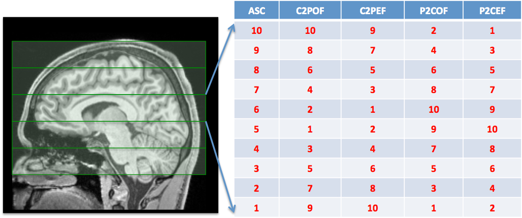
Figure 1. Illustration of alternative slice acquisition orders for high-resolution whole brain MB-EPI PCASL imaging with a total of 60 slices and an multi-band factor 6: ASC represents traditional ascending slice acquisition order; C2POF central-to-peripheral slice acquisition order with odd slice acquired first; C2PEF central-to-peripheral slice acquisition order with even slice acquired first; P2COF peripheral-to-central slice acquisition order with odd slice acquired first; and P2CEF peripheral-to-central slice acquisition order with even slice acquired first.