4945
Combined estimation of dispersion and macrovascular signal in multi-PLD pCASL data using a two-component model1C.J.Gorter Center for high field MRI, Department of Radiology, Leiden University Medical Center, Leiden, Netherlands, 2Wellcome Centre for Integrative Neuroimaging, FMRIB, Nuffield Department of Clinical Neurosciences, University of Oxford, Oxford, United Kingdom, 3Institute of Biomedical Engineering, Research Council UK (EP/P012361/1), University of Oxford, Oxford, United Kingdom
Synopsis
In pCASL a well-defined, box-shaped bolus is created at the labeling plane and for quantification this shape is assumed to be preserved, however, in reality this shape will be dispersed. With multi-timepoint data, the effects of dispersion can be observed in the macrovascular component, which can be separated from the tissue component using a two-component model. In this study the combined estimation of dispersion and macrovascular signal was investigated. When a gamma distribution dispersion kernel was incorporated into the two-component model, a significant decrease in CBF values was found, while a significant increase in macrovascular signal was observed.
Introduction
In pCASL a well-defined, box-shaped bolus is created at the labeling plane and for quantification this shape is assumed to be preserved. However, in reality this shape will be dispersed due to laminar flow profiles in large arteries and diffusion of the labeled water molecules within the blood.1 Including a dispersion kernel in the kinetic model for tissue quantification of arterial spin labeling(ASL) signal can improve the accuracy of the perfusion estimation and will generally lead to higher CBF values, since it corrects for spins in the tail that did not arrive yet in the imaging volume.2,3 Estimation of dispersion relies on the availability of multi-timepoint data, allowing to estimate the dispersion while the label traverses the vascular tree. However, to isolate the tissue perfusion signal from this data, intravascular and tissue components should be separated, e.g. by employing a two-component model that estimates the macrovascular component, otherwise the CBF would be overestimated.4 The ability of the two-component model to separate macrovascular from perfusion signal, could potentially be improved by the inclusion of a dispersion model (better description further into the vascular tree), but it could also lead to underestimation of CBF (perfusion signal interpreted as dispersed macrovascular signal). The goal of this study was to investigate the combined estimation of dispersion and macrovascular signal. Moreover, it was studied whether the temporal resolution affects the balance between these two effects and how they influence the CBF estimation.Methods
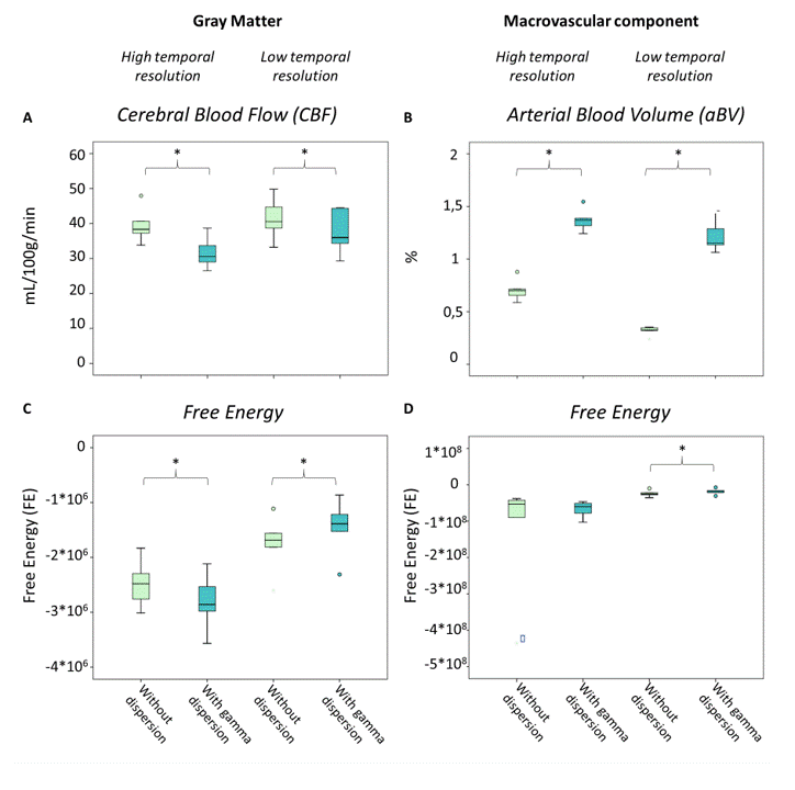
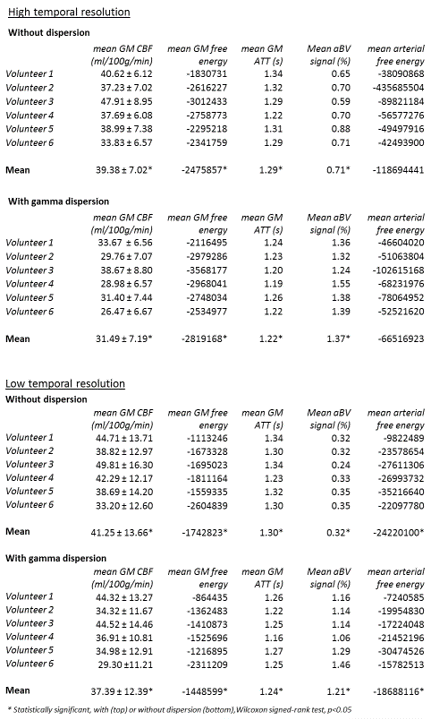
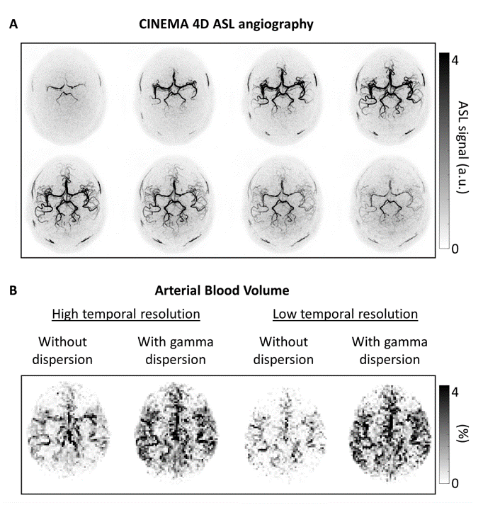
Six volunteers (age 22-54y, 4f/2m) were scanned using a 32-channel headcoil on a 3T-scanner (Achieva, Philips, Netherlands). All volunteers provided informed consent and the study was approved by the local IRB. Densely sampled multi-timepoint (28 timepoints) ASL was acquired by combining time-encoding with a Hadamard-8 matrix and a Look-Locker readout.5 Lower temporal resolution data was acquired by eliminating the Look-Locker readout (7 timepoints, 90 degree flip-angle). The BASIL toolkit of the Oxford Centre for Functional MRI of the BRAIN (FMRIB)’s software library(FSL) was used to quantify the ASL signal with a two-component model within a probabilistic analysis approach.4,6,7 Within this framework a gamma distribution dispersion kernel was included1 and the macrovascular contribution to the ASL signal was fitted resulting in arterial blood volume (aBV) and arterial transit time (ATT) maps. The variance on the perfusion values and the negative free energy(FE) were calculated. FE combines the accuracy of the model’s fit with a penalty for the number of free parameters. For comparison of the aBV maps, a 4D ASL angiography scan (CINEMA) with eight timepoints was acquired in three of the six subjects.8 See table 1 for all acquisition parameters. A Wilcoxon signed-rank test (p<0.05) was used to test for differences between the data with and without dispersion modeling.Results
Figure 1 shows an example of the CBF and aBV maps with and without modeling dispersion of one subject for the high and low temporal resolution scans. The mean cerebral blood flow (CBF) and ATT values in the gray matter (GM) were found to be significantly decreased when a gamma dispersion model was incorporated in the two-component model (Table 2, Figure 2). Significant increase in the macrovascular signal was indicated. For the low temporal resolution scan, the mean negative FE for both GM and the arteries were significant increased, which implies a better model fit when including this dispersion kernel. For the high temporal resolution scan, significant decrease was found for the mean GM negative FE(Figure 2). Figure 3 shows the maximum intensity projections for the CINEMA-scan and the aBV maps of one subject. These aBV maps show a global increase of the aBV signal and voxels more distally in the vasculature show higher intensities.Discussion and Conclusion
When including a gamma distribution dispersion kernel in a two-component model, the CBF values were found to be significantly lower and the macrovascular signal significantly higher. Moreover, the aBV maps show not only a global increase over the complete arterial tree, but especially in more distal arteries implying that signal is fitted deeper into the arterial tree. This can also be concluded when comparing the aBV-maps with the 4D ASL angiography data: without dispersion the aBV map resembles phase three, with dispersion more phase five/six. For the high temporal resolution scan, the FE didn’t improve upon including the gamma dispersion, which could indicate that the gamma dispersion kernel does not sufficiently describe the dispersion process as encountered in pCASL. Based upon the overall improved identification of macrovascular signal and the fact that this probably extends further into the arterial tree, we conclude that the combined estimation of dispersion and macrovascular effects in the kinetic model will improve the quantification of tissue perfusion.Acknowledgements
This work is part of the research programme Innovational Research Incentives Scheme Vici with project number 016.160.351, which is financed by the Netherlands Organisation for Scientific Research (NWO).References
1. Chappell MA,
Woolrich MW, Kazan S, Jezzard P, Payne SJ, MacIntosh BJ. Modeling dispersion in
arterial spin labeling: Validation using dynamic angiographic measurements. Magn
Reson Med. 2013;69(2):563-570. doi:10.1002/mrm.24260.
2. Hrabe J,
Lewis DP. Two analytical solutions for a model of pulsed arterial spin labeling
with randomized blood arrival times. J Magn Reson. 2004;167(1):49-55.
doi:10.1016/j.jmr.2003.11.002.
3. Gallichan D,
Jezzard P. Modeling the effects of dispersion and pulsatility of blood flow in
pulsed arterial spin labeling. Magn Reson Med. 2008;60(1):53-63.
doi:10.1002/mrm.21654.
4. Chappell MA,
MacIntosh BJ, Donahue MJ, Gunther M, Jezzard P WM. Separation of Intravascular
Signal in Multi-Inversion Timw Arterial Spin Labelling MRI. Magn Reson Med.
2010;63(5):1357-1365.
5. Merlijn C.E.
van der Plas, Wouter M. Teeuwisse, Sophie Schmid and MJ van O. More and faster:
multi-timepoint ASL at 150ms time-resolution with whole brain coverage by
combining time-encoding, Look-Locker, Multi-Band and flip-angle sweep. In: In
Proceedings 24th Scientific Meeting, ISMRM, Honolulu,2017. P0676.
6. Chappell MA,
Groves AR, Whitcher B WM. Variational Bayesian inference for a non-linear
forward model. IEEE Trans Med Imaging. 2009;57(1):223-236.
7. A.R. Groves,
M.A. Chappell MWW. Combined Spatial and Non-Spatial Prior for Inference on MRI
Time-Series. Neuroimage. 2009;45(3):795-809.
8. Uchino H,
Ito M, Fujima N, et al. A novel application of four-dimensional magnetic
resonance angiography using an arterial spin labeling technique for noninvasive
diagnosis of Moyamoya
Figures