4922
Improvement of Reproducibility in Quantitative Susceptibility Mapping (QSM) and Transverse Relaxation Rates (R2*) after Physiological Noise Correction1Department of Electrical and Computer Engineering, Seoul National University, Seoul, Korea, Republic of, 2Department of Radiology, Seoul Saint Mary's Hospital, Seoul, Korea, Republic of, 3Division of Biomedical Engineering, Hankuk University of Foreign Studies, Yongin, Korea, Republic of
Synopsis
Respiration-induced local magnetic field variation makes artifacts in gradient echo based images and reduces reproducibility of QSM and R2*. This study investigated reproducibility after respiration-induced error correction. The results showed a significantly improved reproducibility in QSM and R2* mapping.
Introduction
Several studies have found that abnormal accumulation of magnetic susceptibility sources is related to the pathogenesis of neurological diseases.1-4 The magnetic susceptibility is typically measured by two approaches; measuring R2* and QSM using a multi-echo GRE sequence. For the clinical use, reproducibility is an important feature. However, accurate QSM and R2* mapping are difficult to achieve due to various sources such as respiration-induced magnetic field fluctuations. Magnetic field fluctuations during data acquisition induce phase shift and resulting fitting error.5 A few studies reported the reproducibility of QSM6-8 and R2* measurement.9,10 However, the effects caused by respiration on reproducibility were not investigated. In this study, we investigated the improvement of reproducibility with respiration-induced error correction in QSM and R2* mapping.Methods
MR scans: To test intra-session reproducibility, the modified 3D multi-echo GRE scan was acquired twice from 10 healthy controls using 3T (Siemens Healthcare, Erlangen). In the modified sequence, the navigator echo was acquired for the respiration-induced magnetic field fluctuation correction (Fig. 1). Scan parameters were: resolution = 1.2 × 1.2 × 1.2 mm3, TR = 54 ms, TE = 9 to 48.1 ms (5 echoes), GRAPPA = 3, flip angle = 17°, and scan time = 6 min 4 sec. A T1-weighted (MPRAGE) images were also acquired for generating brain tissue segmentation.
Respiration-induced magnetic error correction: The corrected signal value at spatial location kx and TE is represented as follows,5,11
$$$S_N^c(kx,TE) = S_{N}(kx,TE)\cdot e^{-i\cdot TE\cdot (\phi_{N} - \phi_{1} )/ TE_{nav}}$$$ [Eq.1]
where $$$S_{N}(kx,TE)$$$ is the acquired signal intensity of Nth phase encoding line in the in the k-space. TEnav is the echo time of the navigator echo. ϕN and ϕ1 are the mean phases of the Nth and 1st navigator echo, respectively. To calculate the mean phase, 1D inverse Fourier Transform was applied to the navigator echo data along the readout direction (kx) and the mean phase was calculated. The correction was performed calculating the difference between ϕN and ϕ1 and multiplying $$$S_{N}(kx,TE)$$$ by $$$e^{-i\cdot TE\cdot (\phi_{N} - \phi_{1} )/ TE_{nav}}$$$ as shown in Eq. 1. The correction was applied to each phase encoding step. The same process was repeated for all phase encoding steps and acquired data.
QSM and R2* reconstruction: QSM reconstruction was performed using a MEDI software package12-14 after PDF.15,16 For R2* estimation, the voxel-wise R2* values were estimated by weighted nonlinear fitting of a mono-exponential decay function.17
Mask generation: T1-weighted MPRAGE image was aligned to the GRE image using FSL. Three brain region masks (whole brain, cortical gray matter (GM), and white matter (WM) masks) were generated from the T1-weighted MPRAGE image segmented by FSL. Globus pallidus, putamen, and caudate nucleus were manually defined based on the reconstructed QSM map (Fig. 2).
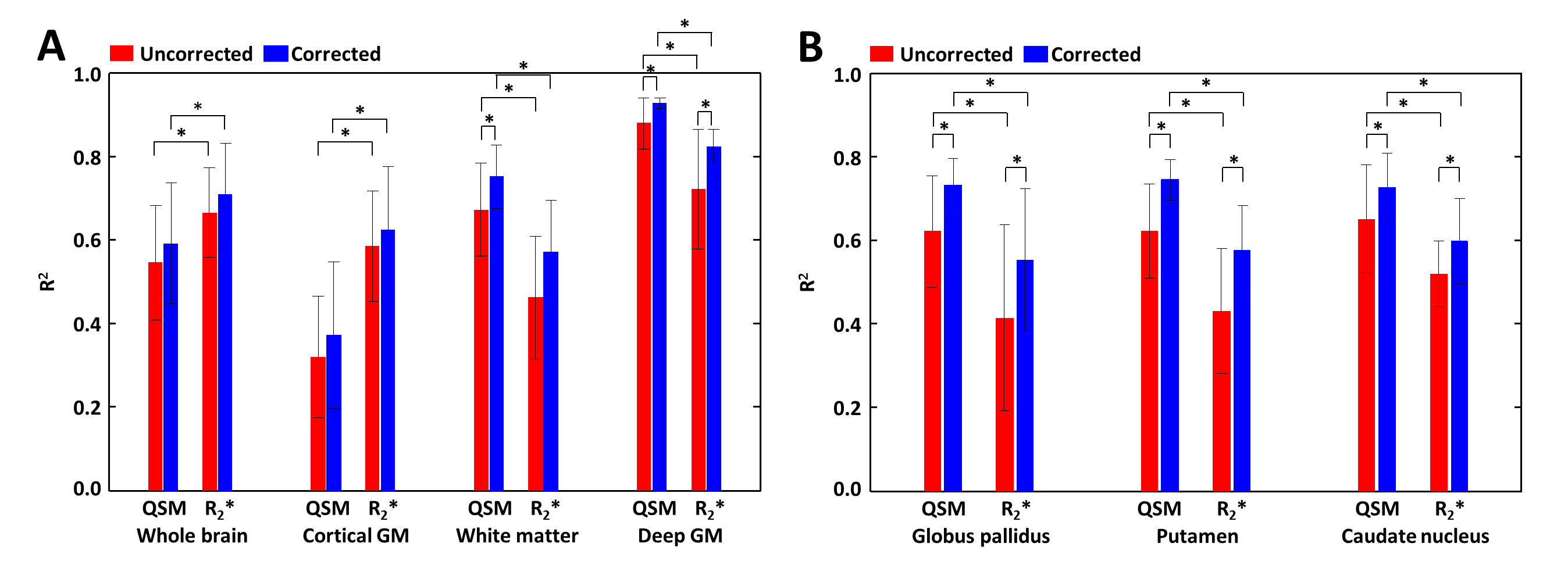
Data analysis: Image SNR and signal ratio of the ghost signal (= $$$\frac{\mu_{G}}{\sigma}$$$, where $$$\mu_{G}$$$ and $$$\sigma$$$ represent mean signal values in the outside of the brain which includes ghost artifacts and noise standard deviation in the background) were measured using GRE magnitude images. To assess reproducibility, a square of voxel-wise correlation (R2) between 1st and 2nd scans of the QSM maps and R2* maps were calculated. A paired t-test was performed to evaluate a statistical significance.
Results
In GRE (Fig. 3), SNR was improved by 13% after the correction (before: 240±34 and after: 271±19, p=0.026). Signal ratios of the ghost signal were decreased by 19% after the correction (before: 16±4 and after: 13±3, p<0.001). Compared to the uncorrected results, the corrected maps demonstrated reduced ghost artifacts in the QSM map (Fig. 4I and J) and the R2* maps (Fig. 4K and L). In R2* map (Figs. 4E and F), the standard deviations of R2* values in globus pallidus were reduced after the correction (before: 4.3Hz and after: 3.8Hz). In all ROIs, R2 values increased after the correction (Fig. 5). R2 values from WM (QSM: 11.89%, p=0.009 and R2*: 23.38%, p=0.024) and deep GM (QSM: 5.50%, p=0.024 and R2*: 13.96%, p=0.019) increased significantly with the correction. In deep GM (Fig. 5B), all R2 values significantly increased after the correction in QSM (16.23%, p<0.05) and R2* (26.72%, p<0.05).Discussion and Conclusion
In this study, we demonstrated the importance of respiration-induced magnetic field variation correction for clinical use of QSM and R2* mapping. The corrected results provide improved reproducibility and help to obtain more reliable QSM and R2* mappings. This feature makes QSM and R2* mapping appealing for clinical neuroimaging applications in which susceptibility measurement is necessary, such as Alzheimer’s disease, Parkinson’s disease, and multiple sclerosis.
Acknowledgements
This work was supported by the National Research Foundation of Korea (NRF) grant funded by the Korea government (MSIT) (NRF-2018R1A2B3008445) and by NRF grant funded by MSIT (NRF-2017R1C1B1008345).References
[1] Bush AI. The metallobiology of Alzheimer's disease. Trends Neurosci 2003;26(4):207-214.
[2] Berg D, Hochstrasser H, Schweitzer KJ, Riess O. Disturbance of iron metabolism in Parkinson's disease -- ultrasonography as a biomarker. Neurotox Res 2006;9(1):1-13.
[3] Bartzokis G, Lu PH, Tishler TA, et al. Myelin breakdown and iron changes in Huntington's disease: pathogenesis and treatment implications. Neurochem Res 2007;32(10):1655-1664.
[4] Ropele S, de Graaf W, Khalil M, et al. MRI assessment of iron deposition in multiple sclerosis. Journal of magnetic resonance imaging : JMRI 2011;34(1):13-21.
[5] Wen J, Cross A, DA. Y. On the role of physiological fluctuations in quantitative gradient echo MRI: Implications for GEPCI, QSM, and SWI. Magnetic resonance in medicine 2014.
[6] Deh K, Eskreis-Winkler S, Spincemaille P, Nguyen T, Wang Y. Repeatability and reproducibility of brain quantitative susceptibility mapping. The 22nd Annual Meeting ISMRM 2014.
[7] Lin PY, Chao TC, Wu ML. Quantitative Susceptibility Mapping of Human Brain at 3T: A Multisite Reproducibility Study. AJNR Am J Neuroradiol 2014.
[8] Santin MD, Didier M, Valabregue R, et al. Reproducibility of R2 * and quantitative susceptibility mapping (QSM) reconstruction methods in the basal ganglia of healthy subjects. NMR in biomedicine 2017;30(4).
[9] Govindarajan ST, Cohen-Adad J, Sormani MP, Fan AP, Louapre C, Mainero C. Reproducibility of T2 * mapping in the human cerebral cortex in vivo at 7 tesla MRI. Journal of magnetic resonance imaging : JMRI 2015;42(2):290-296.
[10] Peran P, Hagberg G, Luccichenti G, et al. Voxel-based analysis of R2* maps in the healthy human brain. Journal of magnetic resonance imaging : JMRI 2007;26(6):1413-1420.
[11] Nam Y, Kim DH, Lee J. Physiological noise compensation in gradient-echo myelin water imaging. NeuroImage 2015;120:345-349.
[12] Liu T, Wisnieff C, Lou M, Chen W, Spincemaille P, Wang Y. Nonlinear formulation of the magnetic field to source relationship for robust quantitative susceptibility mapping. Magnetic resonance in medicine 2013;69(2):467-476.
[13] Liu J, Liu T, de Rochefort L, et al. Morphology enabled dipole inversion for quantitative susceptibility mapping using structural consistency between the magnitude image and the susceptibility map. NeuroImage 2012;59(3):2560-2568.
[14] Schweser F, Sommer K, Deistung A, Reichenbach JR. Quantitative susceptibility mapping for investigating subtle susceptibility variations in the human brain. NeuroImage 2012;62(3):2083-2100.
[15] de Rochefort L, Liu T, Kressler B, et al. Quantitative susceptibility map reconstruction from MR phase data using bayesian regularization: validation and application to brain imaging. Magnetic resonance in medicine 2010;63(1):194-206.
[16] Liu T, Khalidov I, de Rochefort L, et al. A novel background field removal method for MRI using projection onto dipole fields (PDF). NMR in biomedicine 2011;24(9):1129-1136.
[17] Coleman T, Li Y. An interior trust region approach for nonlinear minimization subject to bounds. SIAM J Optim 1996;6(2):418-445.
Figures