4916
Reproducibility of inhomogeneous magnetization transfer (ihMT): a test-retest, multi-site study1Baoji Center Hospital, Baoji, China, 2GE Healthcare China, Beijing, China
Synopsis
Derived from conventional magnetization transfer, inhomogeneous
magnetization transfer (ihMT) has been shown to be a promising method for
myelin imaging in recent studies. In the present study, the test-retest
reproducibility and multi-site variability of ihMT in measuring major white
matter fibers were evaluated. Good test-retest reproducibility and multi-site
agreements were obtained. These findings support the use of ihMT measurements as biomarkers in
multicenter and/or longitudinal studies.
Introduction
The ihMT has been reported to feature superior
sensitivity and specificity for myelin imaging1. However, the reproducibility on ihMT has yet been rarely investigated
up to date. The purpose of the present study is to assess the multi-center
reproducibility and test-retest variability of ihMT in central nervous system.Methods
Five
healthy young volunteers (age from 24 to 33 years, 2 females) without a history
of neurological diseases were recruited and scanned twice on three 3T MRI system
(Discovery MR750, GE Healthcare, Waukesha, USA) equipped with an 8 channels
head coil. All subjects received 3D T1 and ihMT scan. The ihMT images were
acquired using a 3D SPGR sequence with different MT preparation pulses.
Detailed parameters are as following: TR/TE = 10.3ms/2.1ms, 23cm field of view,
matrix 96×96, sagittal plane, 56 slices, flip angle 8°. The original acquired
ihMT images are processed with a post-processing software provided by GE
Healthcare. The maps of quantitative ihMT (qihMT) and ihMT ratio (ihMTR) for
each scan were calculated. Voxel based
analysis then was performed to generate qihMT and ihMTR values for major white matter fibers (Genu of corpus callosum, Body of corpus callosum,
Splenium of corpus callosum, Corticospinal tract, Cerebral peduncle, Limb of
internal capsule, Corona radiata, Posterior thalamic radiation, Cingulate gyrus
and Hippocampus). All data is expressed as mean ± standard deviation (SD). The intra- and inter-scanner reliability
and reproducibility was assessed with intraclass correlation coefficients
(ICCs). Bland-Altman
method was used to show the level of agreement between two measurement types.
Paired t-test and one-way ANOVA test were also used to compare the difference
between inter- and intra-scanner, respectively. All statistical
analysis was performed with software (SPSS v. 19.0, Chicago, IL) and p < 0.05 was set as statistical
significant.
Results
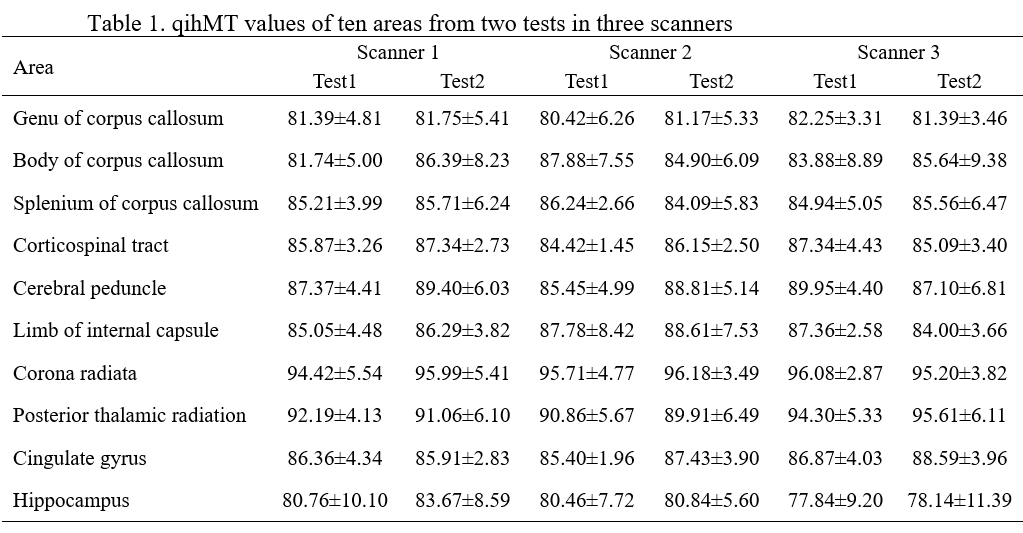
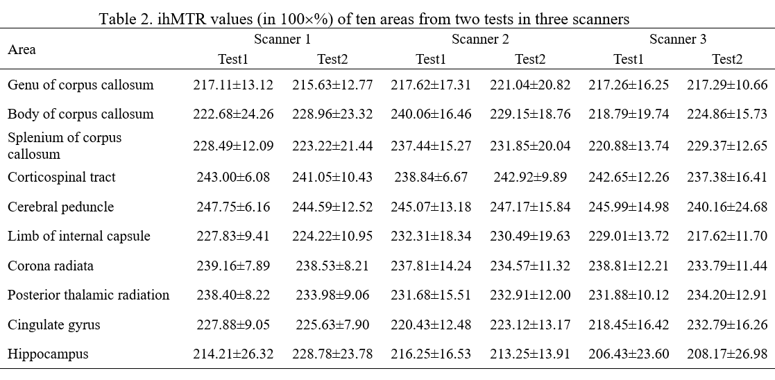
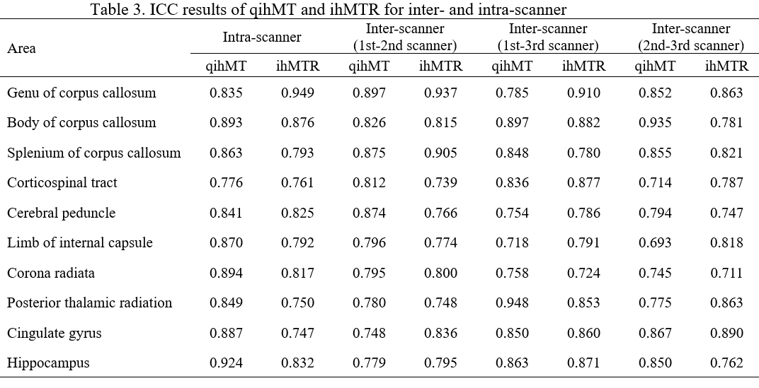
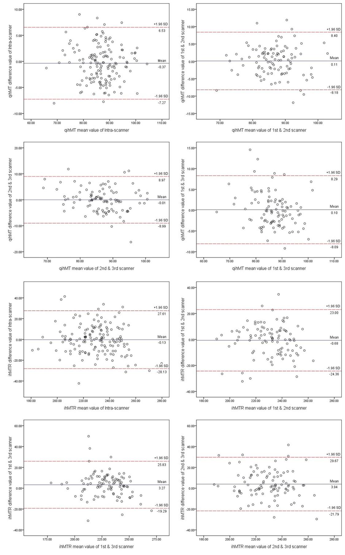
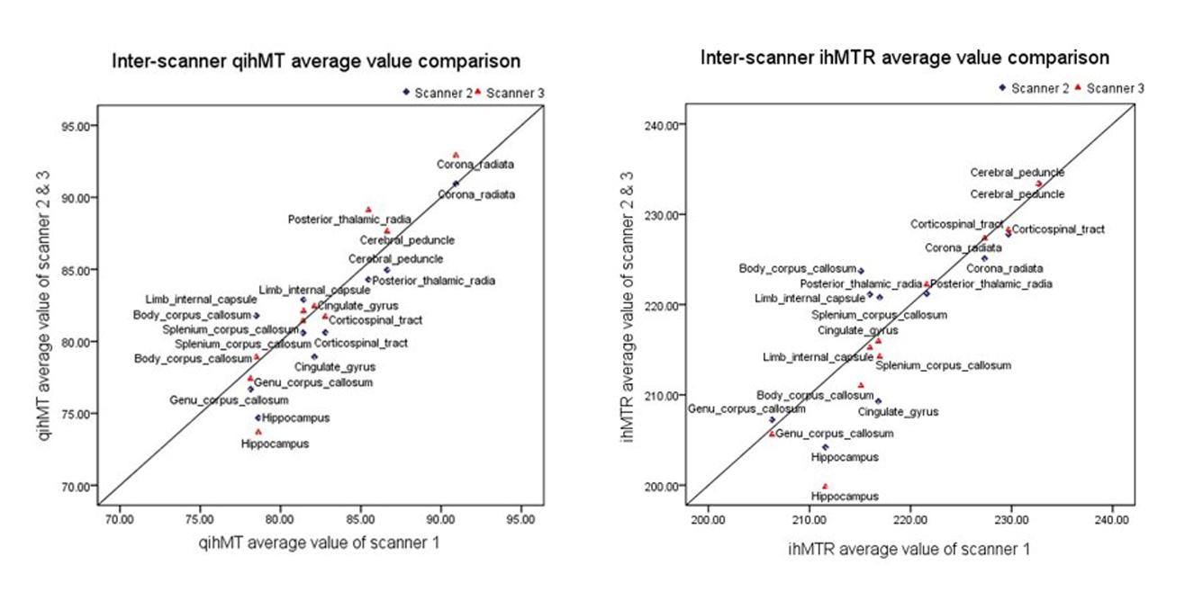
A summary of the mean value and standard deviation (SD) of qihMT and ihMTR of ten areas were shown in Tables 1 and 2. The ICCs of the same areas of qihMT and ihMTR values between intra- and inter-scanner were listed in Tables 3. In the 10 major white matter tracts areas, the ICCs indicated high intra- and inter-scanner measurement reliability and reproducibility. The Bland-Altman plots together with 95% confidence interval (CI) across all ROIs in the five volunteers (Figure 1), and the scatter plot of average values of qihMT and ihMTR on three scanners (Figure 2) also demonstrated good repeatability. No significant inter- and intra-scanner differences were found in Paired t-test and one-way ANOVA tests.Discussion
Derived from conventional magnetization transfer, ihMT has been shown to be a promising method for myelin imaging in recent studies1, 2. Compared to MT, ihMT allows imaging of the specific MT effects arising from inhomogeneously broadened components of the NMR spectrum, which is assumed to be mainly contributed by myelin3. In vivo experiments have demonstrated the sensitivity and specificity of ihMT in discriminating between myelinated and other tissues1. The assessment of myelin content, whether for brain development or disease progression, often involves multiple center collaboration or longitudinal data collection. Several reproducibility studies have been reported on myelin imaging techniques to date. Sandra et al. reported a 0.76 ICC for T2 based myelin water fraction imaging5. Magnetization transfer imaging was reported to have ICCs ranging from 0.572 to 0.962 in different brain region of interest6. In this study, inter-scanner ICCs were found to be higher than 0.693 in both qihMT and ihMTR for all ten measured tracts. Intra-scanner ICCs in our study range from 0.776 to 0.924 for qihMT, and 0.747 to 0.949 for ihMTR. The Bland–Altman plots showed no bias of one scan over the other. In general, these plots revealed good agreement inter- and intra-scanners, showing only a small discrepancy between measurements.Conclusion
Good inter- and intra-scanner reliability and reproducibility of ihMT measurements were observed in this study. These findings support the use of ihMT measurements as biomarkers in multicenter and/or longitudinal studies.Key words
ihMT; reproducibility; myelin imagingAcknowledgements
No acknowledgement found.References
1. Girard OM, Callot V, Prevost VH, et al. Magnetization transfer from inhomogeneously broadened lines (ihMT): improved imaging strategy for spinal cord applications. Magn Reson Med 2017;77:581–591.
2. Girard OM, Prevost VH, Varma G, et al. Magnetization transfer from inhomogeneously broadened lines (ihMT): experimental optimization of saturation parameters for human brain imaging at 1.5 Tesla. Magn Reson Med 2015;73:2111–2121.
3. Varma, G., Duhamel, G., de Bazelaire, C., et al. Magnetization transfer from inhomogeneously broadened lines: A potential marker for myelin. Magn Reson Med 2015;73, 614-622.
4. Meyers SM, Vavasour IM, Mädler B, et al. Multicenter measurements of myelin water fraction and geometric mean T2: intra- and intersite reproducibility. J Magn Reson Imaging. 2013;38:1445–1453.
5. Zou KH, Du H, Sidharthan S, et al. Statistical evaluations of the reproducibility and reliability of 3-tesla high resolution magnetization transfer brain images: a pilot study on healthy subjects. Int J Biomed Imaging. 2010:618747.
Figures