4914
Investigating the Influence of Adipose Fat on the Inhomogeneous Magnetization Transfer (ihMT) Images1Department of Radiology, UT Southwestern Medical Center, Dallas, TX, United States, 2Beth Israel Deaconess Medical Center, Harvard Medical School, Boston, MA, United States, 3Advanced Imaging Research Center, UT Southwestern Medical Center, Dallas, TX, United States, 4Philips Healthcare, Gainesville, FL, United States
Synopsis
Inhomogeneous Magnetization Transfer (ihMT) imaging is a novel enhanced magnetization transfer technique. In this study, we investigated the influence of fat (i.e. adipose tissue) and echo time on the ihMT ratio through simulation, phantom, and in vivo studies. A substantial variation on the ihMTR values in the presence of fat is illustrated, depending on the echo times used.
Introduction
Inhomogeneous Magnetization Transfer (ihMT) is a novel contrast which originates from the non-averaged residual dipolar couplings within tissues1-4. Recent studies have shown that ihMT can be tuned to enhance the contrast from a range of tissues (from brain white matter (WM) to muscles) through dipolar relaxation time (T1D) filtering5-6. This opens new avenues for ihMT applications, particularly for quantitative imaging of nerves as well as human skeletal muscles. IhMT applications outside of the human brain will involve close proximity to adipose tissue (near nerves and muscles). Although ihMT effect is expected to be low in adipose lipids due to their relatively lower motion restriction compared to membrane lipids (e.g. myelin), the presence of adipose tissue is expected to still influence the ihMT ratio (ihMTR) because of the normalization term in the calculation, M0, which contains signal from both water and fat (adipose lipids) pools. In this study, we investigated the influence of fat on ihMTR through simulation, phantom, and in vivo studies and compared different fat suppression methods.Methods
Theory: IhMTR can be calculated by taking the difference between single and dual off-resonance frequency applications:
\[ihMTR=\frac{MT^{+}+MT^{-}-MT^{+-} -MT^{-+}}{M_{0}}\] [1]
Where MT+, corresponds to the signal obtained using a single, positive off-resonance frequency RF saturation, MT-, a single negative off-resonance frequency RF saturation, MT+-, simultaneous dual (by alternating between positive and negative) frequency off-resonance RF saturation, MT-+, simultaneous dual (alternating between negative and positive) frequency off-resonance RF saturation, and M0, a reference condition with no saturation respectively. To illustrate the fat influence on the ihMTR, we considered three pools: a free-water pool, a bound-water pool with dipolar order, and a fat pool. For a voxel which contains fat and water, the MRI signal can be expressed as:
\[S=S_{w}+S_{f}e^{-2\pi i\Delta f_{wf}TE}\] [2]
where Δfwf, Sw, and Sf are the frequency difference between water and fat, signal from the water, and fat pools, respectively. Assuming that fat does not exchange magnetization with either free or bound-water pool,7-10 the subtraction in equation [1] eliminates the fat terms, Sf, from the numerator, whereas fat still contributes to the signal in the denominator (M0):
\[ihMTR=\frac{{MT^{+}_{w}}+{MT^{-} _{w}}-MT^{+-}_{w} -MT^{-+}_{w}}{M_{0w}+M_{0f}{e}^{-2\pi i \Delta f_{wf}TE}}\] [3]
Simulations: The ihMT signal was modeled using the single-bound-pool Morrison model6. To evaluate the influence of fat, in-phase and out-of-phase conditions were simulated while considering a frequency difference of 440 Hz between water and fat, at 3T. Simulations included three pools: free-water, bound water with dipolar order, and fat. The fat and free-water pool fractions were changed from 0 to 1 to investigate the influence of fat on the observed ihMTR signal.
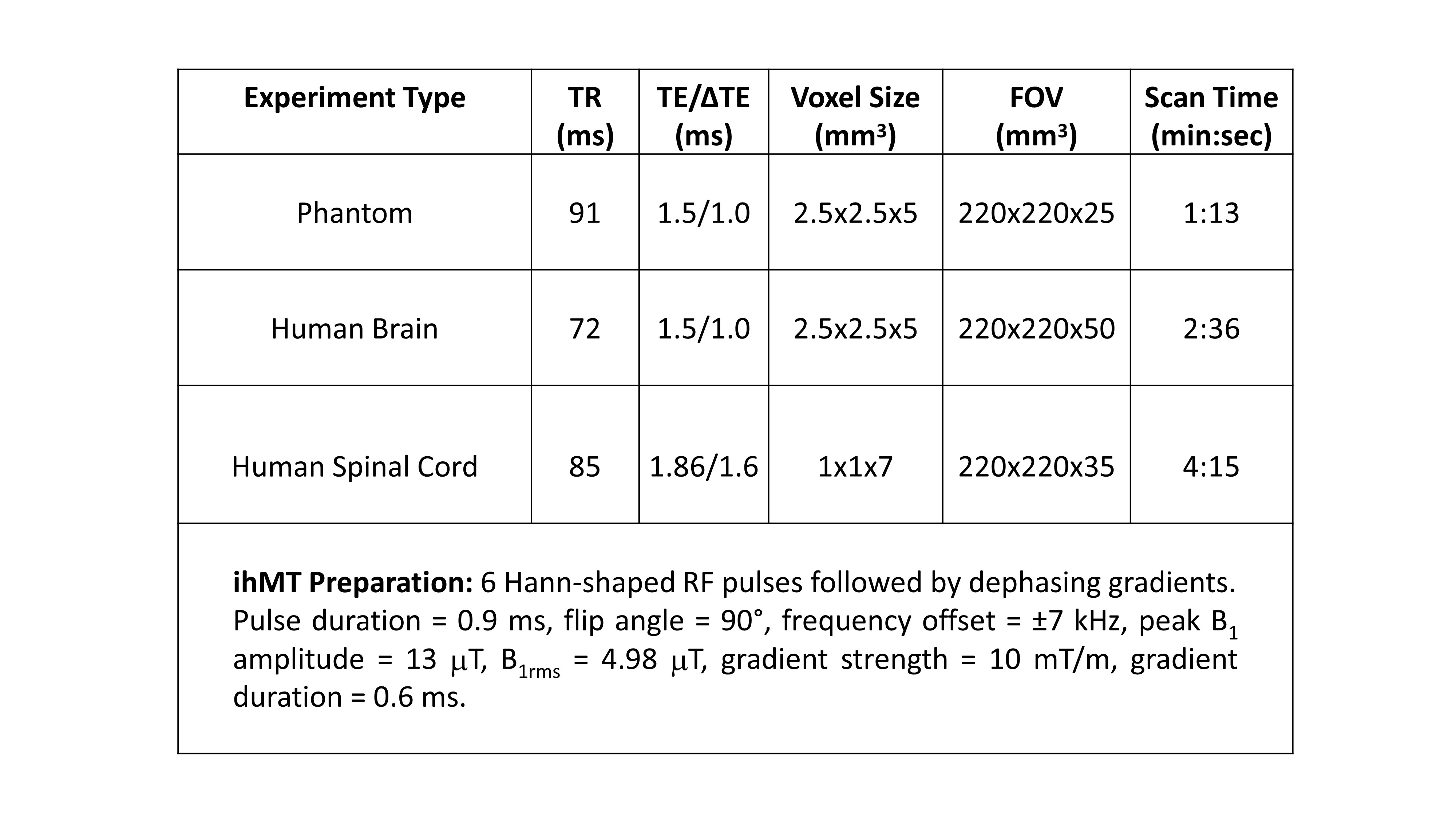
Phantom and in-vivo studies: A phantom consisting of hair conditioner (HC) at the bottom part of a container and oil at the top part of a container, was prepared to create an immiscible HC and oil interface. HC mimics the lamellar structure of myelinated tissues. The phantom was used to evaluate the influence of fat on the ihMTR. Following that, eight healthy volunteers (3 female, age: 27 ± 2 years) were scanned on a 3T Ingenia Philips MRI scanner. A 3D steady-state ihMT gradient echo sequence was applied for ihMT acquisitions (Table1). Fat suppression influence on ihMTR was compared between a three-point Dixon acquisition vs. SPIR. Mean ihMTR values were calculated for each scan and averaged within regions-of-interest drawn in the human brain and spinal cord WM.
Results and Discussion
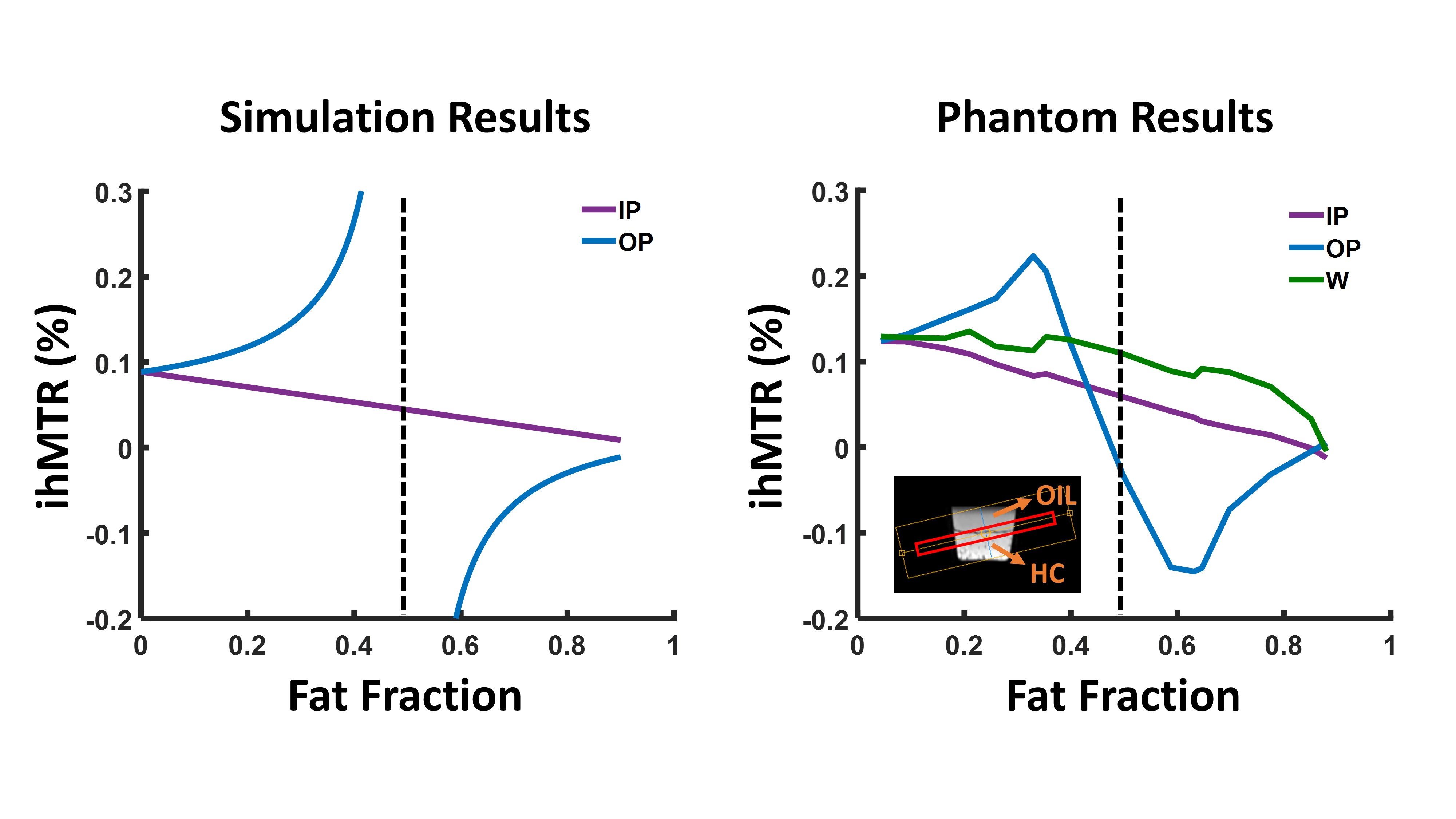
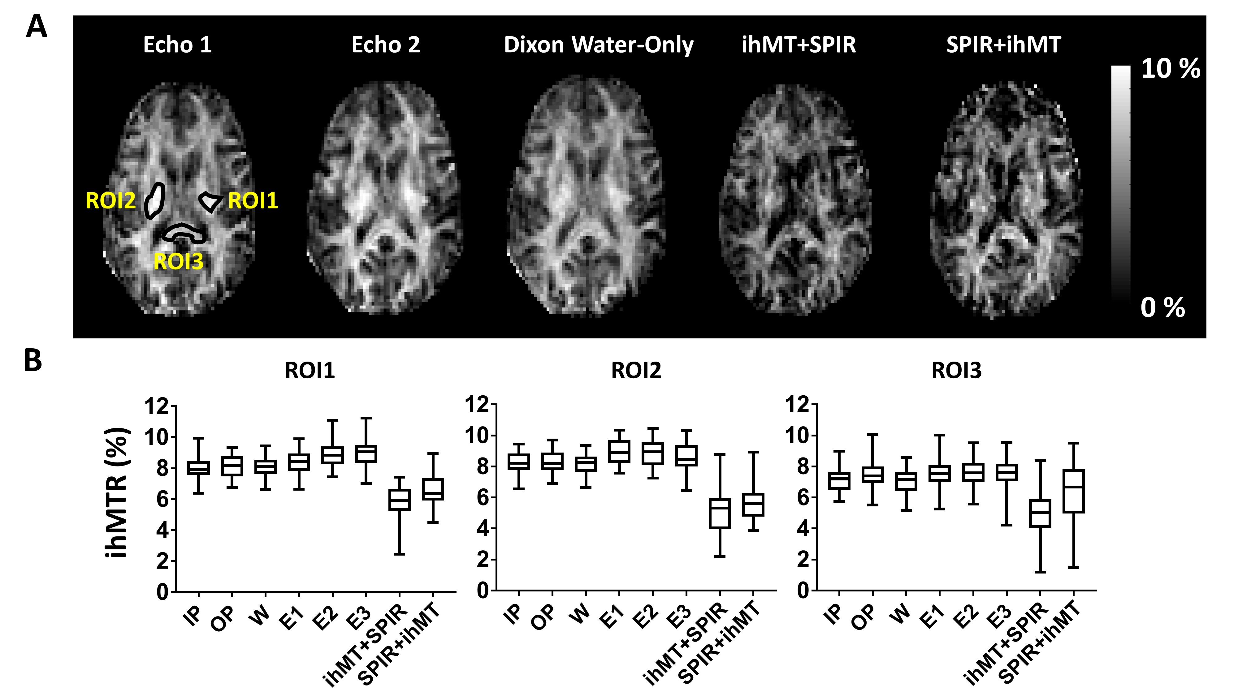
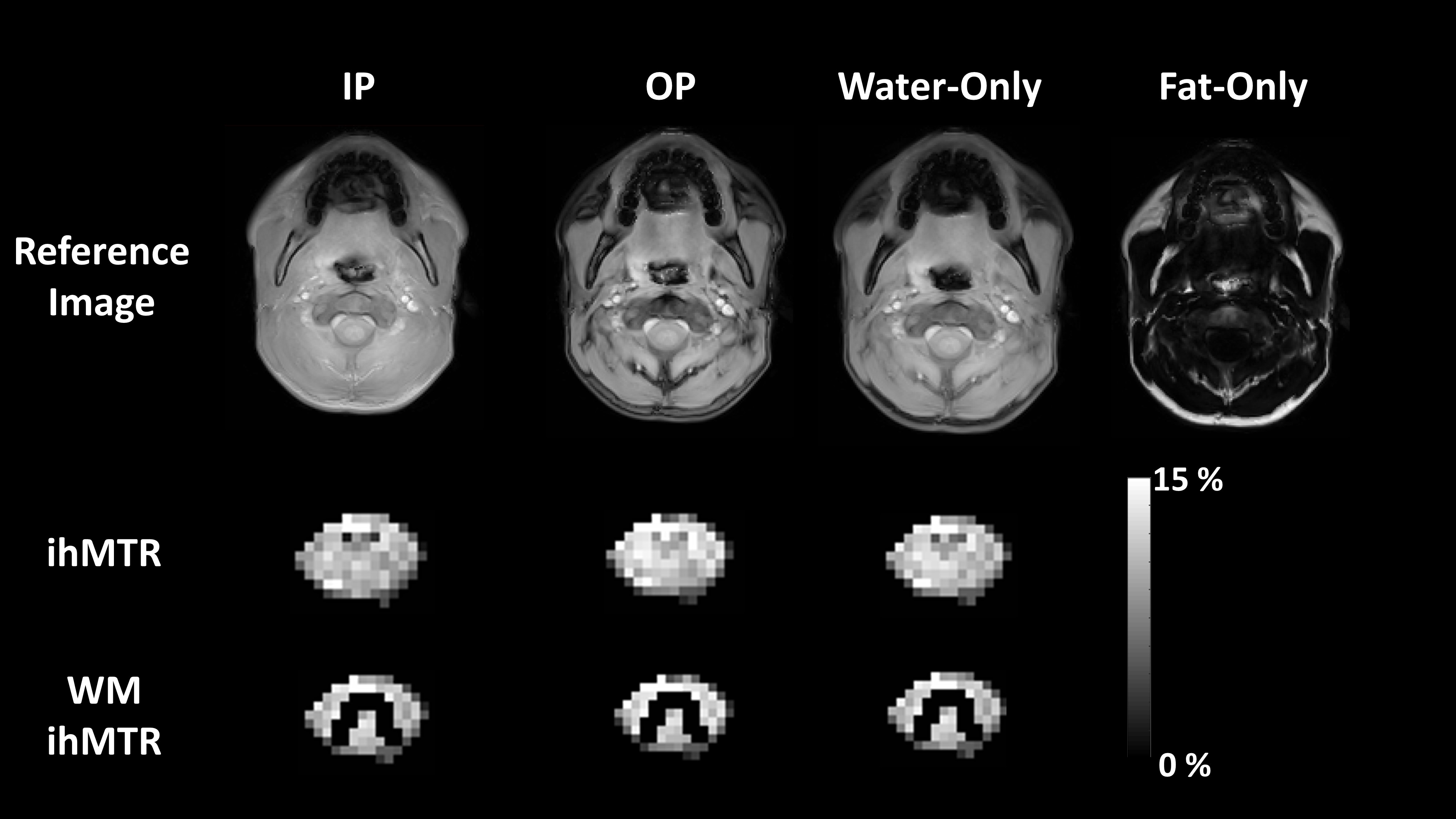
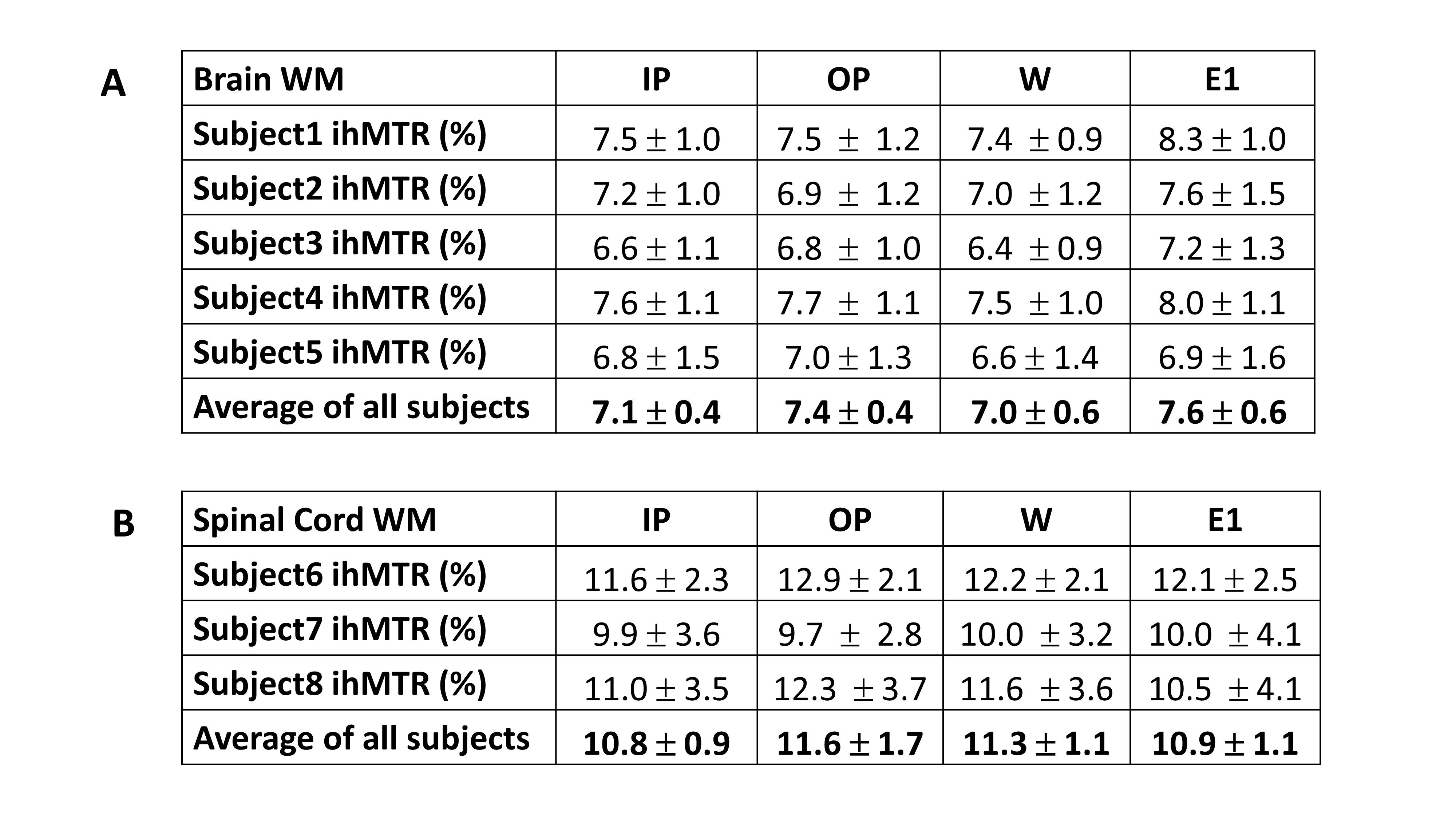
The results from simulation and phantom studies show evidence of a substantial variation on the ihMTR values in the presence of fat, depending on the echo times used (Figure1). Although fat pool has been shown not to contribute to the observable MT effect from water pool9, the fat content within a voxel and the choice of echo time was previously shown to similarly impact the observed MTR value7-8. Dixon method leads to water-only and fat-only images. In a region without adipose tissue contribution (e.g. human brain), the ihMTR values obtained using Dixon water-only images were close to the ihMTR values obtained without any fat suppression. In contrast, ihMTR values obtained using SPIR methods were significantly different (Figure2). The ihMTR values obtained with the Dixon method were consistent among volunteers in brain and spinal cord and were similar to those reported from similar regions in previous publications11-12 (Table2, Figure3).Conclusions
We show that the presence of fat within the voxel affects the ihMTR calculations depending on the echo times used. The ihMT-Dixon method can provide effective fat suppression, outperforming SPIR. Therefore, it is recommended for applications in regions which are in proximity to adipose tissue such as nerves and muscles.Acknowledgements
The authors would like to thank Kelli Key and Trevor Wigal for helping with subject recruitment and MRI scans.References
1. Varma G, Duhamel G, de Bazelaire C, Alsop DC. Magnetization transfer from inhomogeneously broadened lines: A potential marker for myelin. Magn Reson Med 2015;73(2):614-22.
2. Varma G, Girard OM, Prevost VH, Grant AK, Duhamel G, Alsop DC. Interpretation of magnetization transfer from inhomogeneously broadened lines (ihMT) in tissues as a dipolar order effect within motion restricted molecules. J Magn Reson 2015;260:67-76.
3. Girard OM, Prevost VH, Varma G, Cozzone PJ, Alsop DC, Duhamel G. Magnetization transfer from inhomogeneously broadened lines (ihMT): Experimental optimization of saturation parameters for human brain imaging at 1.5 Tesla. Magn Reson Med 2015;73(6):2111-21.
4. Swanson SD, Malyarenko DI, Fabiilli ML, Welsh RC, Nielsen JF, Srinivasan A. Molecular, dynamic, and structural origin of inhomogeneous magnetization transfer in lipid membranes. Magn Reson Med 2017;77(3):1318-1328.
5. Prevost VH, Girard OM, McHinda S, Varma G, Alsop DC, Duhamel G. Optimization of inhomogeneous magnetization transfer (ihMT) MRI contrast for preclinical studies using dipolar relaxation time (T1D ) filtering. NMR Biomed 2017;30(6).
6. Varma G, Girard OM, Prevost VH, Grant AK, Duhamel G, Alsop DC. In vivo measurement of a new source of contrast, the dipolar relaxation time, T1D , using a modified inhomogeneous magnetization transfer (ihMT) sequence. Magn Reson Med 2017;78(4):1362-1372.
7. Li K, Dortch RD, Kroop SF, Huston JW, Gochberg DF, Park JH, Damon BM. A rapid approach for quantitative magnetization transfer imaging in thigh muscles using the pulsed saturation method. Magn Reson Imaging 2015;33(6):709-17.
8. Li W, Wang X, Miller FH, Larson AC. Chemical Shift magnetization transfer magnetic resonance imaging. Magn Reson Med 2017;78(2):656-663.
9. Chen JH, Le HC, Koutcher JA, Singer S. Fat-free MRI based on magnetization exchange. Magn Reson Med 2010;63(3):713-8.
10. Chen JH, Sambol EB, Decarolis P, O'Connor R, Geha RC, Wu YV, Singer S. High-resolution MAS NMR spectroscopy detection of the spin magnetization exchange by cross-relaxation and chemical exchange in intact cell lines and human tissue specimens. Magn Reson Med 2006;55(6):1246-56.
11. Ercan E, Varma G, Madler B, et al. Microstructural correlates of 3D steady-state inhomogeneous magnetization transfer (ihMT) in the human brain white matter assessed by myelin water imaging and diffusion tensor imaging. Magn Reson Med 2018.
12. Ercan et al. 3D Steady-State Inhomogeneous Magnetization Transfer (ihMT) Gradient Echo Sequence for spinal cord imaging at 3T. In: Proceedings of the ISMRM. Paris, France; 2018: p5502.
Figures