4913
Myelin-sensitive imaging of the optic chiasm and optic nerve at 3T using inhomogeneous magnetization transfer (ihMT) with high B1 pulses1Department of Radiology, UT Southwestern Medical Center, Dallas, TX, United States, 2Advanced Imaging Research Center, UT Southwestern Medical Center, Dallas, TX, United States, 3Philips Healthcare, Gainesville, FL, United States, 4Beth Israel Deaconess Medical Center, Harvard Medical School, Boston, MA, United States
Synopsis
Inhomogeneous
magnetization transfer (ihMT) imaging is a novel enhanced magnetization transfer
contrast, which has been shown to originate from long-lived dipolar couplings
in the tissue (e.g. dipolar couplings between the methylene molecules of the
myelin phospholipid bilayer). In this study, we optimized an ihMT scan protocol
for imaging the optic nerve and chiasm for the first time. This method may
potentially be used for quantitative evaluation of patients with multiple
sclerosis (MS), as well as other diseases affecting the visual pathway.
Introduction
Optic nerve involvement (optic neuritis) is a common presentation in Multiple Sclerosis (MS) causing visual impairment and affecting daily function1. Quantitative imaging of the visual pathway is therefore desirable for evaluation and management of patients with optic neuritis. Previous quantitative MRI studies of the optic nerve and chiasm have shown differences between MS patients and healthy controls, however lacked specificity to determine the underlying pathological changes1-5. Inhomogeneous magnetization transfer (ihMT) is a promising novel contrast method which can provide myelin-specific information6-9. ihMT imaging of the visual pathway may provide insights into demyelinating pathologies of the optic nerve, and could have potential applications for investigating new therapies. However, quantitative imaging of these structures is challenging due to susceptibility artifacts near tissue interfaces, low SNR, as well as the small size (3-4 mm diameter) and mobility of these structures1,10. In this study, we address the challenges associated with quantitative imaging of the optic nerve and chiasm, and propose an ihMT sequence with high B1 pulses to obtain myelin-sensitive information in vivo.Methods
Four healthy volunteers (three females; age: 26 ± 3 years) were scanned on a 3T Ingenia Philips MRI scanner equipped with a 32-channel receive head-coil. The study adhered to the local Institutional Review Board guidelines and written informed consent was obtained. A 3D steady-state ihMT sequence with gradient echo acquisition was optimized and applied for optic pathway acquisitions. In order to minimize eye motion, participants were instructed to focus their vision on a marker positioned in front of them. Mean ihMTR values were calculated using the following formula:
\[ihMTR=\frac{MT^{+}+MT^{-}-MT^{+-} -MT^{-+}}{M_{0}}\]
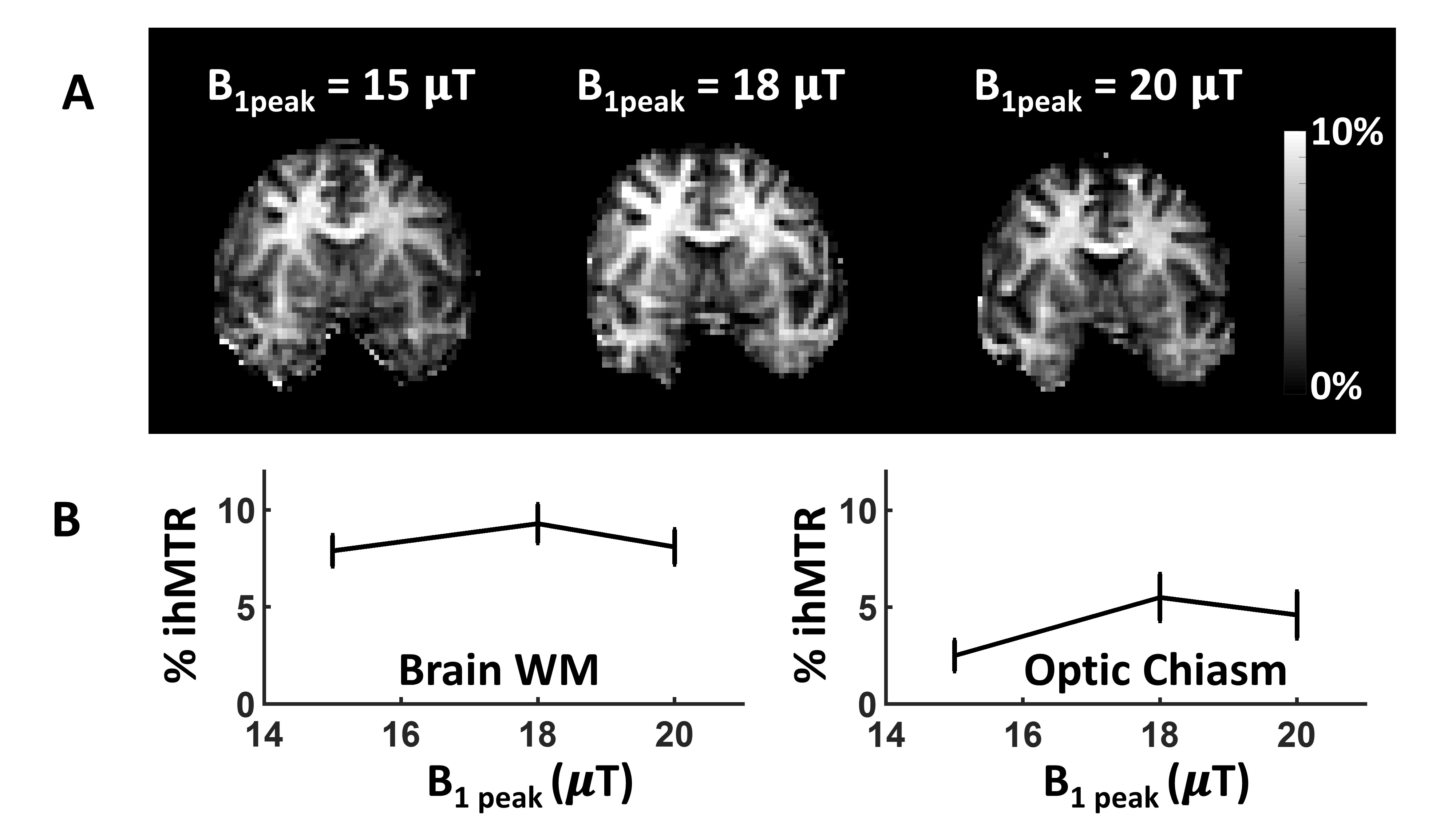
where MT+, MT-, MT+-, MT-+, and M0, correspond to the signal obtained using a single positive off-resonance RF saturation, a single negative off-resonance RF saturation, simultaneous dual (positive and negative alternating) off-resonance RF saturation, simultaneous dual (negative and positive alternating) off-resonance RF saturation, and a reference condition with no saturation, respectively. The optimum peak power of the ihMT preparation RF pulses (leading to the highest ihMTR) was found using low in-plane resolution (2.5 x 2.5 mm2) scans, while varying the peak power and evaluating the ihMTR from regions-of-interest (ROIs) in the cerabral white matter and the optic chiasm (Figure1). The optimized ihMT protocol employed 6 Hanning pulses (pulse angle: 90°, pulse duration: 0.66 ms, off-resonance frequency: +-7kHz) with high B1 peak power (18 μT) and B1rms (6.2 μT) for ihMT preparation. A concentrated energy deposition scheme was used as previously described11-12. This scheme uses high B1 power ihMT preparation pulses followed by a long TR and has been shown to reduce the B1-dependence of ihMT12. Coronal slices were acquired at the level of the optic nerve. Adaptive RF shimming was used to reduce B1+ field inhomogeneities. The imaging parameters for the optimized protocol were the following: flip angle: 7°, TR/TE = 95/1.72 ms, FOV (FH, RL, AP): 220 x 148.5 x 100 mm3, acquired voxel size (FH, RL, AP): 1.5 x 1.5 x 5 mm3. Total scan time was 8 minutes.
Results and Discussion
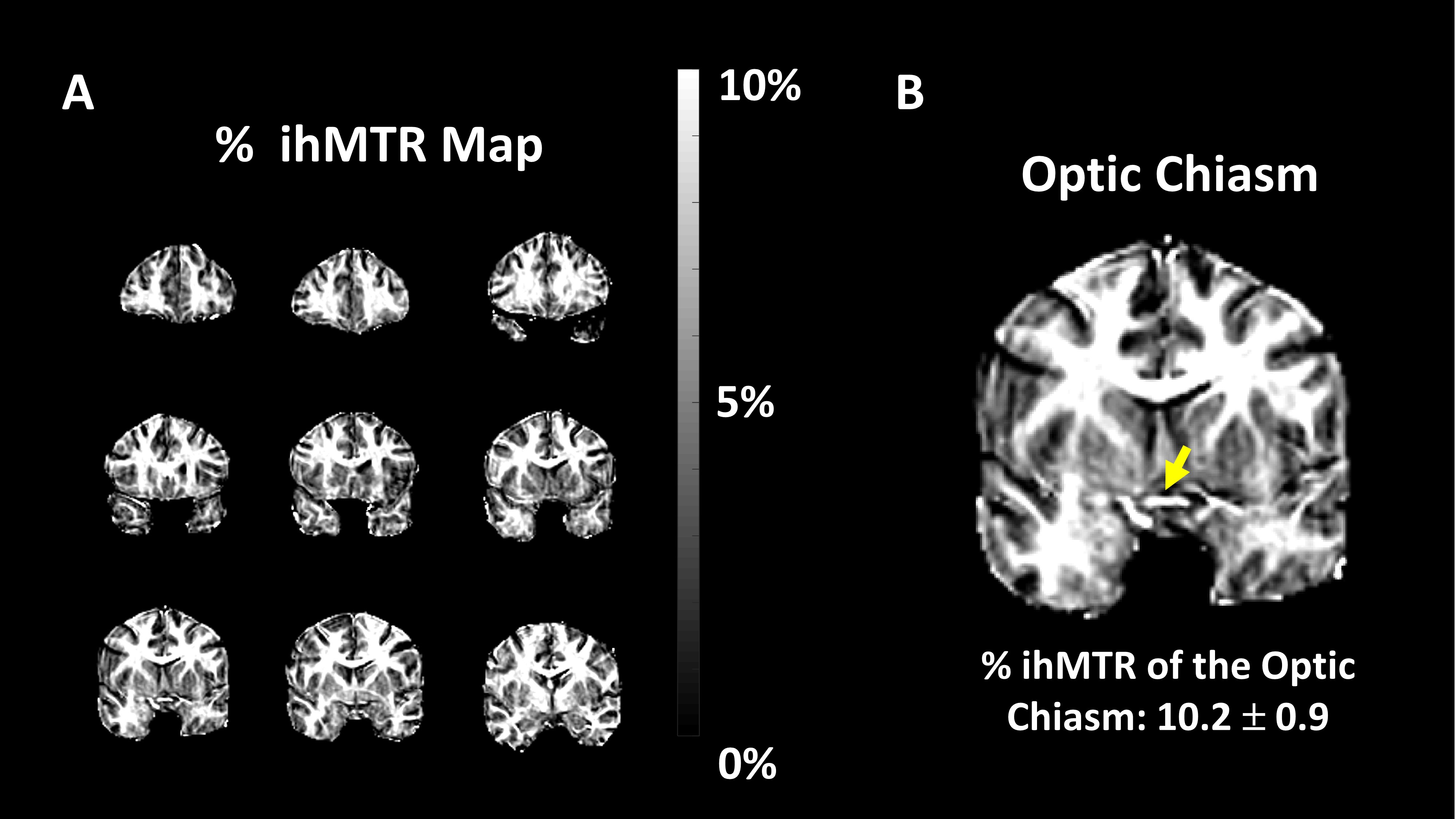
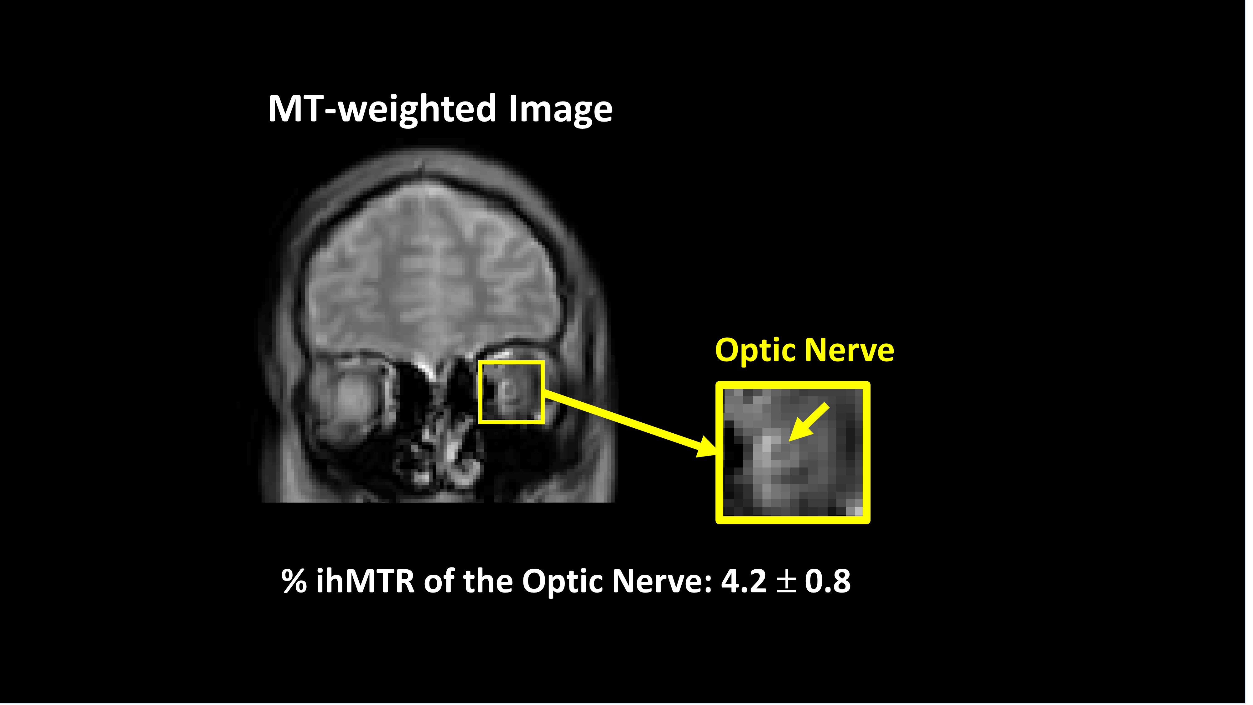
Figure1 shows the % ihMTR maps from Volunteer #1 obtained using different B1peak amplitudes. The ihMTR values increased with an increasing B1peak amplitude before reaching a plateau (and slight decrease) and this dependence was observed in all the volunteers. The observation is consistent with the concentrated energy deposition scheme behavior shown previously in the human brain11-12. Figure2 demonstrates the % ihMTR map obtained from the brain of the Volunteer #2 using the optimized protocol with higher in-plane resolution (1.5 x 1.5 mm2). Mean % ihMTR values obtained with the optimized protocol from the optic chiasm of three volunteers were 8.7 ± 2.9 %. The specific % ihMTR values ranged from 5% to 10%. The variation might be due to the partial volume effect and possible mismatch between the ROIs of different subjects. Figure3 shows the MT-weighted image at +7kHz off-resonance, as well as the observable mean % ihMTR from the optic nerve of the Volunteer #3.Conclusion
This study shows the feasibility of using the ihMT method to obtain myelin-sensitive semi-quantitative information from the human optic nerve and chiasm in vivo. The concentrated energy deposition scheme for ihMT preparation applied together with adaptive RF shimming, higher resolution, and visual fixation led to an observable ihMT effect from both optic nerve and chiasm. This new method could be useful for evaluating demyelinating changes due to optic neuritis, as well as optic nerve pathologies. Work-in-progress focuses on using smaller slice thickness to decrease the partial volume effect as well as higher in-plane resolution for better characterization of the optic nerve and other nerves.Acknowledgements
The authors would like to thank Kelli Key and Trevor Wigal for helping with subject recruitment and MRI scans.References
1. Kolappan M, Henderson AP, Jenkins TM, Wheeler-Kingshott CA, Plant GT, Thompson AJ, Miller DH. Assessing structure and function of the afferent visual pathway in multiple sclerosis and associated optic neuritis. J Neurol 2009;256(3):305-19.
2. Hickman SJ, Toosy AT, Jones SJ, Altmann DR, Miszkiel KA, MacManus DG, Barker GJ, Plant GT, Thompson AJ, Miller DH. Serial magnetization transfer imaging in acute optic neuritis. Brain 2004;127(Pt 3):692-700.
3. Inglese M, Ghezzi A, Bianchi S, Gerevini S, Sormani MP, Martinelli V, Comi G, Filippi M. Irreversible disability and tissue loss in multiple sclerosis: a conventional and magnetization transfer magnetic resonance imaging study of the optic nerves. Arch Neurol 2002;59(2):250-5.
4. Trip SA, Schlottmann PG, Jones SJ, Li WY, Garway-Heath DF, Thompson AJ, Plant GT, Miller DH. Optic nerve magnetization transfer imaging and measures of axonal loss and demyelination in optic neuritis. Mult Scler 2007;13(7):875-9.
5. Filippi M, Rocca MA. Magnetization transfer magnetic resonance imaging of the brain, spinal cord, and optic nerve. Neurotherapeutics 2007;4(3):401-13.
6. Varma G, Duhamel G, de Bazelaire C, Alsop DC. Magnetization transfer from inhomogeneously broadened lines: A potential marker for myelin. Magn Reson Med 2015;73(2):614-22.
7. Varma G, Girard OM, Prevost VH, Grant AK, Duhamel G, Alsop DC. Interpretation of magnetization transfer from inhomogeneously broadened lines (ihMT) in tissues as a dipolar order effect within motion restricted molecules. J Magn Reson 2015;260:67-76.
8. Girard OM, Prevost VH, Varma G, Cozzone PJ, Alsop DC, Duhamel G. Magnetization transfer from inhomogeneously broadened lines (ihMT): Experimental optimization of saturation parameters for human brain imaging at 1.5 Tesla. Magn Reson Med 2015;73(6):2111-21.
9. Swanson SD, Malyarenko DI, Fabiilli ML, Welsh RC, Nielsen JF, Srinivasan A. Molecular, dynamic, and structural origin of inhomogeneous magnetization transfer in lipid membranes. Magn Reson Med 2017;77(3):1318-1328.
10. Barker GJ. Technical issues for the study of the optic nerve with MRI. J Neurol Sci 2000;172 Suppl 1:S13-6.
11. Mchinda S, Varma G, Prevost VH, et al. Whole brain inhomogeneous magnetization transfer (ihMT) imaging: Sensitivity enhancement within a steady-state gradient echo sequence. Magn Reson Med 2018;79(5):2607-2619.
12. Mchinda S, et al. Whole brain inhomogeneous Magnetization Transfer (ihMT) imaging at 3T: concentrating RF power to mitigate RF inhomogeneities effects. In: Proceedings of the ISMRM. Paris, France; 2018: p786.
Figures