4896
Silent Myelin Imaging with a dipolar-coupled/inhomogeneous MT-Prepared ZTE Radial Sequence1Neuroimaging, King's College London, London, United Kingdom, 2ASL Europe, GE Healthcare, Munich, Germany
Synopsis
We generated myelin-specific contrast in a silent radial ZTE sequence using a dipolar-coupled MT-prep module. This sequence has great potential for visualising myelin in patient cohorts that do not tolerate the noise from standard MRI, such as infants.
Introduction
Myelin is both a crucial part of a healthy nervous system and fortuitously the major contributor to MR contrast between great and white matter. It is a complex, layered, semi-solid structure that exhibits many interesting physical phenomena including Magnetization Transfer (MT).
The MT-weighting of myelin can be enhanced by irradiating with dual-frequency RF pulses compared to the traditional single-frequency saturation pulse1. This dipolar-coupled MT (dcMT, previously referred to as inhomogeneous or ihMT in the literature) effect appears to be specific to myelin within the brain2.
Recently Mchinda et al showed that the dcMT effect can be increased by significantly increased by using a preparation module and segmented gradient-echo readout3. The Radial Ultra-Fast Imaging Sequence (RUFIS)4 is a variety of highly efficient gradient-echo sequence that is essentially silent, avoiding the high acoustic noise associated with fast cartesian readouts. We combined a dcMT preparation module with RUFIS for greater patient comfort, and then optimised the acquisition parameters to maximise the dcMT effect and image quality.
Methods
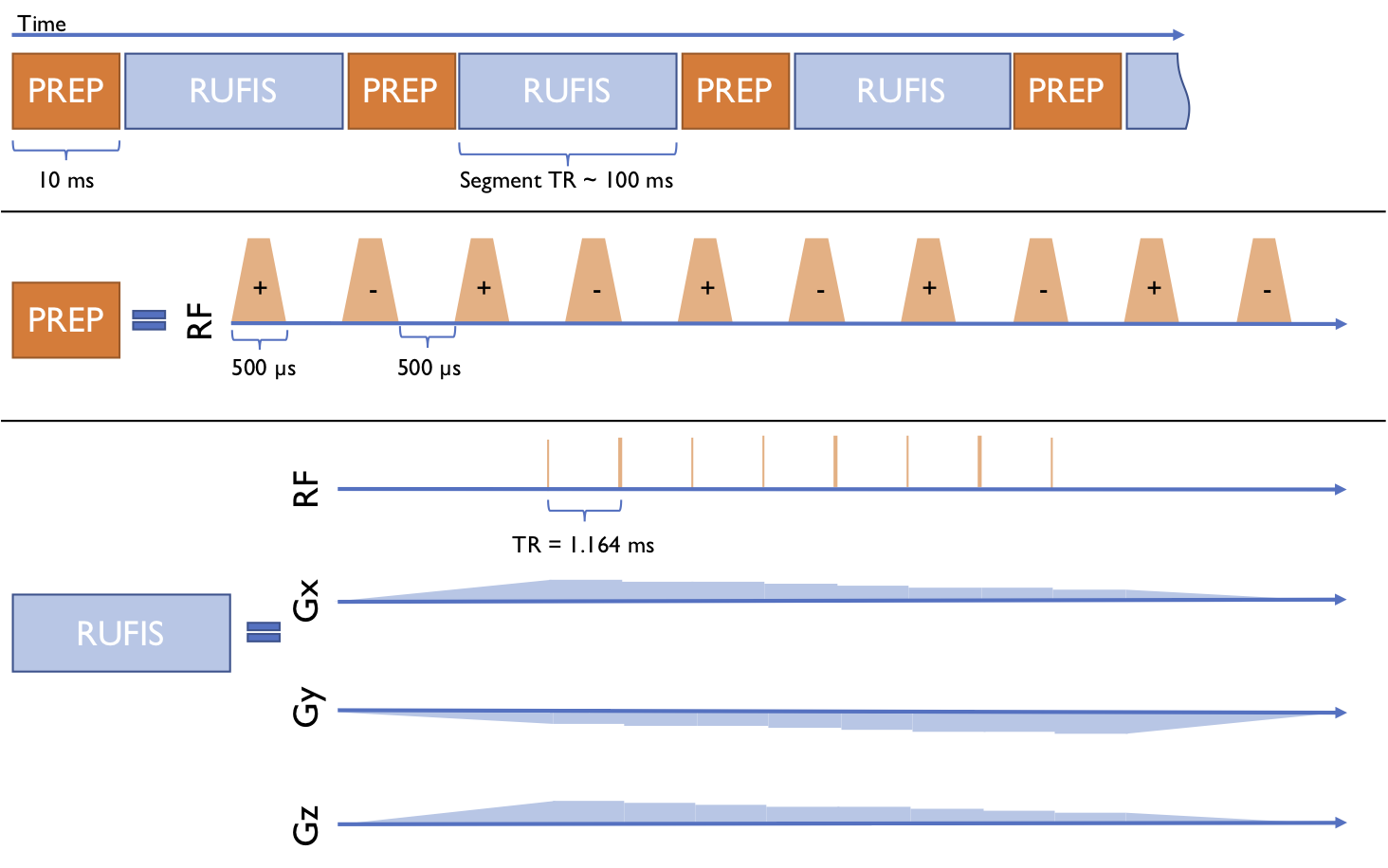
An MT-prep module was implemented in a segmented RUFIS sequence, shown in figure 1, and used to scan a healthy male volunteer with a GE MR750 3T scanner and 12-channel head coil. Images were acquired at 2mm isotropic resolution, 220mm FoV, and receive bandwidth 31.25kHz, corresponding to a TR of 1.164ms.
The MT-prep had 10 Tukey pulses, width 0.5 ms, separation 0.5 ms, with an RMS B1 of 8.5 μT. Dual-frequency irradiation was achieved by alternating the sign of the frequency offset for each pulse. Images were acquired in sets: one with no saturation power (REF), one with +7 kHz saturation (P), one with -7kHz (M), and two with dual-frequency saturation (PM & MP). Each set was motion corrected using mcflirt5 and smoothed with a Gaussian FWHM 1mm. The MT Ratio (MTR) was calculated as $$$1 - (M+P)/(2*REF)$$$, the enhanced MTR as $$$1 - (MP + PM)/(2*REF)$$$ and the dcMTR was calculated as $$$eMTR - MTR$$$.
Sets of images were acquired with varying flip-angles and Spokes Per Segment (SPS) to investigate their effect on dcMTR. As TR is fixed, SPS determines the length of time between prep modules (Segment TR), which determines the overall saturation duty cycle. The flip-angle determines the available SNR and T1-weighting of the sequence, which can compete with the MT-weighting. The flip-angle was 1,2 or 3° and SPS was 32, 64 or 128, for 9 sets in total. These values of SPS gave Segment TRs of 62.4, 99.6 & 174.1 ms corresponding to duty-cycles of 7%, 4.5% & 2.7%.
SPS also determines the acquisition time due by changing the total number of prep modules. The time for each set was 2m23s, 1m59s and 1m56s for 32, 64 & 128 SPS respectively. The acoustic noise for RUFIS and a comparable gradient echo sequence was measured with an MR-compatible microphone.
Results and Discussion
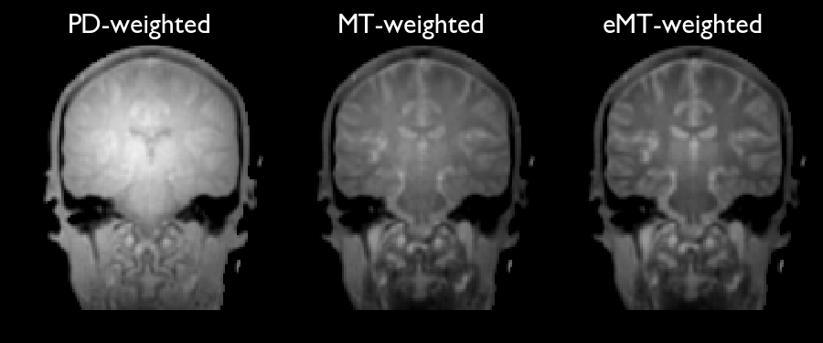
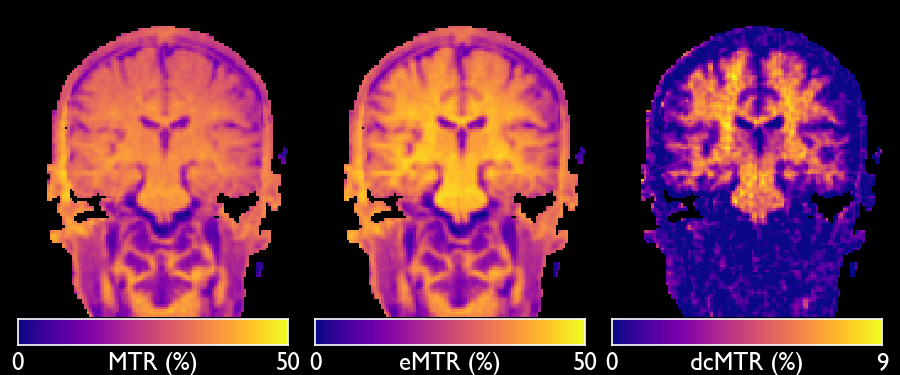
Figure 2 shows examples of the PD-, MT-, and eMT-weighted images for FA=2° and SPS=32. Figure 3 then shows the MTR, eMTR and dcMTR. It is clear that the dual-frequency irradiation enhances the MT-weighting and that this effect is specific to white matter.
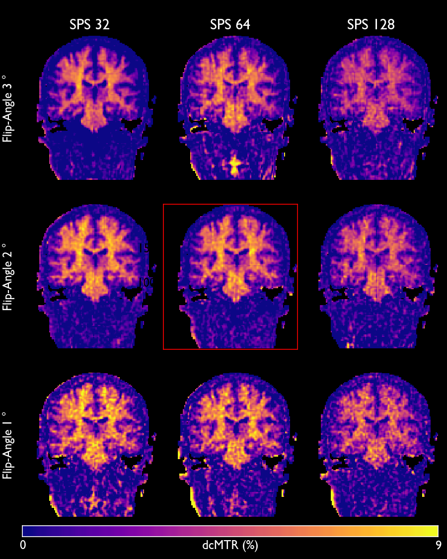
Figure 4 shows the dcMTR calculated for each image set. The dcMTR was lowest for SPS=128 FA=3°, and highest at SPS=32 FA=1° which corresponds to the highest duty-cycle and lowest T1-weighting, with values of between 8.5% and 9% in ascending white matter tracts. dcMTR at SPS=64 was around 1% less, for a scan-time reduction of 17%. In addition, image quality was noticeably improved with FA=2° due to increased SNR. Hence we found that SPS=64, FA=2° was the optimal combination of parameters tested.
The values of dcMTR obtained with the RUFIS sequence are lower than those reported by Mchinda et al3 despite a comparable saturation scheme. The most likely explanation for this is T1-recovery during the segment. Because each RUFIS spoke starts at the center of k-space, every spoke in a segment will contribute to the image contrast and a "centric" k-space ordering to capture the maximum saturation directly after the prep module is not possible.
The measured highest SAR observed was approximately 2.5 W/kg during saturation volumes, which is well below the manufacturer's limit for the head of 3.2 W/kg over 10 minutes. The acoustic noise was measured as 72 dB for the RUFIS sequence, compared to 69 dB background level and 109 dB for a cartesian sequence.
Conclusion
We have demonstrated how to generate myelin-specific contrast from dipolar-coupled MT in a silent ZTE radial sequence. This has potential to allow easy tracking of the myelination process in sleeping infants, or to track demyelination in patient populations who do not normally tolerate MRI.Acknowledgements
This study represents independent research part funded by the NIHR-Wellcome Trust King's Clinical Research Facility and the National Institute for Health Research (NIHR) Biomedical Research Centre at South London and Maudsley NHS Foundation Trust and King’s College London. The views expressed are those of the author(s) and not necessarily those of the NHS, the NIHR or the Department of Health and Social Care. Funding was also received from General Electric Healthcare.References
1. Gopal Varma, Guillaume Duhamel, Cedricde Bazelaire, and David C. Alsop. Magnetization transfer from inhomogeneously broadened lines: A potential marker for myelin. Magnetic Resonance in Medicine, 73(2), February 2014.
2. Alan P Manning, Kimberley L Chang, Alex L MacKay, and Carl A Michal. The physical mechanism of “inhomogeneous” magnetization transfer MRI. Journal of Magnetic Resonance, 274(Supplement C):125–136, January 2017.
3. Samira Mchinda, Gopal Varma, Valentin H. Prevost, Arnaud Le Troter, Stanislas Rapacchi, Maxime Guye, Jean Pelletier, Jean-Philippe Ranjeva, David C. Alsop, Guillaume Duhamel, and Olivier M. Girard. Whole brain inhomogeneous magnetization transfer (ihMT) imaging: Sensitivity enhancement within a steady-state gradient echo sequence. Magnetic Resonance in Medicine, September 2017.
4. David P Madio and Irving J Lowe. Ultra-fast imaging using low flip angles and fids. Magnetic Resonance in Medicine, 34(4):525–529, 1995.
5. Mark Jenkinson, Peter Bannister, Michael Brady and Stephen Smith. Improved Optimization for the Robust and Accurate Linear Registration and Motion Correction of Brain Images. NeuroImage, 17(2):825-841, 2002
Figures