4894
Inversion Recovery Pointwise Encoding Time Reduction with Radial Acquisition (IR-PETRA) for Direct Myelin Imaging in Human Brain1Department of Radiology, University of California San Diego, San Diego, CA, United States, 2GE Healthcare, San Diego, CA, United States, 3Radiology Service, VA San Diego Healthcare System, San Diego, CA, United States
Synopsis
Due to very low proton density and rapid signal decay (T2*<300µs at 3T), it is challenging to directly image myelin in the white matter of the brain using MRI. The literature demonstrates that direct myelin imaging is feasible using inversion recovery (IR) preparation followed by dual echo ultrashort echo time (UTE) MRI, allowing direct capture of the rapidly-decaying myelin signal with greatly improved dynamic range. In this study, we show the efficacy of IR prepared Pointwise Encoding Time Reduction with Radial Acquisition (IR-PETRA) for direct myelin imaging in the human brain.
Introduction
Due to very low proton density and rapid signal decay (T2*<300µs at 3T), it is challenging to directly image myelin in white matter of the brain with MRI. Recent studies have shown that direct myelin imaging is feasible using inversion recovery (IR) preparation followed by dual echo ultrashort echo time (UTE) MRI, allowing direct capture of the rapidly-decaying myelin signal with greatly improved dynamic range1–4. In this study, we explore the efficacy of IR prepared Pointwise Encoding Time Reduction with Radial Acquisition (IR-PETRA) for direct myelin imaging in the human brain.Methods
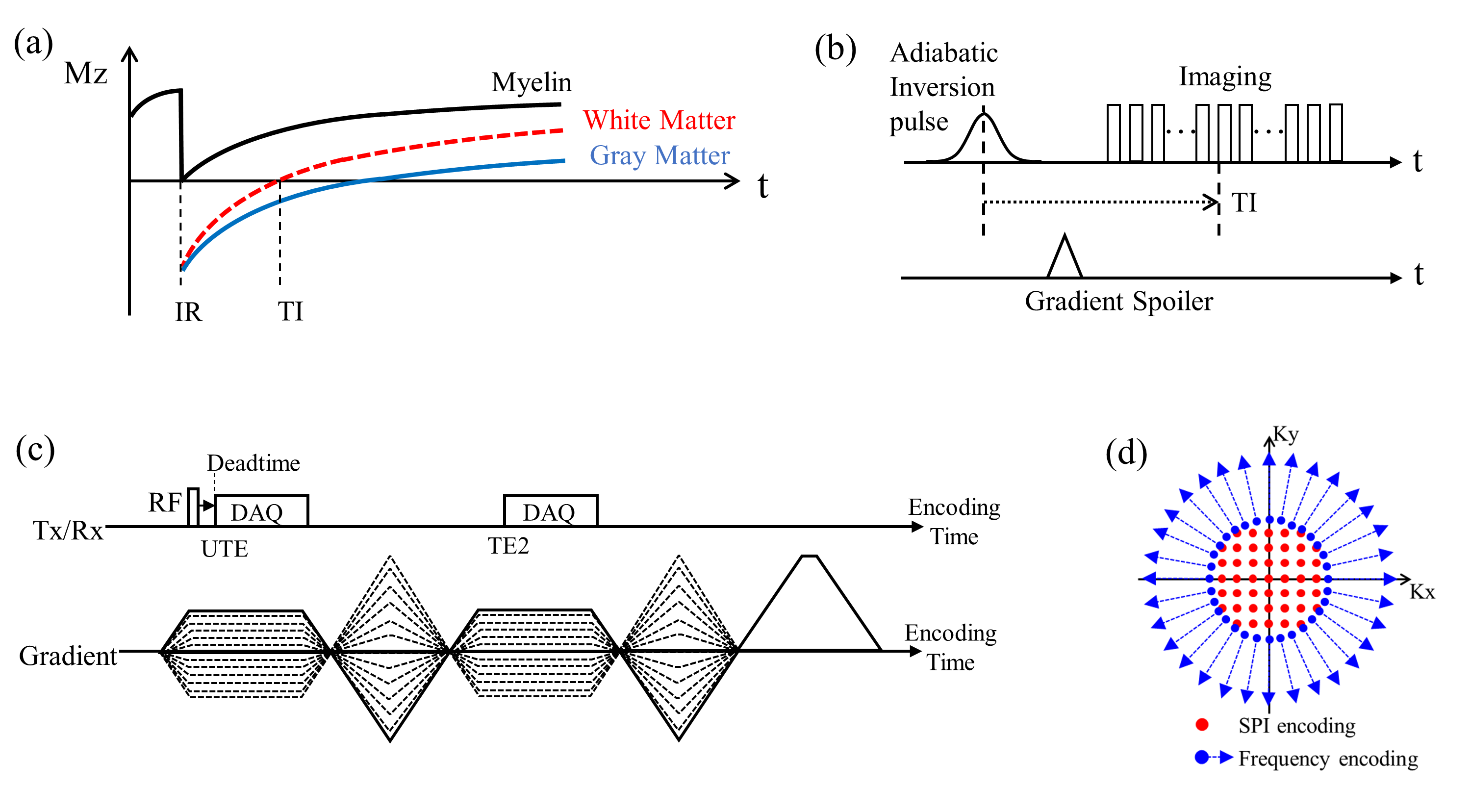
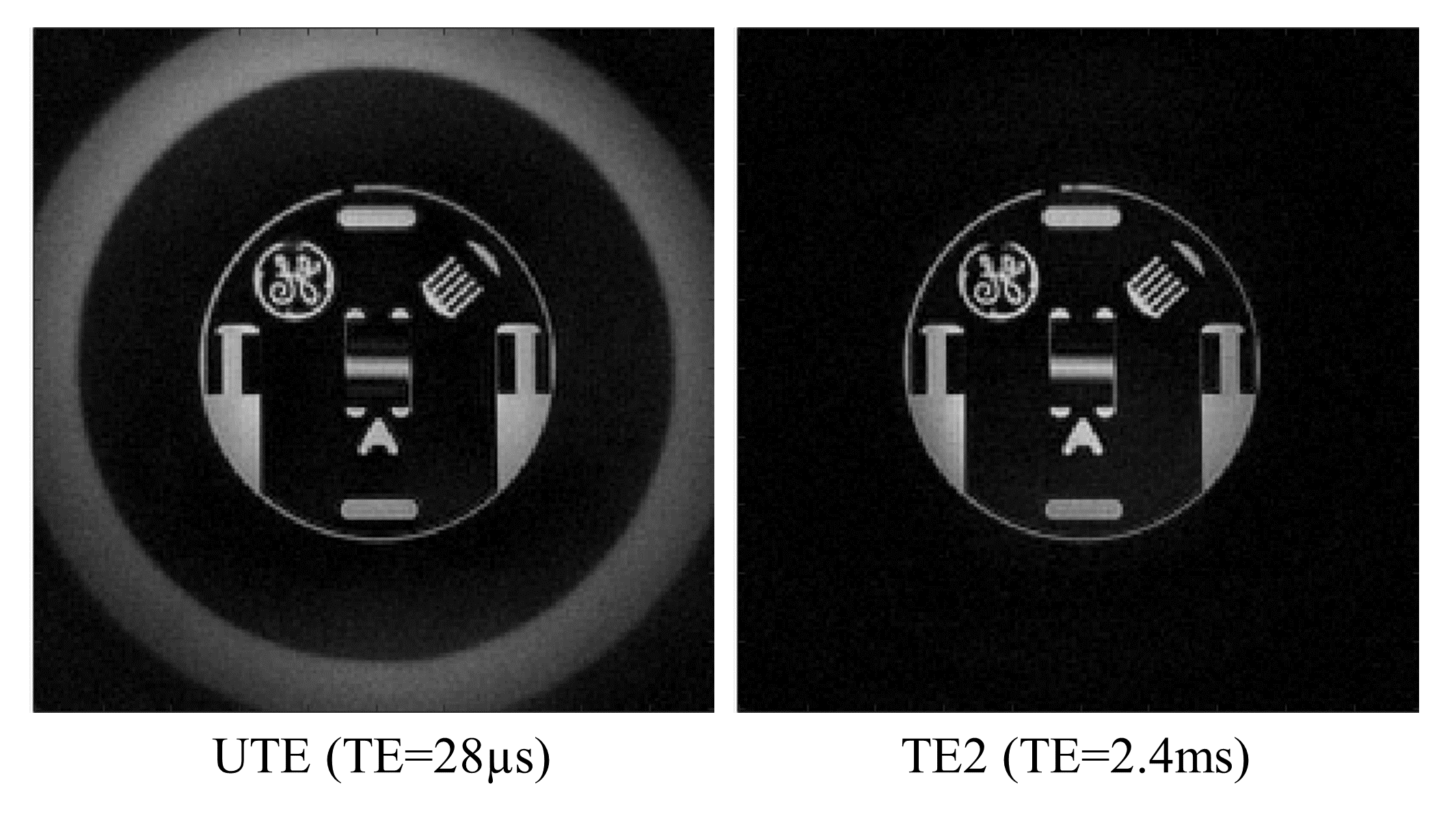
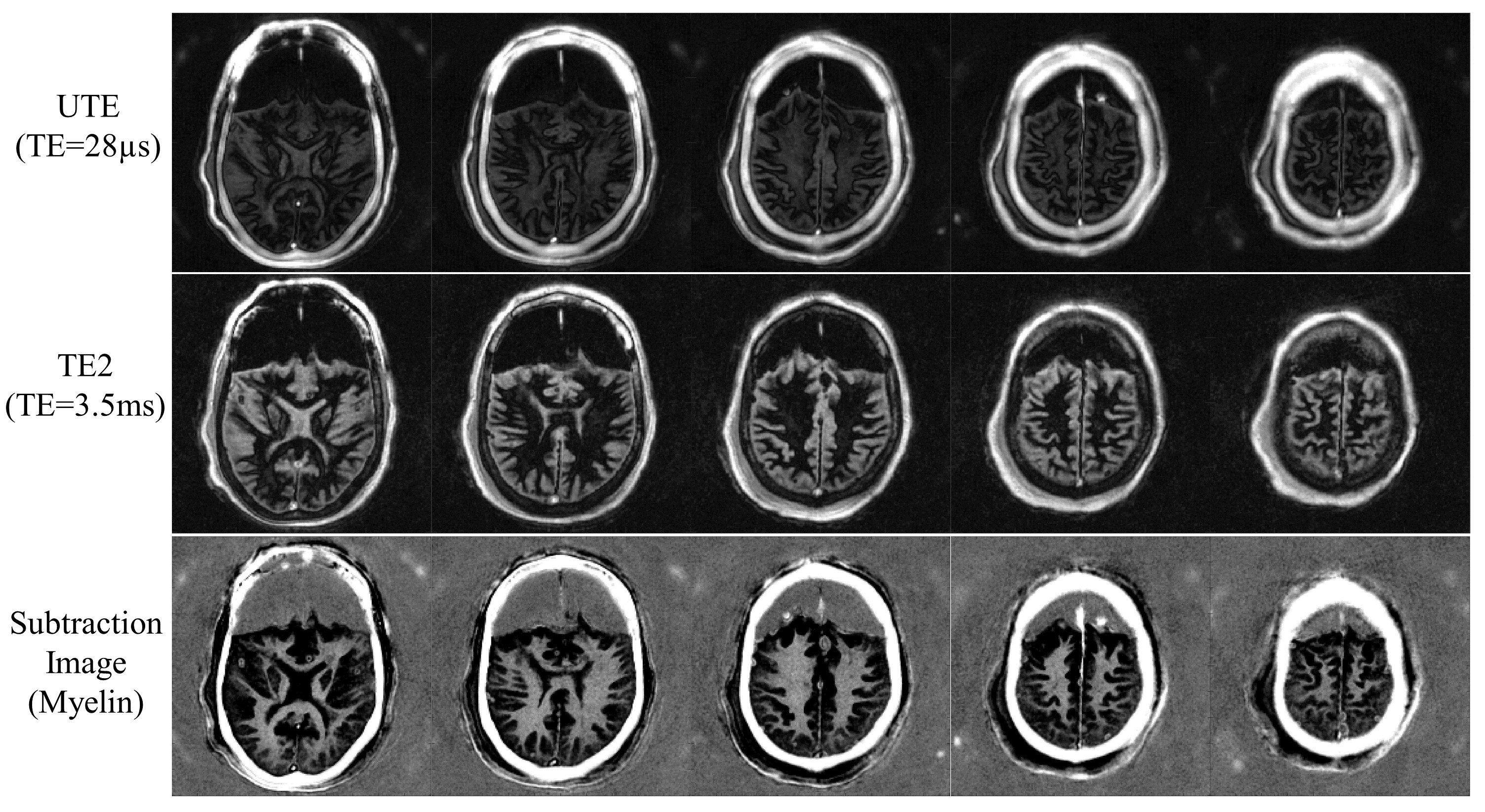
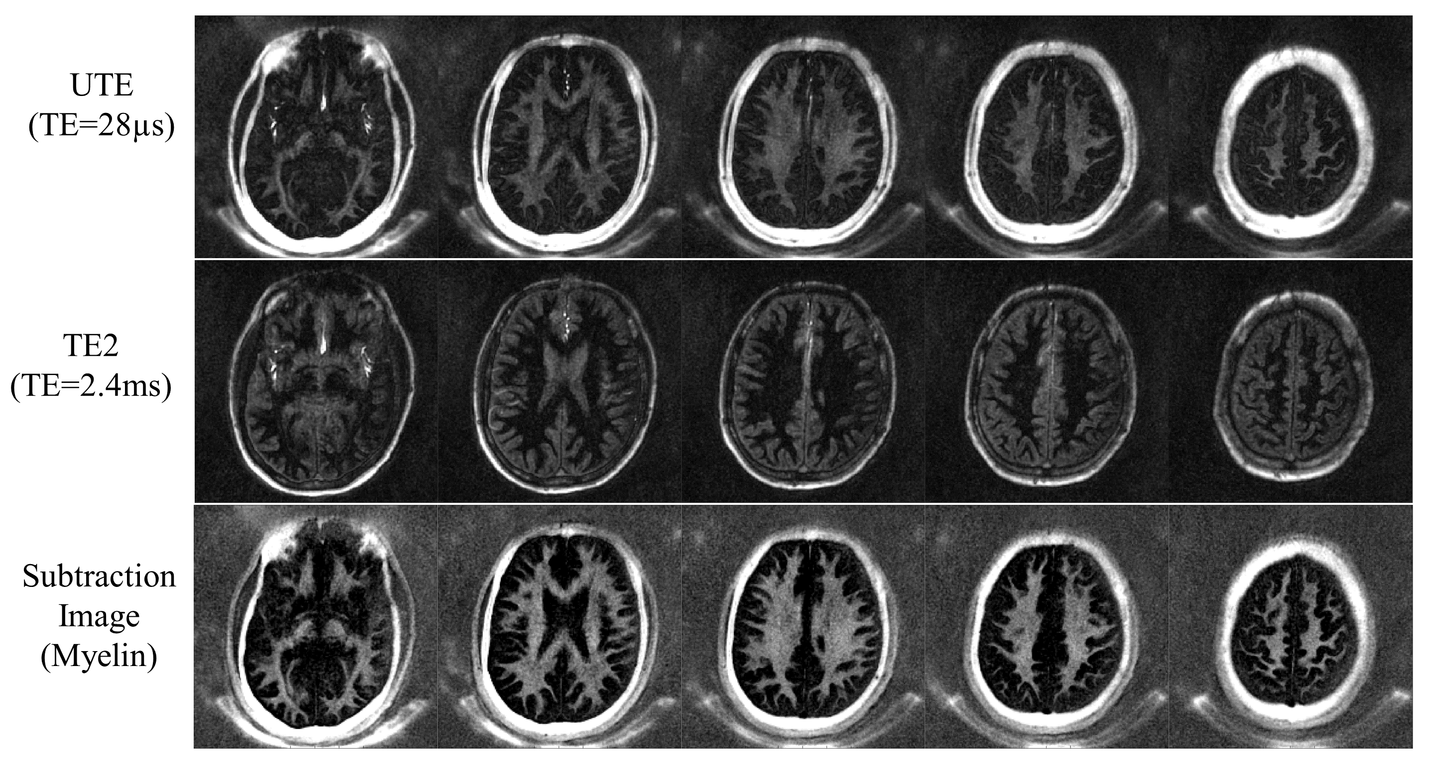
To directly image myelin in MRI, the following strategies are applied: First, an adiabatic IR pulse preparation is applied to provide robust suppression of signals from the long T2 components in white matter, which are mainly water. Second, dual echo imaging is performed to suppress the remaining long T2 gray matter signal by subtracting the second echo from the first one. Since the primary short T2 tissue in white matter is myelin, the remaining signal reflects myelin content. Figure 1-a shows typical inversion recovery curves for tissues in the human brain (i.e., myelin, white matter, and gray matter). After the adiabatic inversion preparation, UTE imaging is performed, as shown in Figure 1-b. Note that multiple spokes are acquired after each IR preparation to reduce the scan time, centered on the optimal inversion time (TI) adjusted to invert and null the longitudinal magnetizations of long T2 white matter. For the imaging, dual echo PETRA5 is performed as illustrated in Figure 1-c, where single point imaging (SPI) encoding6 is utilized to acquire the missing central k-space data due to RF coil deadtime during UTE imaging as shown in Figure 1-d. Note that the second echo (TE2) is also acquired using SPI—despite the fact that there is no RF deadtime and therefore no missing k-space data—in order to keep the sampling pattern consistent between the UTE and TE2, therefore achieving a more reliable subtraction image. To evaluate the feasibility and efficacy of IR-PETRA for direct myelin imaging, we performed a phantom experiment with a GE resolution phantom, performed ex vivo imaging with a cadaveric brain (56-year old female donor), and performed in vivo imaging with three healthy volunteers (36-, 30-, and, 36-year old males). The experiments were performed on a 3T GE-MR750 scanner using a 12-ch receive-only HNU coil. The imaging parameters for the phantom experiment are as follows: a hard pulse with flip angle (FA)=4° (pulse width=20µs), readout BW=±31.25kHz, matrix size=200x200x40, FOV=220x220x160mm3, inter-spoke TR=8ms, TE=28µs/2.4ms, # of SPI encoding=544, # of radial frequency encoding=16944, and scan time=2min 10sec. The ex vivo experiment was performed with the parameters matched above, except for the following parameters: adiabatic inversion pulse applied (Silber Hoult pulse, pulse width=8.64ms), TR=1000ms, TI=310ms, TE=28µs/3.5ms, # of radial frequency encoding=33856, # of spokes per IR preparation=16, inter-spoke TR=10ms, and scan time=35min 54sec. In vivo experiment was performed using the same parameters as in the ex vivo experiment except for TI=325ms, TE=28µs/2.4ms, # of radial frequency encoding=16944, # of spokes per IR=30, inter-spoke TR=8ms, and scan time=11min 18sec. All images were reconstructed using online reconstruction based on GE Orchestra-SDK (v1.7.1).Results
Figure 2 shows the dual echo PETRA images obtained with a GE resolution phantom. As demonstrated, there are no imaging artifacts manifested in either the UTE or TE2 images with the given imaging parameters, which assures the quality of the dual echo PETRA images without IR. Figure 3 shows selected slices from IR-PETRA imaging of the ex vivo cadaveric brain. As seen in the UTE image, myelin signal is selectively detected with long T2 white matter signal suppressed near-completely, verified by the fact that no signal remains in the white matter region in the second echo (myelin signal already decayed to near-zero). By subtracting the second echo from UTE, a highly specific myelin image is obtained, as shown in Figure 3. Figure 4 shows images from the in vivo experiment, which shows the myelin image with high contrast.Discussion and Conclusion
In this study, we demonstrated the feasibility and efficacy of IR prepared dual echo PETRA for direct myelin imaging. We showed high contrast myelin contrast with the ex vivo and in vivo protocols, which suggests potential for IR-PETRA to image myelin for in vivo applications such as multiple sclerosis (MS) and traumatic brain injury (TBI). In future work, we will compare IR-PETRA with other possible approaches such as radial UTE, cones UTE, zero echo time (ZTE) imaging, and ramped hybrid encoding (RHE)7 in myelin imaging in MS and TBI.Acknowledgements
The authors acknowledge research support from GE Healthcare, NIH (R01NS092650), and VA Clinical Science and Rehabilitation R&D Awards (I01CX001388 and I01RX002604).References
1. Du J, Ma G, Li S, et al. Ultrashort echo time (UTE) magnetic resonance imaging of the short T2 components in white matter of the brain using a clinical 3T scanner. Neuroimage 2014;87:32–41 doi: 10.1016/j.neuroimage.2013.10.053.
2. Sheth V, Shao H, Chen J, et al. Magnetic resonance imaging of myelin using ultrashort Echo time (UTE) pulse sequences: Phantom, specimen, volunteer and multiple sclerosis patient studies. Neuroimage 2016;136:37–44 doi: 10.1016/j.neuroimage.2016.05.012.
3. Sheth VR, Fan S, He Q, et al. Inversion recovery ultrashort echo time magnetic resonance imaging: A method for simultaneous direct detection of myelin and high signal demonstration of iron deposition in the brain – A feasibility study. Magn. Reson. Imaging 2016;38:87–94 doi: 10.1016/j.mri.2016.12.025.
4. Waldman a, Rees JH, Brock CS, Robson MD, Gatehouse PD, Bydder GM. MRI of the brain with ultra-short echo-time pulse sequences. Neuroradiology 2003;45:887–92 doi: 10.1007/s00234-003-1076-z.
5. Grodzki DM, Jakob PM, Heismann B. Ultrashort echo time imaging using pointwise encoding time reduction with radial acquisition (PETRA). Magn. Reson. Med. 2012;67:510–8 doi: 10.1002/mrm.23017.
6. Emid S, Creyghton JHN. High resolution NMR imaging in solids. Phys. B+C 1985;128:81–83 doi: 10.1016/0378-4363(85)90087-7.
7. Jang H, Wiens CN, McMillan AB. Ramped hybrid encoding for improved ultrashort echo time imaging. Magn. Reson. Med. 2016;76:814–825 doi: 10.1002/mrm.25977.
Figures