4862
Improving the Quality of Synthetic FLAIR Images with Deep Learning Using a Conditional Generative Adversarial Network for Pixel-by-Pixel Image Translation1Radiology, The University of Tokyo Hospital, Tokyo, Japan, 2Radiology, Juntendo University Hospital, Tokyo, Japan, 3Radiology, National Institute of Radiological Sciences, Chiba, Japan, 4Neurology, Juntendo University Hospital, Tokyo, Japan, 5Radiology, Hopital Saint-Joseph, Paris, France
Synopsis
Synthetic FLAIR images are of lower quality than conventional FLAIR images. Here, we aimed to improve the synthetic FLAIR image quality using deep learning with pixel-by-pixel translation through conditional generative adversarial network training. Forty patients with MS were prospectively included and scanned to acquire synthetic MRI and conventional FLAIR images. Acquired data were divided into 30 training and 10 test datasets. Using deep learning, we improved the synthetic FLAIR image quality by generating FLAIR images that have contrast that is closer to that of conventional FLAIR images and fewer granular and swelling artifacts, while preserving the lesion contrast.
Introduction
Synthetic MRI technique can be used to create any contrast-weighted image, based on R1 and R2 relaxation rates and proton density.1 However, synthetic FLAIR image quality is generally inferior to that of conventional FLAIR images.2,3 Further, synthetic FLAIR images reportedly show more artifacts than other contrasts,3 including hyperintensity on the brain-CSF interface with apparently swollen brain parenchyma,4,5 and granular hyperintensities in the CSF, neither of which have been reported for conventional FLAIR.3
Even though deep learning (DL) is a potential candidate to improve synthetic FLAIR, it often generates artificial objects in empty space or eliminates real objects.6 To solve these issues, herein, we propose using a pixel-by-pixel neural network function, which ensures that the two identical input pixel values become the same output pixel value, as a generator to improve the synthetic FLAIR image quality.
Methods
Participants and MRI
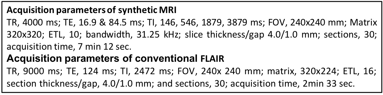
40 patients with multiple sclerosis (MS) were included in this study, and scanned by 2D axial synthetic MRI7 and conventional FLAIR on a 3T MR scanner (Discovery 750w, GE). Scan parameters are shown in Table 1. The first consecutive 30 subjects were used as a training set and the last 10 subjects were used as a test set. The SyMRI software version 8.0 (SyntheticMR, Sweden) was used to create synthetic FLAIR with post-processing TR, 15000 ms; TE, 100 ms; TI, 3000 ms.
DL Framework
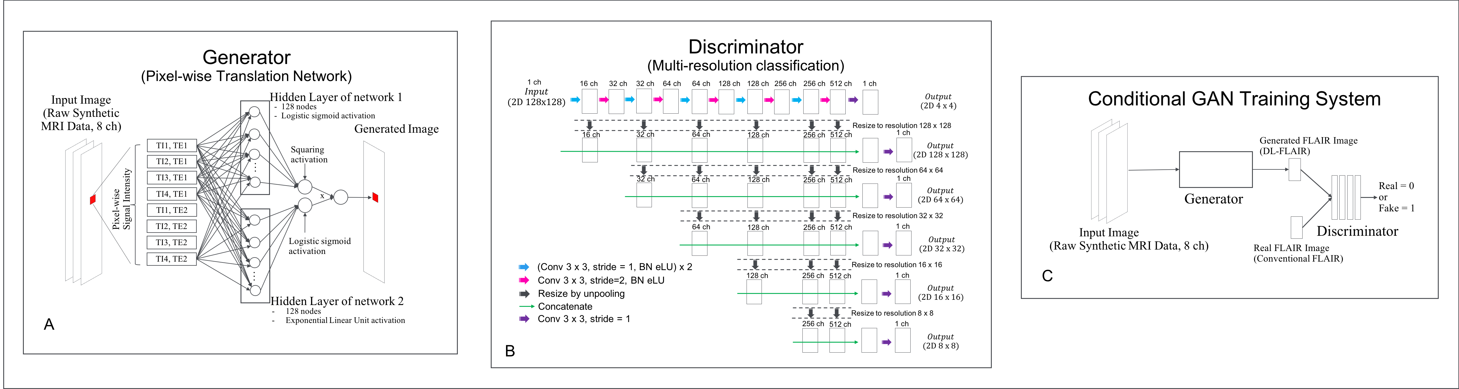
We designed a pixel-wise translation network that receives raw data (8 complex images per slice) of synthetic MRI and outputs FLAIR by translating input signal intensities into FLAIR using the same function across all pixels. Because conventional FLAIR images, which were used as the target, were acquired separately from the input images, we trained the generator model with a conditional generative adversarial network (cGAN)-type learning system with patches to avoid any adverse misregistration. GAN8 uses an image generator and a discriminator, which are simultaneously trained to transcend each other. A cGAN is a newly proposed technique that conditions the output on the input to render the same underlying information between them, ultimately keeping the generated image realistic.6
Fig 1A–C show the architectures of the generator and the discriminator, and the conceptual description of the cGAN training system, respectively. The program was coded using Python 3.6 with Chainer 3.2.0.
Evaluation of the Model
Normalized root mean squared error (NRMSE) was calculated for synthetic and DL-FLAIR images against conventional FLAIR images for the test set. The dice index of lesion maps acquired using Lesion Segmentation Toolbox 2.0.15 was also calculated between synthetic or DL-FLAIR images and conventional FLAIR images.
Synthetic and DL-FLAIR images of the test set were randomly shown to an neuroradiologist. The neuroradiologist evaluated lesion conspicuity and the existence of surface hyperintensity and granular artifacts on 5-point scales. Further, synthetic and DL-FLAIR images for each patient were simultaneously shown to the neuroradiologist with overlay function for evaluation of parenchymal swelling artifact.
We used the nonparametric Wilcoxon signed-rank test to compare the quantitative and qualitative scores between synthetic and DL-FLAIR images. Significance was set at P < 0.05.
Results
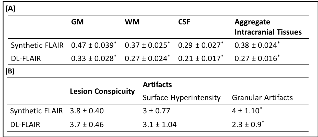
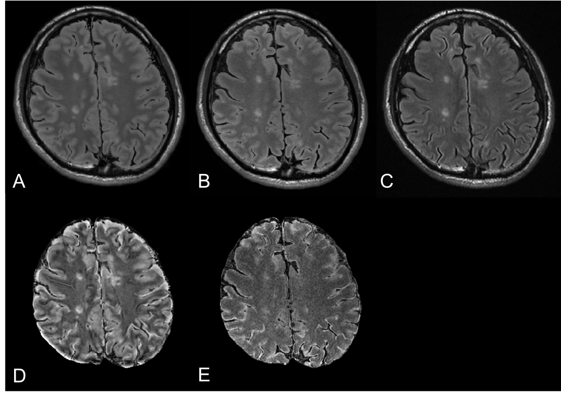
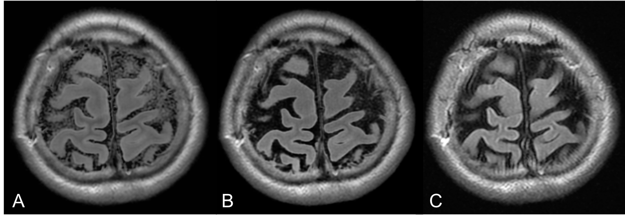
Fig 1 shows images from a representative patient. The NRMSE was significantly lower for DL-FLAIR than synthetic FLAIR images in GM, WM, CSF, and aggregate intracranial tissues (all P<0.001, Table 1). The dice index of lesion maps was comparable between DL-FLAIR (mean±SD, 0.49±0.11) and synthetic FLAIR images (0.47±0.12)(P=0.34). No significant differences in lesion conspicuity or existence of surface hyperintensity artifacts were identified (P=0.59 and 0.78, respectively. Table 2). However, fewer granular artifacts were present in the CSF of DL-FLAIR than synthetic FLAIR images (P=0.003, Fig 2). The neuroradiologist agreed that the brain parenchyma looked grossly swollen on all synthetic FLAIR images compared with the DL-FLAIR images (Fig 3).Discussion
We proposed a DL model to improve the image quality of synthetic FLAIR. The NRMSE were lower in all tissue segments in DL-FLAIR than in synthetic FLAIR images, meaning that DL-FLAIR images more accurately replicated conventional true FLAIR images. Although synthetic FLAIR images rely on the well-established Bloch equation,7 this approach may not be the best for replicating true FLAIR images.
While some perceived surface hyperintensity remained, brain parenchymal swelling artifacts were improved in all patients. The current synthetic MRI technique adopted a monoexponential decay model assuming a homogeneous voxel,7 and may not appropriately produce the FLAIR signal in a voxel with more than one tissue compartment.
Conclusion
We improved the synthetic FLAIR image quality using DL, by creating FLAIR images that have contrast similar to that of conventional FLAIR images, with fewer swelling artifacts and minimal granular artifacts, while preserving lesion contrast.Acknowledgements
No acknowledgement found.References
1. Hagiwara A, Warntjes M, Hori M, et al. SyMRI of the Brain: Rapid Quantification of Relaxation Rates and Proton Density, With Synthetic MRI, Automatic Brain Segmentation, and Myelin Measurement. Invest Radiol 2017;52:647-57
2. Blystad I, Warntjes JB, Smedby O, et al. Synthetic MRI of the brain in a clinical setting. Acta Radiol 2012;53:1158-63
3. Tanenbaum LN, Tsiouris AJ, Johnson AN, et al. Synthetic MRI for Clinical Neuroimaging: Results of the Magnetic Resonance Image Compilation (MAGiC) Prospective, Multicenter, Multireader Trial. AJNR Am J Neuroradiol 2017;38:1103-10
4. Hagiwara A, Hori M, Yokoyama K, et al. Synthetic MRI in the Detection of Multiple Sclerosis Plaques. AJNR Am J Neuroradiol 2017;38:257-63
5. Helmersson T. Evaluation of Synthetic MRI for Clinical Use. Master's Thesis. Department of Medical and Health Sciences: Linköping University; 2010 http://liu.diva-portal.org/smash/record.jsf?pid=diva2%3A389732&dswid=9594 Accessed August 30, 2018:71
6. Isola P, Zhu J, Zhou T, et al. Image-to-Image Translation with Conditional Adversarial Networks. arXiv:161107004 2017
7. Warntjes JB, Leinhard OD, West J, et al. Rapid magnetic resonance quantification on the brain: Optimization for clinical usage. Magn Reson Med 2008;60:320-9
8. Goodfellow I, Pouget-Abadie J, Mirza M, et al. Generative Adversarial Nets. Advances in Neural Information Processing Systems; 2014:2672-80
Figures