4859
Head Movement Detection from Radial k-Space Lines using Convolutional Neural Networks – A Digital Phantom Study1Institute of Medical Engineering, Universität zu Lübeck, Lübeck, Germany, 2Institute for Robotics and Cognitive Systems, Universität zu Lübeck, Lübeck, Germany
Synopsis
Magnetic resonance imaging-guided linear particle accelerators use reconstructed images to adapt the radiation beam to the tumor location. Image-based approaches are relatively slow, causing healthy tissue to be irradiated upon subject movement. This study targets on the use of convolutional neural networks to estimate rigid patient movements directly from few acquired radial k-space lines. Thus, abrupt patient movements were simulated in image data of a head. Depending on the number of acquired spokes after movement, the network quantified this motion precisely. These first results suggest that neural network-based navigators can help accelerating beam guidance in radiotherapy.
Introduction
Image-guided radiotherapy of head tumors uses magnetic resonance (MR) imaging to precisely localize the pathological tissue and guide a linear particle accelerator in order to treat solely the target area.1 Head movements are monitored based on continuously acquired and reconstructed images, preventing healthy tissue to be mistakenly exposed to radiation.2,3 Therefore, fast movement detection is mandatory and challenged by the time-consuming image acquisition, reconstruction and motion evaluation. This digital phantom study uses convolutional neural networks (CNN) to quantify motion extent directly from few radial k-space lines to make shorter reaction times possible.Methods
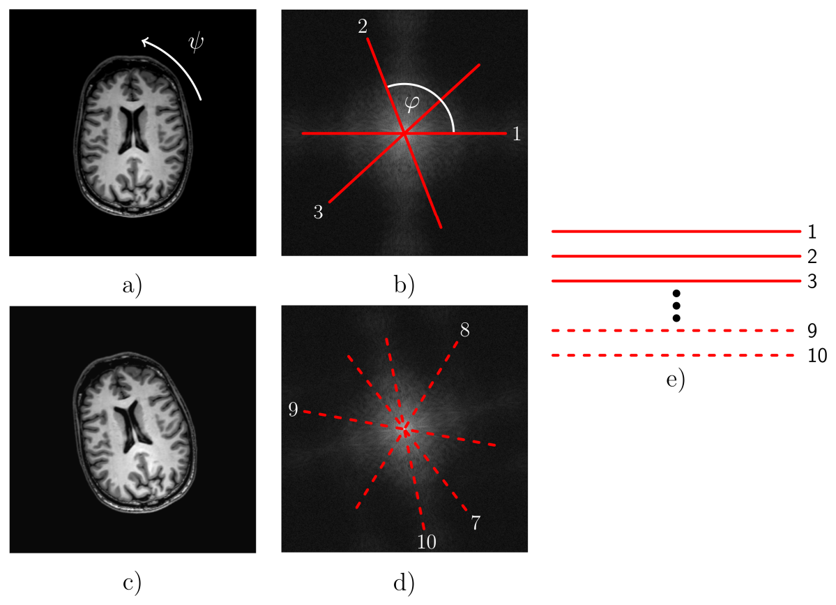
Axial slices of a reconstructed T1-weighted Cartesian 3D turbo FLASH sequence were used as 2D digital phantoms. A golden-angle radial gradient echo sequence was mimicked by converting the slices to their k-space representations and extracting 10 spokes from these k-space data, each containing 240 sampling points, using bilinear interpolation (see Figure 1).5 The images were subjected to translation by $$$(x,y)$$$ using $$$x,y\in [-5.00, 5.00]$$$ voxels, and rotation by $$$\psi\in[-8.00^\circ, 8.00^\circ]$$$ about the center. The image transformations were performed in between two spoke extractions, to mimic discontinuous in-plane movements of the patient's head. The 10 spokes become the rows of a 10$$$\times$$$240 matrix (referred to as 'scan' below) and serve as training data for a CNN. Either the last one, last 2 or last 4 rows are spokes recorded from the moved phantom ('moved spokes').
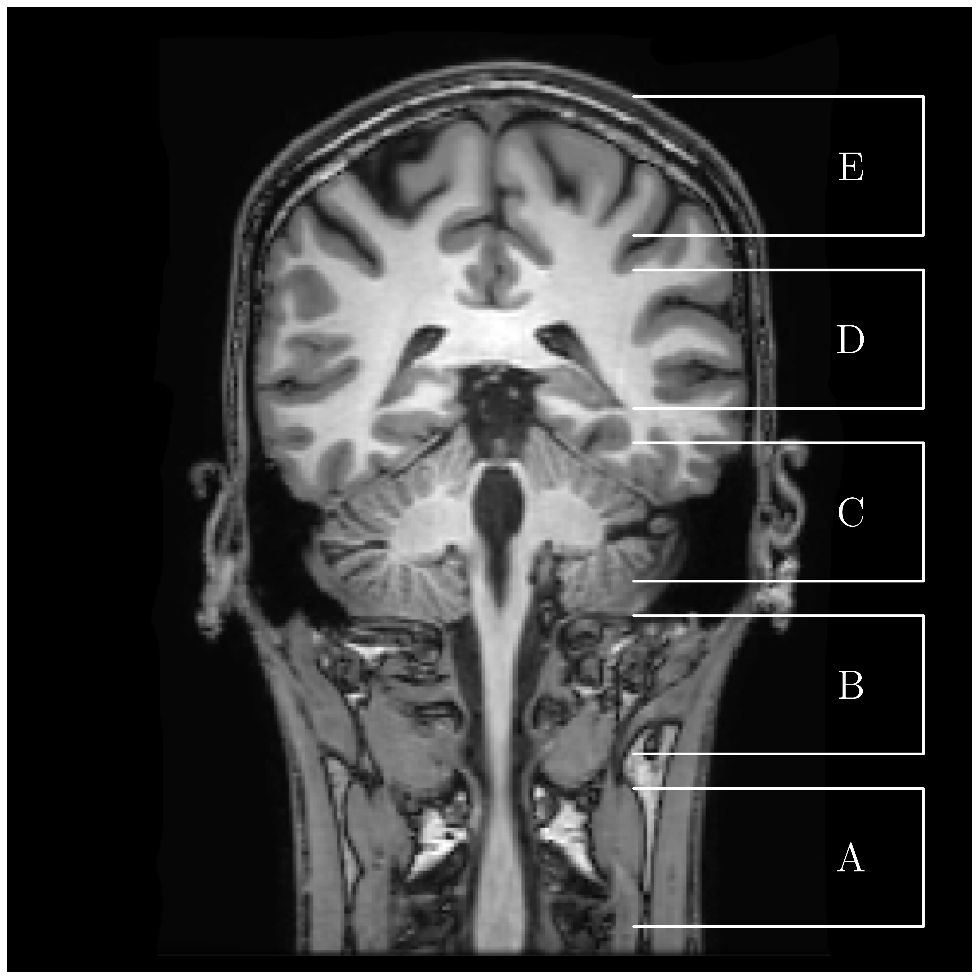
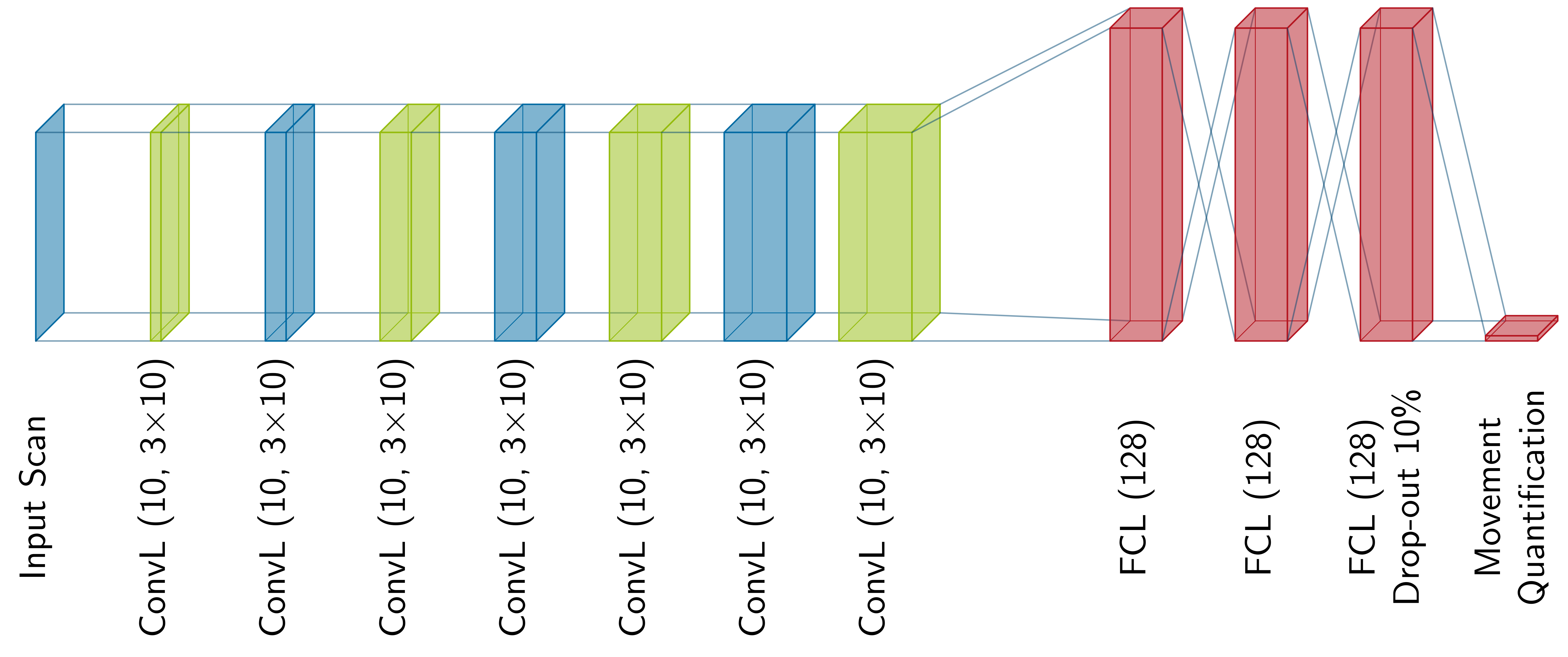
The CNN-based motion quantification was evaluated for two motion types (translation and rotation), three different numbers of 'moved spokes', and for various phantoms, which were bundled into the 5 slice stacks (A, B, C, D and E), see Figure 2. For each combination, a scan set of 5,000 scans was recorded using the settings described above. These scans of one set differ from one another by the degree of movement and the selected slice inside the stack. The CNN model of the architecture described in Figure 3 was trained and tested on these scan sets, respectively. For evaluation, a 5-fold cross-validation (80% training, 20% test) was performed using permutations of the training and test data sets.6 The movement quantification was evaluated by calculating the absolute error between the induced movement and the movement estimated by the CNN.
Results and Discussion
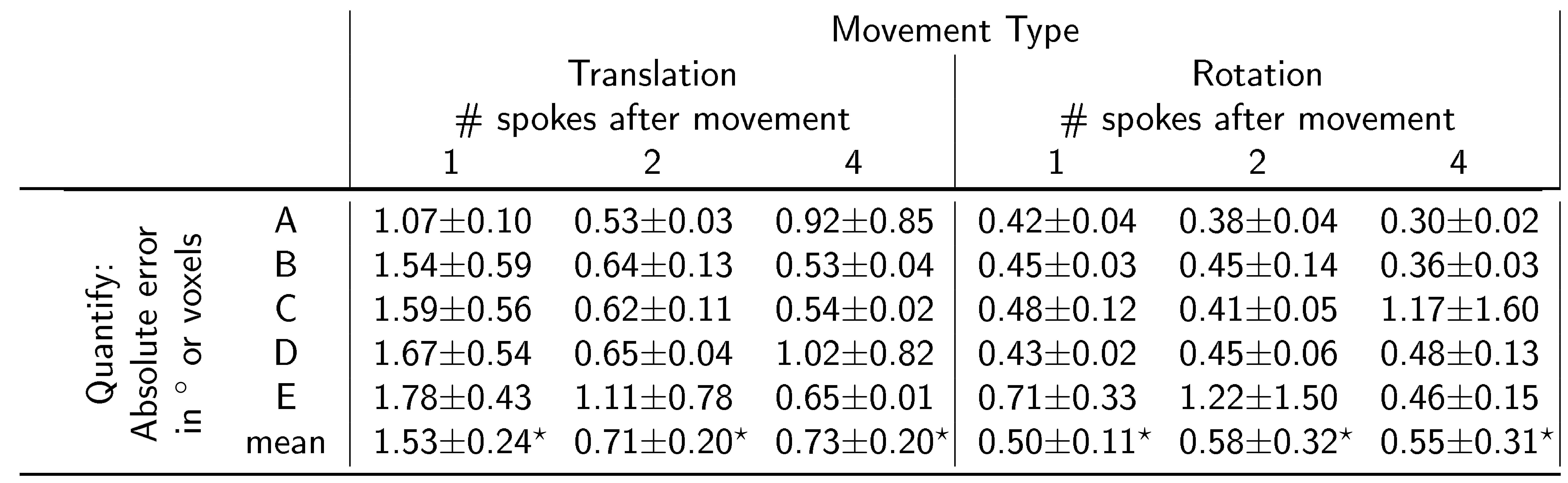
Overall the CNN was able to quantify the movements with an absolute error between 0.30$$$^\circ$$$ and 1.22$$$^\circ$$$ for rotations and between 1.78 and 0.53 voxel (1.29 and 0.38 mm) for translations, indicating that CNN-based movement quantification from few 'moved spokes' is possible (see Table 1). Even though the data with 4 'moved spokes' carries the most information about motion, two 'moved spokes' seem to be enough to provide a satisfying movement quantification. In 4 out of 6 experiments, data set E has the highest mean absolute error, which is likely due to the simpler anatomical structure in image slices of the skullcap.Conclusion
This 2D digital phantom study shows that CNNs can quantify rotational and translational head movements with a high accuracy from very few radial k-space lines. The degree of movement in scans with just a single 'moved spoke' was able to be detected with a promising accuracy. The possibility to track movements from these few spokes has great potential for real-time guidance in radiotherapy as data conversion to the spatial domain becomes unnecessary. The aim of further experiments is to design a CNN-based navigator, which could be fed with data from a continuously acquired radial sequence in order to use the same data for imaging and navigation. In future studies, the influence of imperfect signals from real MR-scanners and 3D out-of-plane movements need to be investigated. Transferring the proposed method on motion sensitive examinations like liver or cardiac will also be targeted. Finally, more advanced network architectures should be taken into account to increase the quantification accuracy even further.Acknowledgements
No acknowledgement found.References
1 BW Raaymakers, IM Jürgenliemk-Schulz, GH Bol, M Glitzner, ANTJ Kotte, B van Asselen, JCJ de Boer, JJ Bluemink, SL Hackett, MA Moerland, et al. First patients treated with a 1.5 T MRI-Linac: clinical proof of concept of a high-precision, high-field MRI guided radiotherapy treatment. Physics in Medicine & Biology, 62(23):L41, 2017.
2 J. Yun, K. Wachowicz, M. Mackenzie, S. Rathee, D. Robinson, and BG Fallone. First demonstration of intrafractional tumor-tracked irradiation using 2D phantom MR images on a prototype Linac-MR. Medical Physics, 40(5), 2013.
3 B. Stemkens, R. H. N. Tijssen, B. D. de Senneville, J. J. W. Lagendijk, and C. A. T. van den Berg. Image-driven, model-based 3D abdominal motion estimation for MR-guided radiotherapy. Phys. Med. Biol., 61(14):5335-5355, 2016.
4 Y. LeCun, Y. Bengio, and G. Hinton. Deep learning. Nature, 521(7553):436, 2015.
5 Z. Zhou, F. Han, L. Yan, D. JJ Wang, and P. Hu. Golden-ratio rotated stack-of-stars acquisition for improved volumetric MRI. Magnetic Resonance in Medicine, 78(6):2290-2298, 2017.
6 J. D Rodriguez, A. Perez, and J. A. Lozano. Sensitivity analysis of k-fold cross validation in prediction error estimation. IEEE Transactions on Pattern Analysis and Machine Intelligence,32(3):569-575, 2010
Figures