4846
Automated slice-to-volume registration between histology and whole-brain post-mortem MRI1Wellcome Centre for Integrative Neuroimaging, FMRIB, University of Oxford, Oxford, United Kingdom, 2Nuffield Department of Clinical Neurosciences, University of Oxford, Oxford, United Kingdom, 3Department of Radiology, University of Chicago, Chicago, IL, United States, 4Institute of Medical Informatics, Universität zu Lübeck, Lübeck, Germany
Synopsis
Validating MRI data against histological ground truth is essential in the process of devising disease-specific imaging biomarkers that are sensitive to early microstructural changes in neurodegeneration. Current MRI–histology registration techniques are too labour- or resource-intensive to be used in large-scale studies. We introduce an automated pipeline for registering sparsely sampled, small (25x30mm) 2D stained histological images with 3D post-mortem MRI of the whole human brain. Our tests indicate sub-voxel (<0.5 mm) precision using simulated data, and <1 mm precision with real data. Implemented in a new, flexible image registration framework (TIRL), the pipeline is adaptable to various research needs.
Introduction
Clinical imaging biomarkers have a great potential to facilitate early diagnosis in neurodegenerative conditions, but require validation1 against histological ground truth to achieve sufficient specificity. Quantitative comparison between histology and MRI data is hindered by the challenges of cross-modality 2D-to-3D image registration. Existing methods typically employ sequential histological sampling2-4 to reconstruct a 3D histological stack, or match manually identified landmarks5. Neither of these scales well to the population level as required by biomarker research. Here we present an automated method to accurately register small, sparsely sampled 2D histological images with whole-brain MRI, aiming to achieve ease of use that would even be compatible with routine neuropathological practice.Methods
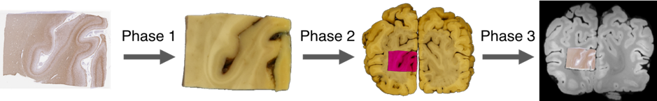
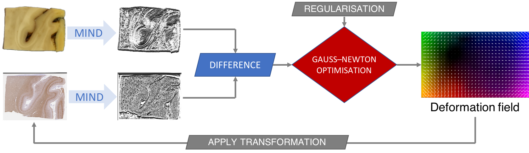
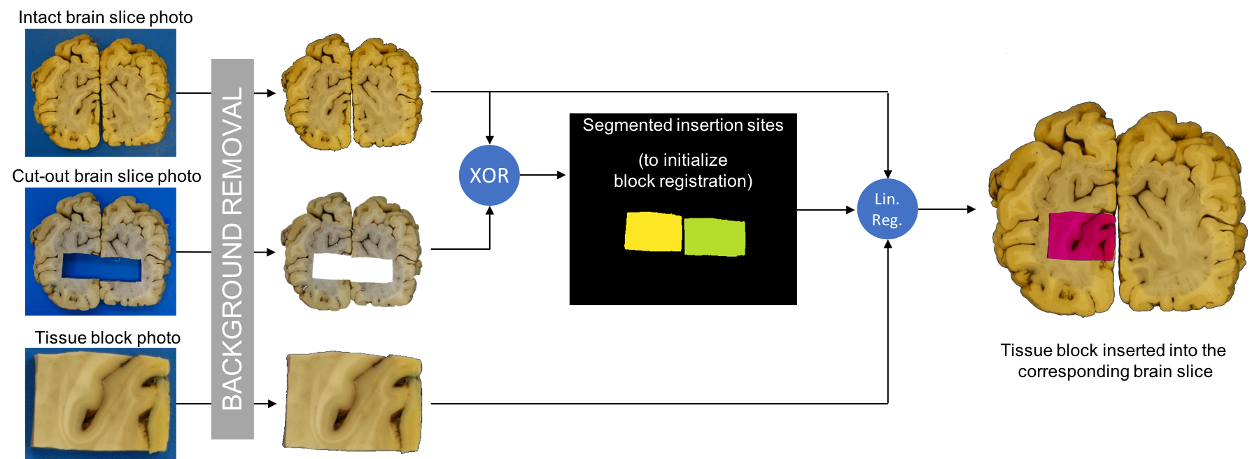
The registration pipeline (Figure 1) handles three types of images6 (histology, photograph, MRI), and performs three consecutive registrations (phase 1, 2, and 3) to establish a forward mapping between the 2D coordinates of a histological image $$$\left(\mathbf{x}\in\mathbb{R}^2\right)$$$ and the 3D coordinates of MRI data $$$\left(\mathbf{x'}\in\mathbb{R}^3\right)$$$. Given this non-linear mapping $$$\phi\left(\mathbf{x}\right)=\mathbf{x'}$$$, MRI data can be interpolated at the chosen resolution of the histological image for localised quantitative comparison of histology- and MRI-derived measures. The three phases of the proposed pipeline were implemented in a novel general-purpose image registration framework (Tensor Image Registration Library, TIRL), that we designed to address the missing software needs for working with different types of images in an integrated, customisable and all-compatible workflow that also allows slice-to-volume registration. During histological processing, the tissue shrinks and deforms, leading to a change in the in-plane geometry compared to the block-face image. These are compensated by a combined affine and non-linear transformation in phase 1 (Figure 2). Registering a small 2D image to a large 3D image is an ill-posed problem with a huge search space. Using photographs of the dissected brain slices as an intermediate modality, the search space can be significantly reduced, since the anatomical pattern of the cut surface almost exactly defines the location of the slice within the brain. Phase 2 (Figure 3) therefore aligns the small tissue block with the corresponding brain slice photograph, that is subject to slice-to-volume registration in phase 3 (Figure 4, A-B). During slice-to-volume registration, the affine alignment is optimised at gradually increasing resolution levels, which is later refined by allowing deformations orthogonal to the slice, finally by allowing in-plane deformations as well (free-form deformation). Transformations are penalised by a membrane energy term in the cost function to avoid sharp bending of the slice photograph during optimisation.Results
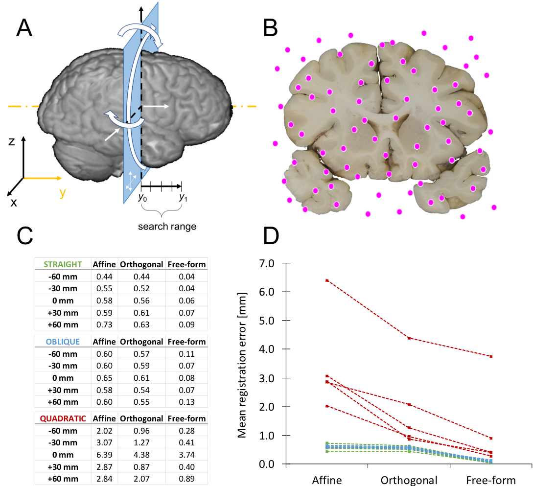
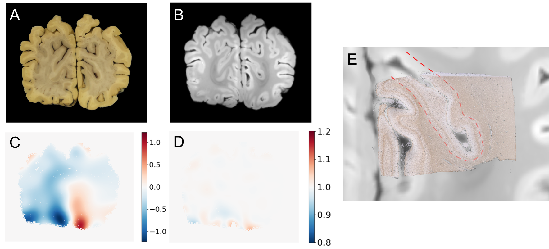
Our previous work7 included a landmark-based semi-automatic slice-to-volume registration. This was the least accurate and only manual step in the pipeline and, therefore, in this work we performed a set of simulations to test the accuracy and robustness of our novel automated slice-to-volume registration. At distances of -6cm, -3cm, 0cm, 3cm, and 6cm from the mid-coronal plane, the MRI was resampled along two perfectly planar surfaces (one coronal and one slightly oblique [Euler angles <20°]) and a quadratic surface. Each of the three simulated images were put through phase 3 (using regular 2-cm thick search ranges) and the resultant insertion coordinates were compared with the ground truth (Figure 4, C-D). As a second test, we registered all 14 coronal brain slice photographs of one brain (real images, not simulated) with the phase 3 method, using 2-cm search ranges (Figure 5, A-D). Finally, as a third test, we registered 5 histological images to MRI using all three phases of the protocol (Figure 5E).Discussion
Experiments with simulated brain slices indicated that the automated slice-to-volume registration algorithm performs accurately, with sub-voxel (<0.5mm) error magnitudes in 4 out of 5 polynomial cases with as few as 32 control points and default parameters (tuned for 2.5mm expected curvature of the cut surface). Further improvement of the results could be achieved by increasing the number of control points at the expense of additional computation time, or by fine-tuning parameters. Although ground truth coordinates are unknown for registration with actual post-mortem images, based on a visual comparison of misaligned gyri, the errors are less than 1mm in most slices. By combining all steps of the pipeline, it was possible to register small histological images to MRI without noticeable misalignment (Figure 5E).Conclusion
An automated MRI–histology registration pipeline was created based on a novel image registration framework (TIRL), that allows customisation of the pipeline for a wide range of applications. Studying the microstructural correlates of MRI signal changes in both healthy and diseased tissue will hopefully enable the design of more specific imaging biomarkers, that facilitate earlier diagnosis in neurodegenerative conditions.Acknowledgements
This work was supported by funding from the Engineering and Physical Sciences Research Council (EPSRC) and Medical Research Council (MRC) [grant number EP/L016052/1]. The Wellcome Centre for Integrative Neuroimaging is supported by core funding from the Wellcome Trust (203139/Z/16/Z). The authors would like to further express their gratitude to the donors and benefactors of the Oxford Brain Bank.References
1. Beach, T. G. A Review of Biomarkers for Neurodegenerative Disease: Will They Swing Us Across the Valley? Neurol. Ther. 6, 5–13 (2017).
2. Alegro, M. et al. Multimodal Whole Brain Registration: MRI and High Resolution Histology. in 2016 IEEE Conference on Computer Vision and Pattern Recognition Workshops (CVPRW) 634–642 (IEEE, 2016). doi:10.1109/CVPRW.2016.85
3. Seehaus, A. et al. Histological validation of high-resolution DTI in human post mortem tissue. Front. Neuroanat. 9, 98 (2015).
4. Mollink, J. et al. Dentatorubrothalamic tract localization with postmortem MR diffusion tractography compared to histological 3D reconstruction. Brain Struct. Funct. 221, 3487–3501 (2016).
5. Gangolli, M. et al. Quantitative validation of a nonlinear histology-MRI coregistration method using generalized Q-sampling imaging in complex human cortical white matter. Neuroimage 153, 152–167 (2017).
6. Pallebage-Gamarallage, M. et al. Dissecting the pathobiology of altered MRI signal in amyotrophic lateral sclerosis: A post mortem whole brain sampling strategy for the integration of ultra-high-field MRI and quantitative neuropathology. BMC Neurosci. 19, 11 (2018).
7. Huszar, I. N. et al. Pipeline for registering histological sections to MRI volumes. in Proceedings of the International Society for Magnetic Resonance in Medicine, electronic poster #3427 (2018).
8. Heinrich, M. P. et al. MIND: Modality independent neighbourhood descriptor for multi-modal deformable registration. Med. Image Anal. 16, 1423–1435 (2012).
9. Fischer, B. & Modersitzki, J. A unified approach to fast image registration and a new curvature based registration technique. Linear Algebra Appl. 380, 107–124 (2004).
Figures