4843
FitLike, a software for the analysis of T1 dispersion for Fast Field-Cycling experimentation1Unit 1205 BrainTech Lab, INSERM, Grenoble, France, 2University of Aberdeen, Aberdeen, United Kingdom
Synopsis
Since early 2000 commercial solutions are available to study the dispersion of T1 with the magnetic field strength. This has generated a growing interest in T1 relaxometry study of sample material and a large amount of data to analyse. Yet data analysis for T1 relaxometry is almost entirely done with homemade software, which makes access to the technology difficult and limits the exchanges between research groups. Here we propose a new tool for the analysis of T1 dispersion profiles, software called FitLike that runs with Matlab.
Introduction
T1 dispersion is a very rich source of information that has been used since the early years of MRI thanks to its ability to access information on molecular dynamics non-invasively1,2. The dispersion of T1 with the magnetic field strength can be measured using Fast Field-Cycling (FFC), a technique that consists in varying the magnetic field strength faster than the T1 value so that one can visualise the magnetisation decay during evolution time for different values of the magnetic field strength. This is an iterative procedure that can be performed by dedicated relaxometers or more recently by dedicated whole-body MRI scanners3. These are called FFC-NMR and FFC-MRI respectively and both produce large amounts of data that are processed in several stages. So far, most research groups in FFC relaxometry use their own software for data analysis and the authors know of only one tool available for advanced processing of dispersion curves4. This is making comparisons difficult and limits the access to this technology to advanced users. To avoid these we designed a modular software able to perform data processing in an intuitive way that also provides easy access for the addition of new models or processing algorithms.Methods
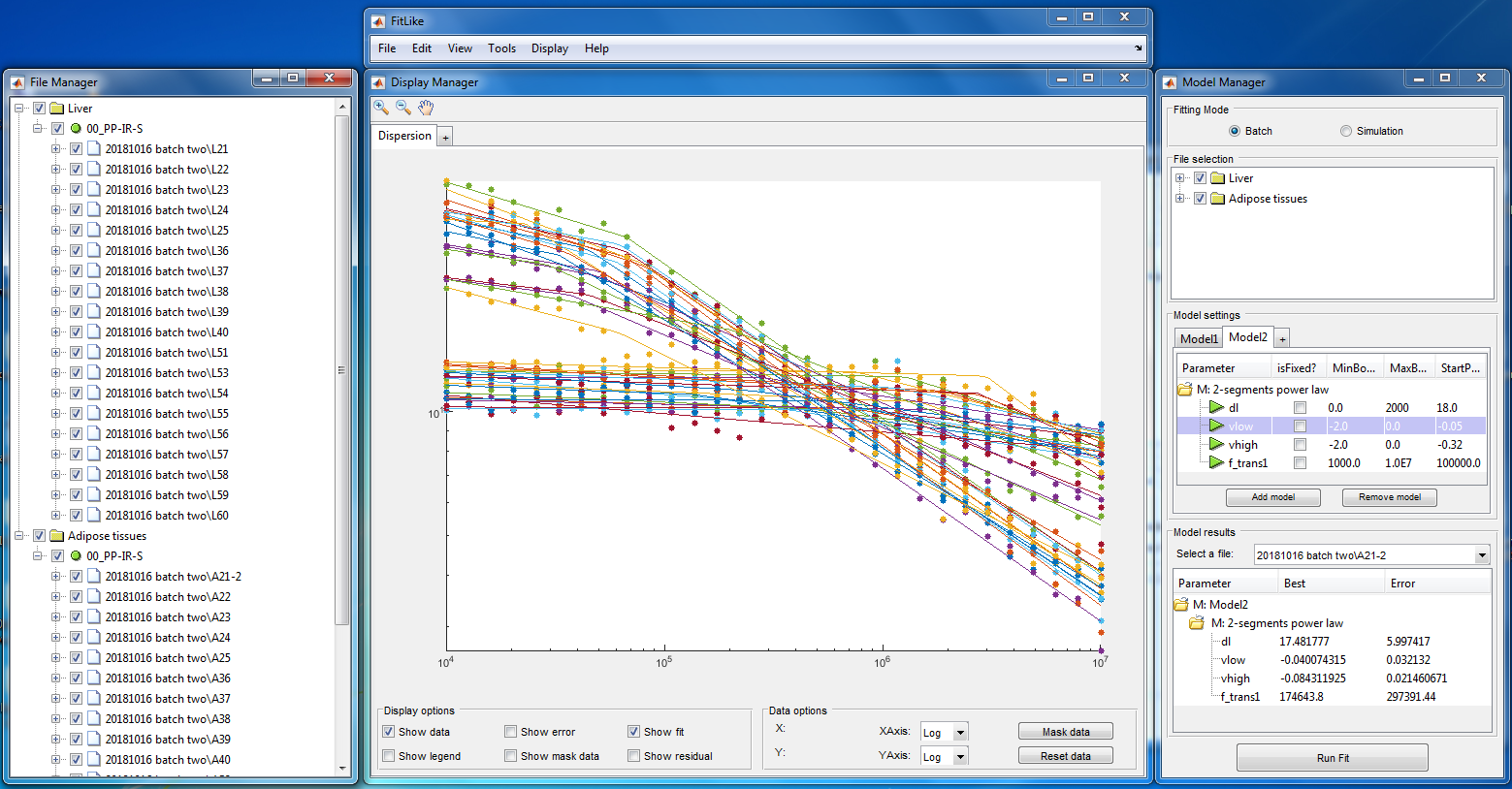
The software proposed here handles relaxometry data using four layers: blocs, zones, dispersions and experiments. Bloc objects hold the raw data obtained during the experiments. These are usually processed in a way to provide the magnetisation of the sample, which is stored in Zone objects. At this stage one typically uses one or more exponential decay model to fit the magnetisation signal, which provides the T1 values that are stored in Dispersion objects. These can then be analysed by curve fitting using adapted models from an extensible bank, and the data obtained can be regrouped into Experiment objects for comparisons. Each processing function is defined by a dedicated object that can be modified or created by the user. These various objects are handled by a graphical interface so that non-experts users can perform analyses without having to write any code. This was coded in Matlab (The Mathworks, Natick, Massachusetts, USA) with versions 2015a, 2016a, 2017b and 2018a. The code was made available on GitHub5 on GPLv3 license for wide distribution.Results
Data are readily imported from commercial relaxometers (Stelar, Mede, Italy) and from the Aberdeen FFC-MRI scanner and import scripts can be added to load custom files into Bloc objects. The GUI (see Figure 1) allows selecting the processing pipeline to change bloc data into Zone and Dispersion objects according to the type of experiment performed. Single and multiexponential models are available and more models can be coded as plug-in using the templates provided. A collection of models is already available to analyse the T1 dispersion curves, typically Lorentzian, power laws, quadrupolar-enhanced cross-relaxation and other models obtained from optical spectroscopy. These models can be added together to process complex samples and the fit is performed in the logarithmic space to avoid the need for weighting in datasets that present large dispersion.Discussion
The GUI interface allows using the code relatively easily but is still immature and evolving rapidly. It is possible to use the Matlab command windows to use the base functions but this requires some knowledge in Matlab coding. The choice of Matlab as a programming language was mainly motivated by the readily available processing tools and the familiarity of the language for the authors, but future developments of FitLike may include a translation to Python for better portability and responsiveness. Yet the code can import and process 100 MBits of data in under one minute, which offers many possibilities for rapid and custom analyses of FFC data.Conclusion
We propose FitLike as a solution for non-specialist users of FFC-NMR or FFC-MRI equipment to get a quick and easy access to data processing and interpretation of data, which is often a tedious task. This tool has been evolving for three years now and is still in developments regarding many aspects but it is being used by students and researchers in its current state. Sharing and helping is also welcome via the GitHub repository.Acknowledgements
This project has received funding from the European Union’s Horizon 2020 research and innovation programme under grant agreement No 668119 (project “IDentIFY”).References
1. Anoardo, E., Galli, G. & Ferrante, G. Fast-field-cycling NMR: Applications and instrumentation. Appl. Magn. Reson. 20, 365–404 (2001).
2. Kimmich, R. & Anoardo, E. Field-cycling NMR relaxometry. Progress in Nuclear Magnetic Resonance Spectroscopy 44, 257–320 (2004). .
3. Lurie, D. J. et al. Fast field-cycling magnetic resonance imaging. Comptes Rendus Physique 11, 136–148 (2010).
4. Sebastião, P. J. The art of model fitting to experimental results. European Journal of Physics 35, 015017 (2014). 5. https://github.com/ManuIdentiFY/FitLike2. Curve fitting toolbox for relaxometry data. (2018).
Figures