4841
Dynamic platform-independent MRI vs. manufacturer’s implementations1MR Physics, Fraunhofer MEVIS, Bremen, Germany, 2MR-Imaging and Spectroscopy, Faculty 01 (Physics/Electrical Engineering), University of Bremen, Bremen, Germany
Synopsis
MR sequence development is usually performed within vendor-specific frameworks, which do not allow for an easy sequence transfer to other manufacturers’ scanners. A platform-independent rapid prototyping environment for MR sequences was presented to allow both, a sequence transfer without code compilation and the generation of dynamic sequences at the scanner. This framework was used to implement a set of standard sequences and modules, which can easily be exchanged or implemented into different sequences. The aim of this work is to show that this approach of vendor-independent sequence development produces same image results as the sequences provided by the manufacturer.
Introduction
MR sequence development is usually performed within vendor-specific frameworks, which do not allow for an easy sequence transfer to other manufacturers’ scanners and thus for the use in multi-center studies. Different approaches [1-5] exist to overcome this problem, but with some drawbacks. Recently, a platform-independent rapid prototyping environment for MR sequences was presented [6-8] to allow both, a sequence transfer without code compilation and the generation of dynamic sequences at the scanner using a self-written graphical user interface. This framework was used to implement a set of standard sequences and modules, which can easily be exchanged or implemented into different sequences (e.g., fat suppression, EPI readout, preparation module for arterial spin labeling, etc.). The aim of this work is to show that this approach of vendor-independent sequence development produces same image results as the sequences provided by the manufacturer.Methods
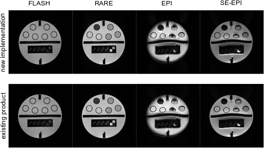
To assess the feasibility of the approach, various sequences were implemented. The results section show images acquired with fast low-angle shot (FLASH [9]), rapid imaging with refocused echoes (RARE [10]), echo-planar imaging (EPI [11]) and spin echo EPI (SE-EPI) sequences. During the design process of those sequences, modules were implemented to be flexibly reusable. For instance, the RARE sequence did not require the implementation of any module that directly relates gradient, RF, ADC, protocol, system or k-space calculation components. It sufficed to use parts of the FLASH and SE-EPI sequence and connect them in an alternative loop and refocusing strategy. The modules that connect device properties and common protocol settings to the rest of the sequence were also shared, such that no extra work was required to provide a seamless configuration experience to the scanner operator.
All measurements were performed on a 3 T whole-body MR scanner (MAGNETOM Skyra, Siemens Healthineers, Erlangen, Germany) with a 16-channels head coil. Sequence protocol parameters to compare the new implementation with the existing product sequence are as follows: FLASH: TR = 50 ms, TE = 5 ms, flip angle = 20°, readout bandwidth = 1000 Hz/px, field-of-view = 256×256 mm2, matrix size = 256×256. RARE: TR = 4 s, TE = 77 ms, flip angles = 90° and 180°, readout bandwidth = 1000 Hz/px, 16 lines/shot, field-of-view = 256×256 mm2, matrix size = 256×256. Single-shot EPI: TE = 65 ms, flip angle = 90°, readout bandwidth = 1955 Hz/px, field-of-view = 256×256 mm2, matrix size = 128×128. Single-shot SE-EPI: Identical to EPI, except for TE = 120 ms and SE flip angle of 180°.
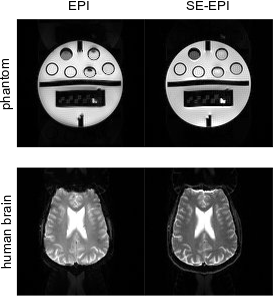
Since the EPI-based product sequences available at our scanner do not allow for multi-shots, segmented EPI images were acquired by a dynamic protocol change for the new implementation only. Here, sequence protocol parameters are changed to: EPI: TR = 4 s, TE = 35 ms, 64 lines/shot. SE-EPI: Identical to EPI, except for TE = 55 ms and SE flip angle of 180°. The images were reconstructed on the manufacturer’s hardware using the manufacturer’s reconstruction framework. Sequence protocol parameters were configured after deployment with no intermediate processing steps.
Results & Discussion
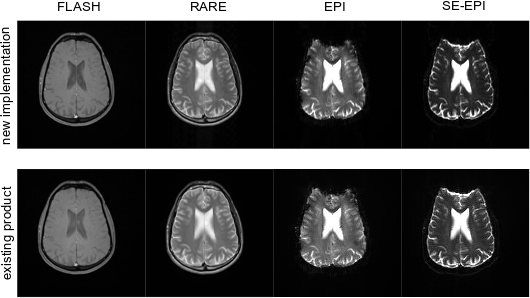
Figure 1 shows a comparison between the new implementations and the existing product sequences using a structure phantom. Measurements with the identical sequences were performed on a healthy volunteer, but with an added fat saturation pulse within EPI and SE-EPI (cf. Figure 2). Image contrasts and quality of both sequence implementations are indistinguishable at same parameters. Slight deviations occur since the sinc-shaped RF pulse was implemented with a higher time-bandwidth product compared to the manufacturer’s implementation to obtain better slice profiles. Additionally, shortest possible gradients with higher slew rates were used to allow for shortest possible echo times, which could result in higher eddy currents with more image distortions at different parameters. Measurements with two segments were performed for EPI-based imaging with the new implementation (cf. Figure 3). The artifacts are less strong than for the single-shot versions due to a shorter echo and readout time. Future work will focus on the driver development for different manufacturers and following comparison studies regarding MR sequences and reconstruction (Gadgetron).Conclusion
It could be demonstrated that this framework provides MR sequences with same image quality and contrast as the product sequences and could therefore be used in multi-center studies at different scanners.Acknowledgements
All funding for this study was provided by the internal Attract (600172) funding program of the German Fraunhofer-Gesellschaft.References
- Jochimsen TH & von Mengershausen M. J Magn Reson 2004;170(1):67-78.
- Stöcker T, Vahedipour K, Pflugfelder D, Shah NJ. Magn Reson Med 2010;64(1):186-93.
- Magland JF, Li C, Langham MC, Wehrli FW. Magn Reson Med 2016;75(1):257-65.
- Layton KJ, Kroboth S, Jia F, Littin S, Yu H, Leupold J, Nielsen JF, Stöcker T, Zaitsev M. Magn Reson Med 2017;77(4):1544-52.
- Nielsen JF & Noll DC. Magn Reson Med 2018;79(6):3128-34.
- Cordes C, Honroth T, Hoinkiss D, Archipovas S, Porter DA, Günther M. Proc Intl Soc Mag Reson Med 2016;3198.
- Cordes C, Konstandin S, Porter D, Günther M. Magn Reson Med (under review).
- Honroth T, Cordes C, Archipovas S, Hoinkiss DC, Günther M, Porter D. Proc Intl Soc Mag Reson Med 2016;3207.
- Haase A, Frahm J, Matthaei D, Hanicke W, Merboldt KD. J Magn Reson (1969) 1986;67(2):258-66.
- Hennig J, Nauerth A, Friedburg H. Magn Reson Med 1986;3(6):823-33.
- Mansfield P. J Phys C 1977;10(3):L55-8.
Figures