4834
A Web-Based Data Management System as a Collaborative Imaging Research Platform1Research and Development, Synaptive Medical, Toronto, ON, Canada
Synopsis
A web-based data management system specifically aimed at imaging researchers is presented as a possible solution to the challenges of systematic data management and processing in a research environment. The system was employed during the development of a head-only MRI for post-processing quality assurance. Extending the use of the system to facilitate training of machine learning algorithms is proposed.
Introduction
Management and organization of images and other data types in research imaging is often challenging due to a lack of effective systems designed for research purposes. As a result, ad hoc storage of data on individual contributors' computers, shared network drives, USB keys, etc., is the de facto practice. Such methods lack important functions necessary in research imaging, such as DICOM connectivity, data sharing and pipeline processing. Occasionally, clinical PACS systems are employed for research purposes, but they too are often a poor fit for research data management, due to a lack of research specific tools and features.
Here we present a dedicated, web-based, image management system aimed specifically at imaging researchers. We propose that such a system could be used to automate routine image management and analysis tasks in a research setting, simplify multi-center research study data management and address the growing need for organized data curation and processing.
Functionality for Imaging Research
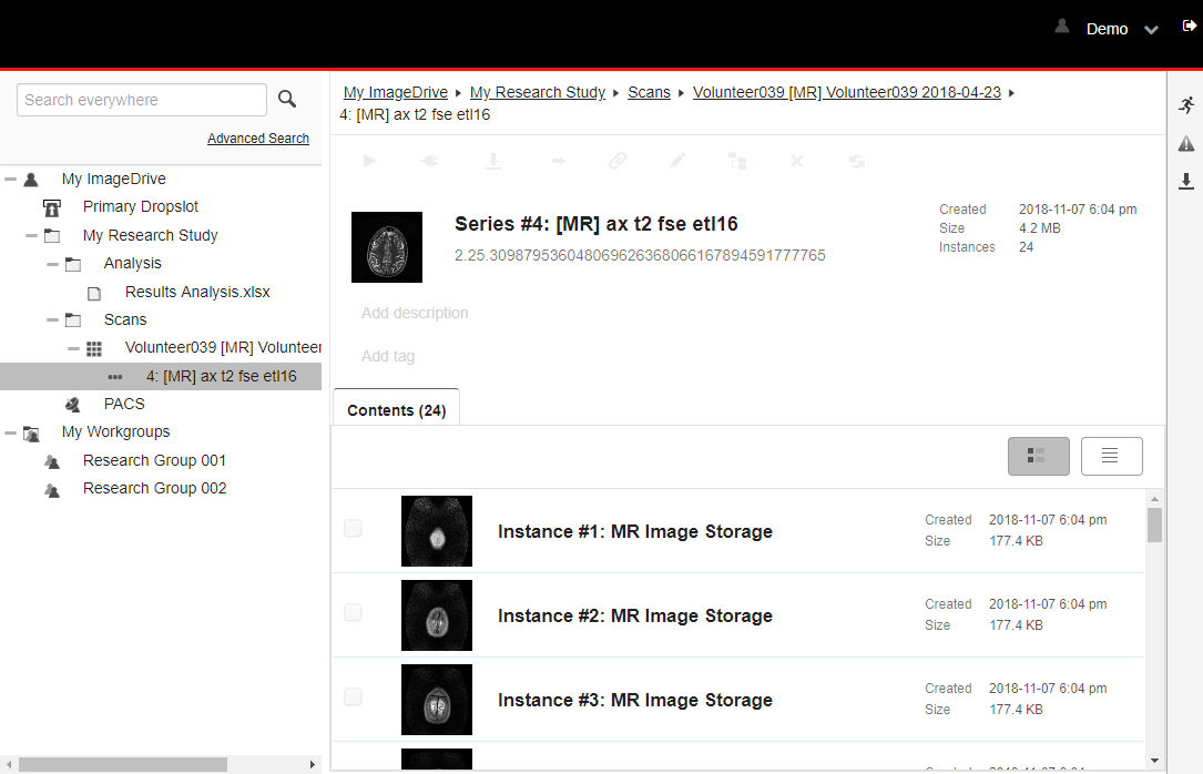
Unlike a traditional PACS which typically only stores DICOM data, the System accepts both DICOM and non-DICOM data in a taggable and searchable Virtual Folder System (VFS) (Fig.1). This was an early design decision that reflected the diversity of file types in imaging research, including documents, spreadsheets, videos and binary data. DICOM data itself is fulsomely supported through the native representation in the VFS of the study-series-image hierarchy, DICOM networking for communication with modalities and PACS, web-based image viewing, and export to NIfTI and NRRD. Research collaboration is made possible using special VFS folders known as Workgroups, which facilitate permissions based data sharing with other users.
A particularly notable capability of the System is its ability to be integrated into an existing ecosystem of internally developed software. Users can invoke an External Application on a selected dataset (Fig.2), which sends a configurable request to an http based listener that in turn can run software that is external to the System. To complete the communication loop, the System also offers a RESTful API that the External Application can use to perform operations such as retrieving data, writing back data, adding tags, etc. This External Application functionality can be further leveraged to create an automated processing pipeline by making use of the System's rules engine. Rules can be defined on VFS folders to automate the processing of DICOM images. By daisychaining folder rules together, a multistage processing pipeline can be created, with each folder representing a different stage in the pipeline. As many research environments have already made significant investments in novel image processing algorithms, the use of External Applications, the RESTful API and folder rules in this manner allows those investments to be leveraged for systematic and automated processing of imaging data.
Usage in MRI Development
The System was recently provided to researchers involved in the development of a new head-only MRI scanner. The System was used to store and tag DICOM images, raw image data files and scan prescription details in the VFS, enabling them to easily find relevant data after scans were complete. Using the External Application functionality, a variety of automated post-processing quality assurance tools were implemented as a series of Python-based External Applications. These included SNR measurements, geometric distortion, image and field uniformity, and image ghosting measurements in accordance to the ACR phantom guidance [1] to replace time consuming manual measurements. The Python applications also leveraged the System’s RESTful API to post quality assurance results for review with plans for adding additional automatically invoked processing tools to evaluate data trends.Application in Machine Learning
There has been much interest recently in applying machine learning (ML) techniques to medical imaging. For this to be effective however, significant numbers of imaging datasets need to be curated and processed in a systematic, automated and repeatable way. The System presented here may represent the infrastructure necessary for such an endeavour. A simple training implementation could be realized in the following manner: 1) training imaging data is fed into a VFS folder 2) a rule is defined on the folder that invokes an External Application 3) the External Application is an ML algorithm which processes the dataset and 4) results are written back to the System. Validation of the algorithm can be achieved in a similar manner simply by feeding the System--and by extension, the trained algorithm--the appropriate validation datasets.Conclusion
We have presented the design, implementation and sample adoption of a web-based data management system which helps address existing issues facing imaging researchers in multi-faceted, collaborative data management.Acknowledgements
No acknowledgement found.References
[1] https://www.acraccreditation.org/modalities/mriFigures