4829
Bloch image simulations of brain pulse sequences using a GPU-installed gaming PC1MRI simulations Inc., Tokyo, Japan
Synopsis
Bloch image simulations for typical brain pulse sequences were performed using a GPU-installed gaming laptop PC and a numerical brain phantom. Artifact-free brain MR images were obtained by the Bloch image simulation using optimized numbers of subvoxels. Because the simulation times were the same order as the imaging time for the experiments, we concluded that the Bloch image simulator installed in an inexpensive gaming PC can be a powerful research tool for many MRI engineers and scientists.
Introduction
Graphics processing units (GPUs) are revolutionary devices for Bloch image simulations because GPUs have made the simulation speed 10-100 times faster comparing with CPUs. We recently reported that 32-bit floating point operation was sufficient for Bloch image simulations if a local isochromat summation technique was properly implemented.1 This technique drastically reduced the cost of GPUs used for Bloch image simulations. In this study, we performed Bloch image simulations of brain pulse sequences using a GPU-installed gaming laptop PC to demonstrate that the Bloch simulator can be a powerful research tool for many MRI researchers.Methods
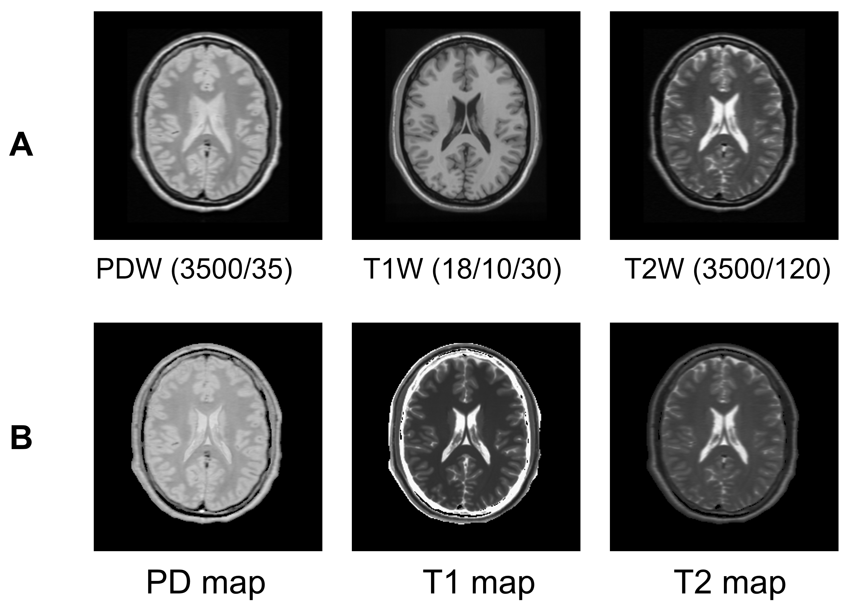
A numerical brain phantom was constructed from PDW, T1W, and T2W brain MR image datasets available on the website (Fig.1).2 The parameter maps were stored in three 2563 voxel matrices with 1 mm cube voxel size and used for Bloch image simulations. MRI pulse sequences were developed for a virtual MRI system with a gradient coil set having a 45 mT/m maximum strength and a 200 mT/m/ms slew rate and a transmitter coil having 30 mT maximum-strength B1 field. 3D RF-spoiled gradient echo, 2D multi-slice spin echo, 2D multi-slice spiral, MPRAGE, and MR fingerprinting (MRF) sequences3,4 were implemented for the numerical brain phantom. The RF pulses were approximated by many hard RF pulses with sufficiently short (10-20μs) duration times.1 For example, the selective excitation pulse (hamming windowed sinc pulse, ±2π, 3.2 ms duration) was approximated using 160 short pulses (20μs duration). A Bloch image simulator developed by our group1,5 was installed in a gaming laptop PC (CPU: core i7-7700HQ, 16 GB memory, GPU: GeForce GTX 1070 for laptop, 2048 CUDA core, NVIDIA) and used for the Bloch image simulations.Results
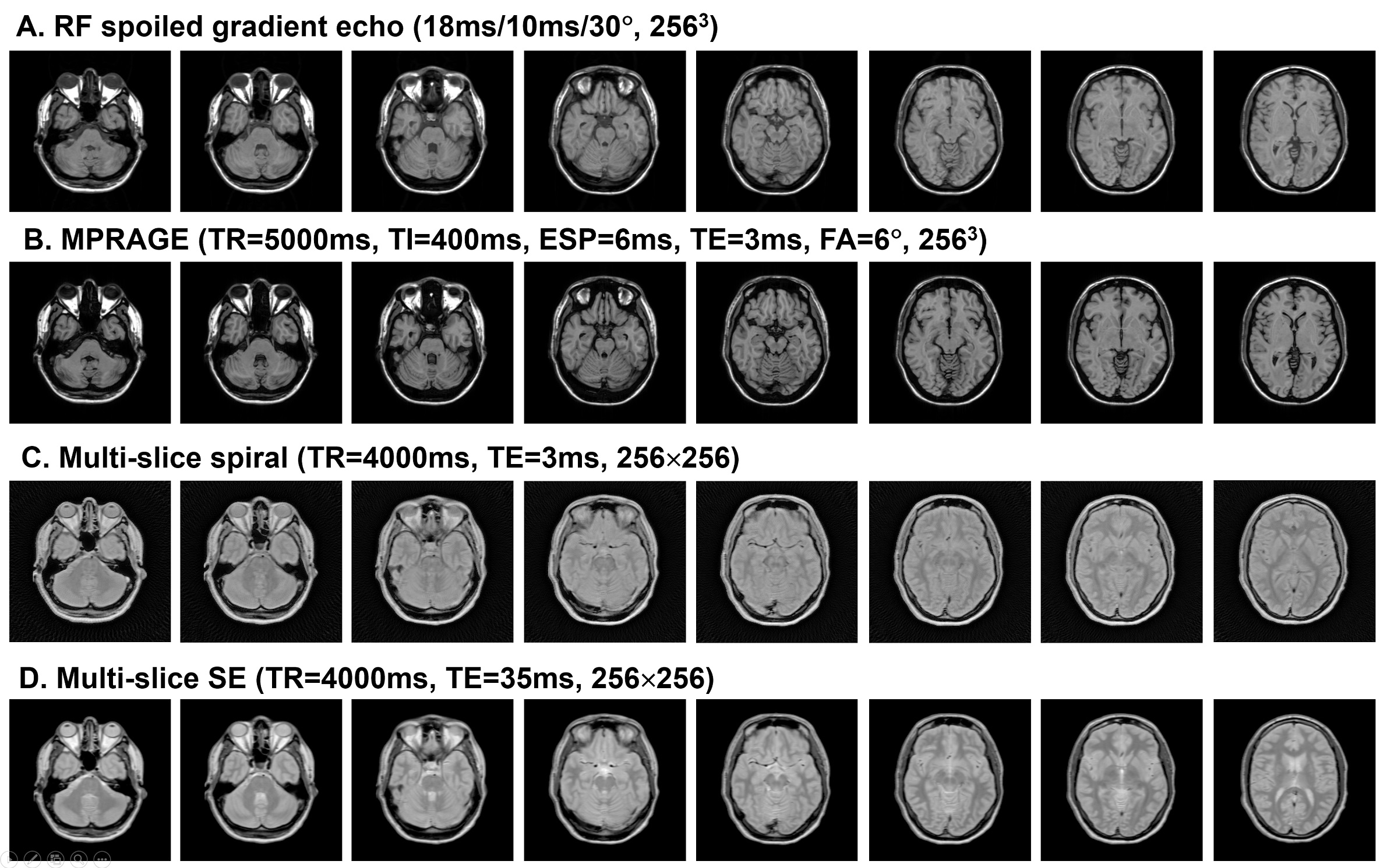
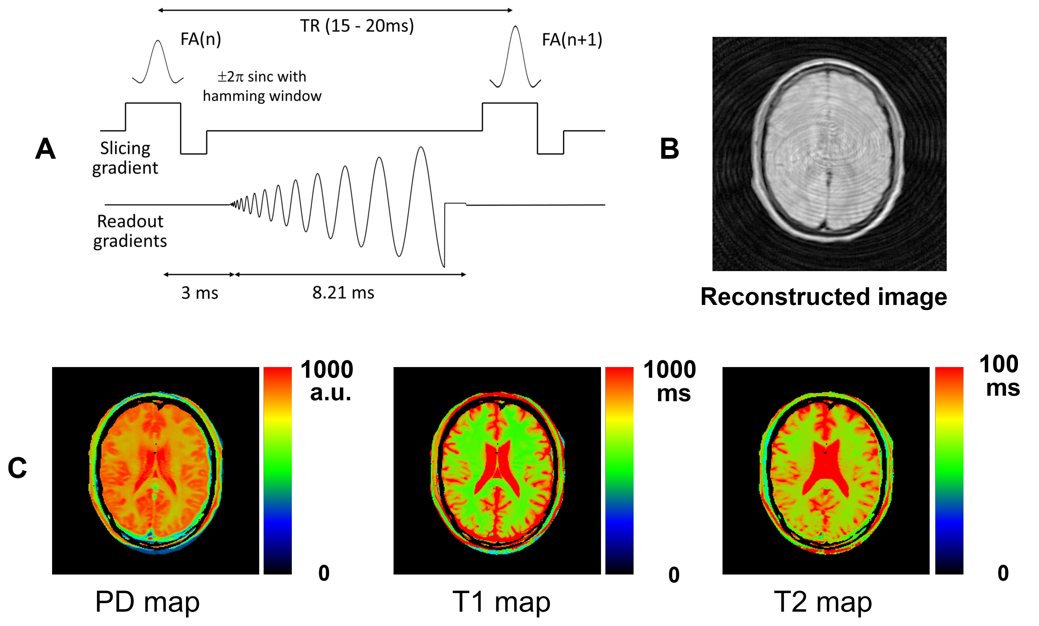
Figure 2 shows cross-sectional MR images obtained by the Bloch image simulations. The numbers of subvoxels, essential to suppression of image artifacts and dominant in the calculation speed, were 17´×1×1, 5×1×1, 1×1×8, and 8×1×2 along the x, y, and z directions for the RF-spoiled GRE, MPRAGE, 2D multi-slice spiral, and 2D multi-slice SE sequences, respectively. Figure 3 shows one TR unit of the MRF sequence, an MR image selected from the MRF image series reconstructed from seven contiguous spiral shots using a sliding window reconstruction technique,6 and NMR parameter maps obtained by a pattern matching. This figure clearly demonstrates that the process of MRF was correctly reproduced by the Bloch image simulation.
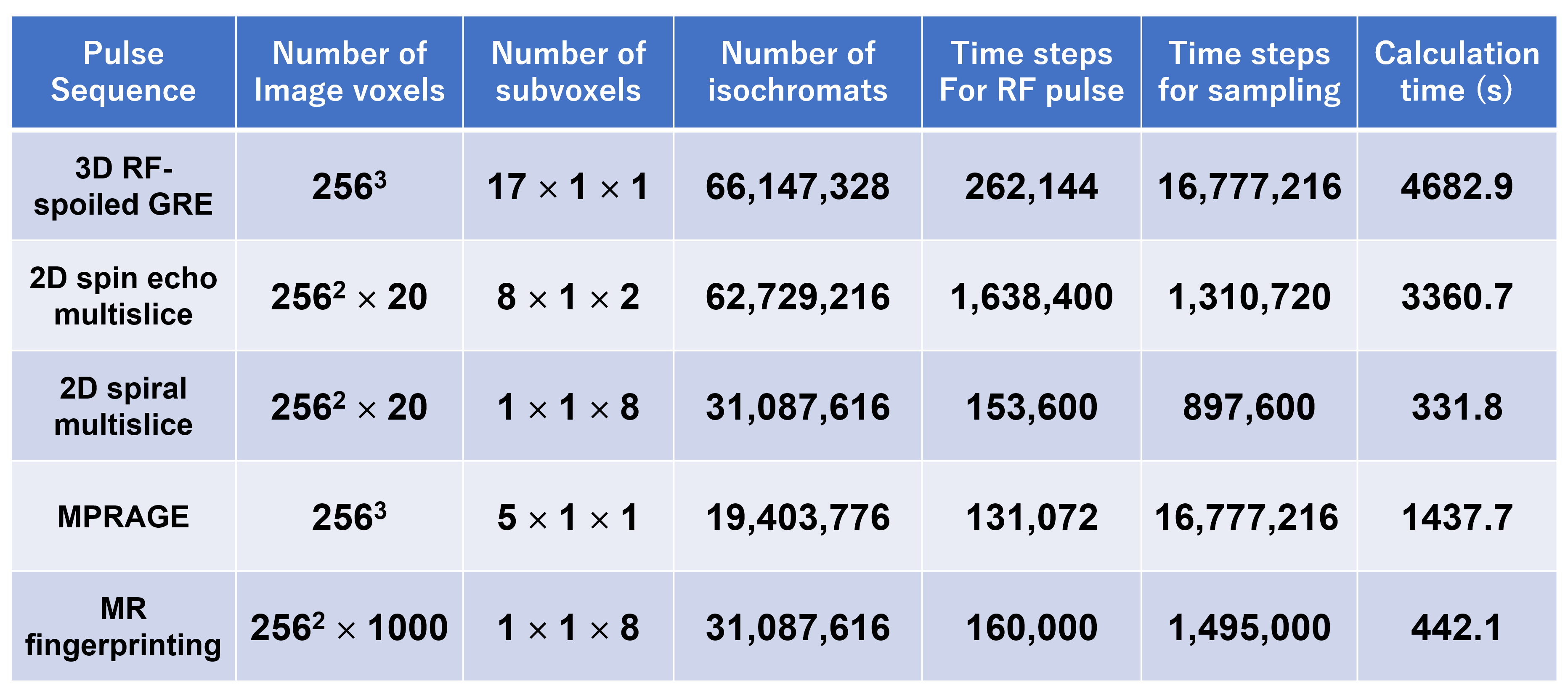
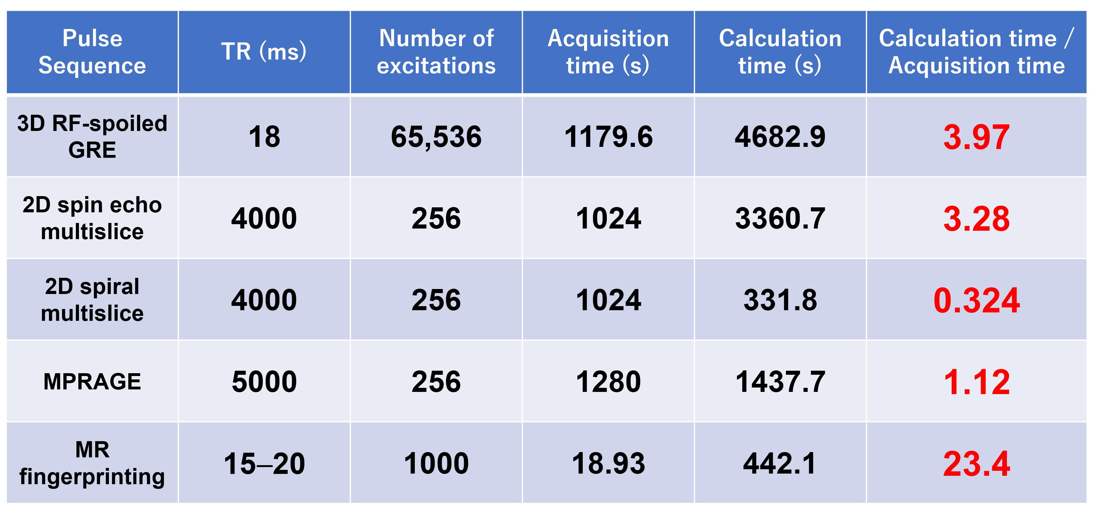
Table 1 summarizes the parameters and calculation times for the Bloch image simulations. Because the calculation times were proportional to the number of isochromats and consisted of the processing times for the RF pulses and those for signal sampling, the processing times for 106 isochromats were estimated from the calculation times. The estimated processing times for one short RF excitation, one signal sampling for the Cartesian trajectory, and one signal sampling for the Non-Cartesian trajectory were 29.7, 3.76, and 6.3-6.8μs, respectively. The processing time for the Non-Cartesian sampling varied according to the number of sampling points for one acquisition. Table 2 summarizes ratios of the calculation times to the acquisition times for the pulse sequences used for the Bloch image simulations. This table clearly shows that the calculation times were the same order as the imaging time in the experiments except the MRF sequence.
Discussion
We demonstrated that artifact-free brain MR images could be obtained using the Bloch image simulator installed in a gaming laptop PC in the comparable time with that of the experiments. The calculation times for the simulation were shown to be composed of the time required for RF excitation and that required for the signal calculation. However, the signal calculation time for the Non-Cartesian sampling was about 1.7 times of that for the Cartesian sampling. This is because in the case of the Cartesian sampling, the rotation factors for the isochromats are constant during the data-acquisition period, whereas in the case of the Non-Cartesian sampling the rotation factors have to be recalculated almost at all points during the data-acquisition period. Since the number of the calculation steps of the GPU required for one signal sampling is 10 steps and its calculation time for 106 isochromats is 3.76μs for the Cartesian sampling, the time for one calculation step for one isochromat is about 0.376×10⁻¹² seconds, which corresponds to an operation speed of about 2.7 TFLOPS. This is a reasonable value from the peak computation speed of 6.7 TFLOPS of the GPU (GTX 1070) used in this study.
In conclusion, this study demonstrated that a GPU-installed gaming laptop PC can perform Bloch image simulations of brain clinical sequences in reasonable processing times and can be a powerful research tool for many MRI researchers.
Acknowledgements
No acknowledgement found.References
1. Kose R, Kose K. BlochSolver: A GPU-optimized fast 3D MRI simulator for experimentally compatible pulse sequences. J Magn Reson 2017;281:51-65.
2. Aubert-Broche B, Evans AC, Collins L. A new improved version of the realistic digital brain phantom. NeuroImage 2006;32:138 – 145.
3. Ma D, Gulani V, Seiberlich N, Liu K, Sunshine JL, Duerk JL, Griswold MA. Magnetic Resonance Fingerprinting. Nature 2013;495:187–192.
4. Jiang Y, Ma D, Seiberlich N, Gulani V, Griswold MA. MR fingerprinting using fast imaging with steady state precession (FISP) with spiral readout. Magn Reson Med 2015;74:1621–1631.
5. Kose R, Setoi A, Kose K. A fast GPU-optimized 3D MRI simulator for arbitrary k-space sampling. Magn Reson Med Sci, accepted for publication in 2018.
6. Cao X, Liao C, Wang Z et al. Robust sliding-window reconstruction for accelerating the acquisition of MR fingerprinting. 2017;78:1579-1588.
Figures