4824
Development of a computer software to quantify bowel motility shown on cine MR imaging by using classical Horn-Schunck approach1Diagnostic Radiology, Tokyo Medical and Dental University, Tokyo, Japan, 2Gastroenterology and Hepatology, Tokyo Medical and Dental University, Tokyo, Japan, 3Gastroenterology and Hepatology, Tokyo Medical and Dental Univerisity, Tokyo, Japan, 4Tokyo Medical and Dental University, Tokyo, Japan
Synopsis
We developed a computer software which quantifies the small bowel motility shown on cine MR imaging using optical flow algorithm with Horn-Schunck approach, by adding a preprocessing step for analyzing cine MR images. A high Pearson’s correlation coefficient was obtained between direct measurement on cine MR and motility map value (r= 0.83 [95% confidence interval: 0.83 – 0.95, P<0.0001]).
Purpose
To develop a computer software which quantifies the small bowel motility shown on cine MR imaging using optical flow algorithm, and to compare the bowel motility value obtained from the software with the direct measurement on cine MR images.Methods
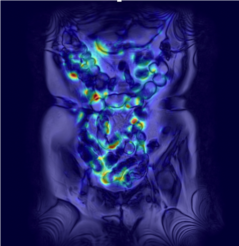
The institutional review board approved this retrospective study. We developed a computer software to present motion vector magnitudes between two consecutive cine MR images as bowel motility maps with classical Horn-Schunck approach(1). This software added a preprocessing step for analyzing cine MR images, and can produce motility maps and color-coded motility maps overlaid onto the cine MR images (Figure 1). Two Crohn disease patients, who underwent MR enterography and had a normal small bowel proven by balloon-assisted enteroscopy, were enrolled. Cine MR imaging was performed by using balanced steady-state free precession sequence in coronal direction, and repeating the acquisition 25 times during 12.8 sec to capture small bowel motility. The direct measurement of small bowel motility on cine MR was performed by using a line region of interests (ROIs) that traces the movement of a point placed on small bowel wall during two consecutive cine MR images, and the measurements were repeated the first 5 phases for five places in each patient. Measurements of motility map value were performed by copying round-shaped ROIs placed on the line ROIs and pasting to the corresponding phase of motility maps. Linear correlation between direct measurement on cine MR and motility map value was calculated using Pearson’s correlation coefficient. A p value < 0.05 was considered statistically significant.Results
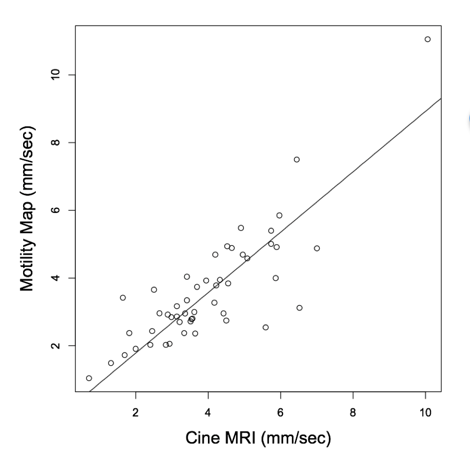
A high positive correlation coefficient was obtained between direct measurement on cine MR and motility map value (r= 0.83 [95% confidence interval: 0.83 – 0.95, P<0.0001])(Figure 2).Discussion
We developed a motility mapping computer software using optical flow algorithm with classical Horn-Schunck approach. In the previous literatures, many studies from a single study group have been reported to evaluate small bowel motilities by using motility mapping tool obtained from the variable brightness optical flow equation with a method of Teng CH et al. and non-rigid registration(2-7). Horn-Schunck approach alone could not provide satisfactory results due to the brightness constancy assumption, however, we have overcome the problem by adding a preprocessing step for cine MR images.
In conclusion, motility map obtained from cine MR images using Horn-Schunck approach was feasible for locally quantifying the bowel motility.
Acknowledgements
No acknowledgement found.References
1. Horn BKP, Schunck BG. Determining optical flow. Artificial Intelligence. 1981;17:185–203.
2. Odille F, Menys A, Ahmed A, Punwani S, Taylor SA, Atkinson D. Quantitative assessment of small bowel motility by nonrigid registration of dynamic MR images. Magn Reson Med. 2012;68:783–793.
3. Menys A, Atkinson D, Odille F, et al. Quantified terminal ileal motility during MR enterography as a potential biomarker of Crohn's disease activity: a preliminary study. Eur Radiol. 2012;22:2494–2501.
4. Menys A, Taylor SA, Emmanuel A, et al. Global small bowel motility: assessment with dynamic MR imaging. Radiology. 2013;269:443–450.
5. Menys A, Helbren E, Makanyanga J, et al. Small bowel strictures in Crohn's disease: a quantitative investigation of intestinal motility using MR enterography. Neurogastroenterol Motil. 2013;25:967–e775.
6. Plumb AA, Menys A, Russo E, et al. Magnetic resonance imaging-quantified small bowel motility is a sensitive marker of response to medical therapy in Crohn's disease. Alimentary Pharmacology & Therapeutics. 2015;42:343–355.
7. Menys A, Makanyanga J, Plumb A, et al. Aberrant Motility in Unaffected Small Bowel is Linked to Inflammatory Burden and Patient Symptoms in Crohn's Disease. Inflamm Bowel Dis. 2016;22:424–432.
Figures