4822
Voxel-based morphometry results in first-episode schizophrenia: a comparison of publicly available software packages1Department of Radiology, Xijing Hospital, Fourth Military Medical University, Xi'an, China, 2GE Healthcare, Xian, China
Synopsis
Investigations of brain structure in schizophrenia using magnetic resonance imaging (MRI) have identified variations in regional grey matter (GM) volume throughout the brain but the results are mixed. This study aims to investigate whether the inconsistent voxel-based morphometry (VBM) findings in schizophrenia are due to the use of different software packages. our data indicate that the GM volume differences between FESZ and HCs depend on which software are used(FSL, SPM), algorithms of GM tissue segmentation and image registration might contribute to these disparate results.
INTRODUCTION
Investigations of brain structure in schizophrenia using magnetic resonance imaging (MRI) have identified variations in regional grey matter (GM) volume throughout the brain but the results are mixed. One factor that contributes to the inconsistency is that some studies recruited both first-episode schizophrenia (FESZ) and chronic schizophrenia,1,2 which are confounded by the effects of chronic illness or antipsychotic treatment, another factor might result from the methodological issues that are related to the analyses of brain structural images.3 This study aims to investigate whether the inconsistent voxel-based morphometry (VBM) findings in schizophrenia are due to the use of different software packages.METHODS
T1 MRI images were obtained from 86 first-episode schizophrenia (FESZ) patients and 86 age- and gender-matched Healthy controls (HCs). VBM analysis was carried out using FMRIB software library (FSL) 5.0 and statistical parametric mapping 8 (SPM8). All images were processed using the default parameter settings as provided by these software packages.RESULTS
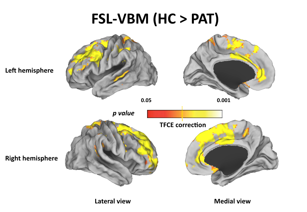
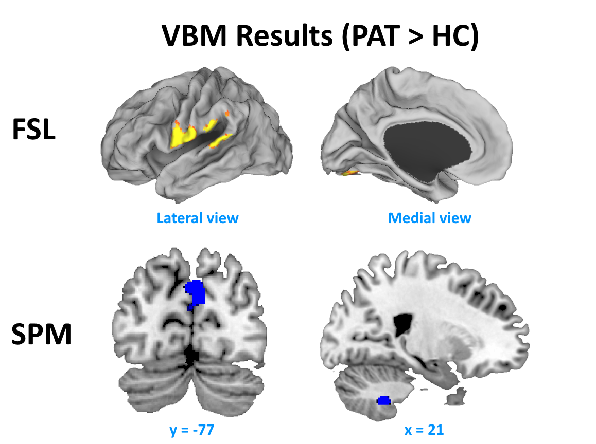
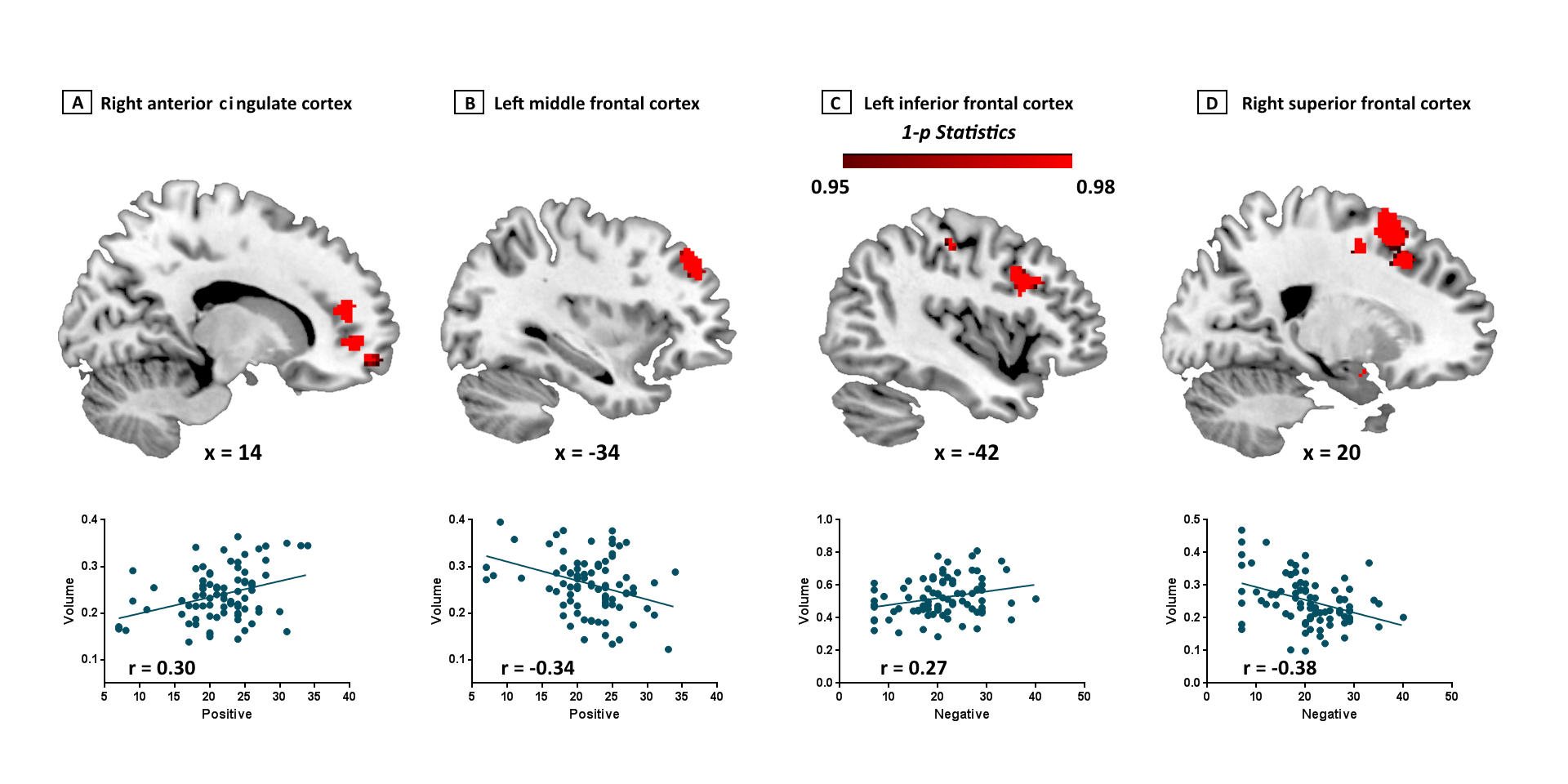
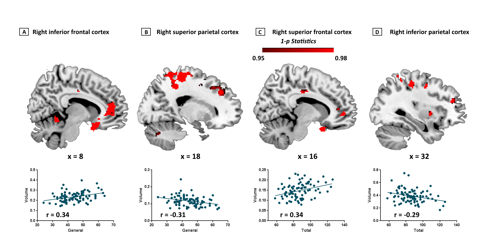
For FSL-VBM, significant differences are found between FESZ patients and HCs, FESZ patients show widespread GM reductions in frontal, temporal, parietal and cingulate cortices, more detailed results are shown in figure 1. However, for SPM-VBM, no significant GM reductions are found in FESZ patients compared with HCs. Both FSL-VBM and SPM-VBM reveal significant increased GM volume in FESZ patients compared with HCs, but the regions are less extensive, detailed results are shown in figure 2. No significant results were found using SnPM statistics. The GM volume of above-mentioned areas are extracted, averaged and regressed with the PANSS Scale, significant correlations were found in brain regions which showed GM reductions including bilateral superior frontal cortex, bilateral inferior frontal cortex, left middle frontal cortex, right superior parietal cortex, right inferior parietal cortex and right anterior cingulate cortex (ACC), detailed results are shown in figure 3 and figure 4.DISCUSSION
In the current study, we explored the GM volume differences between first-episode schizophrenia patients and healthy controls using two commonly used software packages. Marked differences between software packages were observed. Specially, we found that the extensive results obtained by FSL-VBM resemble the findings of most previous structural studies, while SPM-VBM revealed discordant and circumscribed results. Morphometric abnormalities findings reported in FESZ patients might depend on the choice of analysis method.CONCLUSION
our data indicate that the GM volume differences between FESZ and HCs depend on which software are used(FSL, SPM), algorithms of GM tissue segmentation and image registration might contribute to these disparate results. Future studies are needed to determine which algorithms are the most appropriate for processing these types of imaging data.Acknowledgements
This study was financially supported by the National Natural Science Foundation of China under Grant Nos. 81571651, 81601474 and 81801772.References
1. John, J.P., Lukose, A., Bagepally, B.S., Halahalli, H.N., Moily, N.S., Vijayakumari, A.A., Jain, S., 2015. A systematic examination of brain volumetric abnormalities in recent-onset schizophrenia using voxel-based, surface-based and region-of-interest-based morphometric analyses. Journal of Negative Results in Biomedicine,14,1
2. Palaniyappan, L., Maayan, N., Bergman, H., Davenport, C., Adams, C.E., Soares-Weiser, K., 2015. Voxel‐based morphometry for separation of schizophrenia from other types of psychosis in first episode psychosis. John Wiley & Sons, Ltd.
3. Adduru, V.R., Michael, A.M., Helguera, M., Baum, S.A., Moore, G.J., 2017. Leveraging Clinical Imaging Archives for Radiomics: Reliability of Automated Methods for Brain Volume Measurement. Radiology 284(3), 862-869.
Figures