4817
Robust detection of anatomical landmark by combining adaptive boosting and active shape model for automated scan plane planning of spine MRI1Research & Development Group, Hitachi, Ltd., Tokyo, Japan, 2Healthcare Business Unit, Hitachi, Ltd., Tokyo, Japan
Synopsis
Automated scan plane planning is expected to improve MRI scanner usability and provide consistent scan plane prescriptions which are useful for follow-up examinations. However, a landmark degenerated by formation of a lesion such as an intervertebral disc in the case of hernia patient is difficult to detect because shape and properties of tissue greatly deviate from normal cases. In this study, we have proposed combining adaptive boosting and active shape model to detect intervertebral discs robustly for automated scan plane planning of spine MRI.
Purpose
A manual scan plane setup in MRI examination requires careful positioning and repetition of the same task at each examination. Automated scan plane planning is expected to improve MRI scanner usability and provide consistent scan plane prescriptions which are useful for follow-up examinations1. Generally, automated scan plane planning method uses object extraction processing in order to extract anatomical landmarks for scan plane setup. However, a landmark degenerated by formation of a lesion such as an intervertebral disc in the case of hernia patient is difficult to detect because shape and properties of tissue greatly deviate from normal cases. In this study, we have proposed combining adaptive boosting2 and active shape model3 to detect intervertebral discs robustly for automated scan plane planning of spine MRI.Method
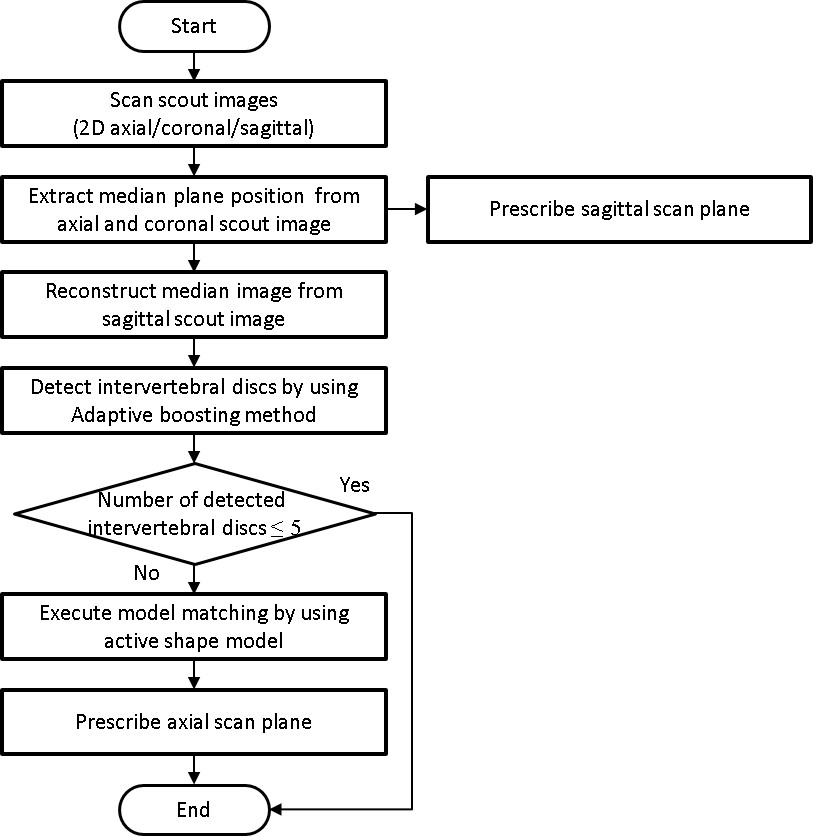
A flow chart of the proposed algorithm is shown in Figure 1. Firstly, 2D scout images of multi-slice orthogonal three-planes (axial, coronal, and sagittal) were scanned. Then, a median plane position, which is defined as left-right symmetry plane, was extracted from axial and coronal scout images by maximizing the normalized cross-correlation of the left and right regions. A sagittal scan plane for diagnosis was prescribed to coincide with the median plane. Furthermore, an image at the median plane position was reconstructed by interpolation from sagittal scout images. Then, intervertebral discs on the reconstructed median plane image were extracted by the adaptive boosting method using LBP (local binary pattern). If the number of detected intervertebral discs was less than five, the processing was terminated. Otherwise, the active shape model matching was performed by using detected intervertebral discs as initial positions. Finally, axial scan planes for diagnosis were prescribed from the matching result.
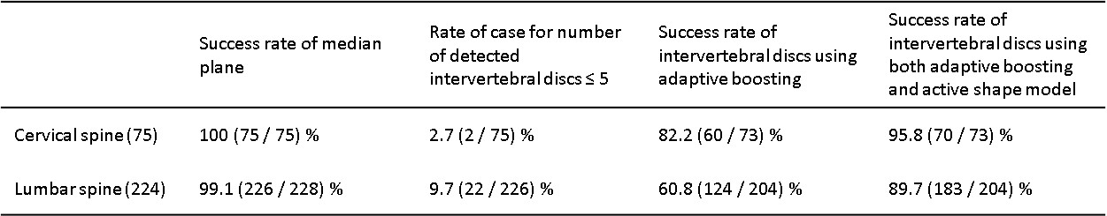
Scout imaging was performed on 75 cervical spine and 228 lumbar spine using 1.5 T and 3 T system. Data were obtained according to the standards of internal review board on Healthcare Business Unit, Hitachi, Ltd. The following success rates of extraction were evaluated: A; median plane for cervical and lumbar spine, B; intervertebral discs form C2 to C7 for cervical spine, C; intervertebral discs form L1 to S1 for lumbar spine. The extraction success rates of intervertebral discs were calculated only when extraction of median plane succeeded and adaptive boosting extracted six or more intervertebral discs. The success rates were compared between a case of using adaptive boosting and a case of using both adaptive boosting and active shape model.
Results
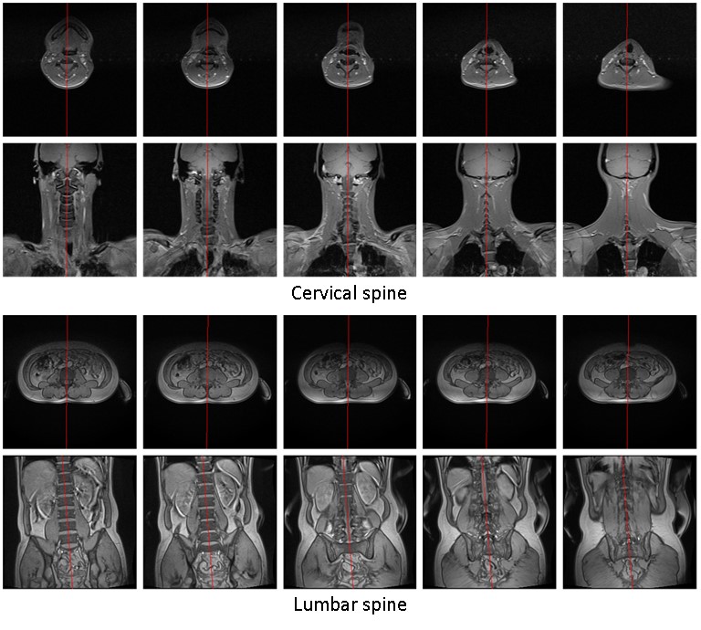
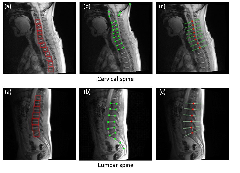
Figure 2 shows an example of automatically prescribed sagittal planes. The sagittal planes coincided with the median plane. Figure 3 shows example of automatically prescribed axial planes ((a) detected intervertebral discs by using adaptive boosting, (b) matching result of active shape model and (c) prescribed axial scan planes). The axial planes coincided with the inclination of intervertebral discs. The results of evaluation are summarized in Table 1. The success rates of extracting median plane were 100 % for cervical spine and 99.1 % for lumbar spine. When the number of detected intervertebral discs were less than five, the rates were 2.7 % for cervical spine and 9.7 % for lumbar spine. The success rate of extracting intervertebral discs using only adaptive boosting were 82.2 % for cervical spine and 60.8 % for lumbar spine. The success rate of extracting intervertebral discs by combining adaptive boosting and active shape model were 95.8 % for cervical spine and 89.7 % for lumbar spine. The processing time was about 2.5 seconds, executing offline processing with CPU: Intel® Core ™ 2 Duo 3.00 GHz calculator.Discussion
The proposed method combining adaptive boosting and active shape model greatly improves the success rate of extracting intervertebral discs when compared with using simple object extraction method (i.e. only adaptive boosting). Adaptive boosting which extract individual intervertebral discs often suffer from errors such as over detection and detection failure. On the other hand, active shape model can reduce such errors to extract intervertebral discs maintaining the positional relationship of the entire vertebral body, but the method’s success rate of matching is affected by the initial position for matching. In our method, combining the two methods makes it possible to complement each other’s weak points and realize robust detection. In this study, approximately half of the failure was caused by the matching errors, and the other half was caused by unclear of intervertebral discs due to large degeneration or motion and susceptibility artifacts.Conclusion
Acknowledgements
No acknowledgement found.References
1. Yokosawa S, Taniguchi Y, Bito Y, et al. Automated scan plane planning for brain MRI using 2D scout images, Proceeding of ISMRM 18, 2010: 3136.
2. Freund Y, Schapire RE, A decision – theoretic generalization of on-line learning and an application to boosting, Journal of computer and system sciences 1887;55(1):119-139.
3. Cootes TF, Taylor CJ, Cooper DH, Graham J, Active Shape Models -Their Training and Application, CVIU 1995;61(1):38-59.
Figures