4805
Machine Learning Techniques for Bone Tumor Segmentation using Diffusion MRI1Center for Biomedical Engineering, Indian Institute of Technology, Delhi, India, New Delhi, India, 2Medical Oncology, IRCH, All India Institute of Medical Sciences, New Delhi, India, New Delhi, India, 3Radio Diagnosis, All India Institute of Medical Sciences, New Delhi, India, New Delhi, India, 4Department of Biomedical Engineering, All India Institute of Medical Sciences, New Delhi, India, New Delhi, India
Synopsis
Automatic and accurate segmentation of osteosarcoma region in MRI images can assist doctor to prepare a feasible treatment plan, hence resulting in improved cure rate. The purpose of this study was to evaluate and compare the performance of automated and semi-automated algorithms that might be effective in segmenting bone tumor in MRI data, with reasonable accuracy, speed and minimal manual input. The results are very conclusive for efficient performance.
Purpose:
Osteosarcoma is the most common bone cancer in children and adolescents1 that primarily affects the long bones of the body. Delineation of tumor and assessment is vital in treatment planning and monitoring. Therefore accurate and non-invasive method for segmentation is needed since manual segmentation is time-consuming and prone to error & operator subjectivity2. To determine the extent of tumor and monitoring treatment response in the patient, digital image analysis is a potential method to automate this process.Method:
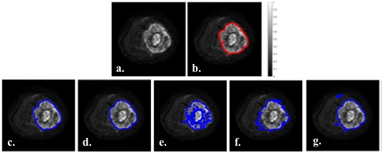
MRI dataset for Twenty patients (n=20;M:F=16:4;15.5±2.6yrs) with Osteosarcoma were acquired on1.5T Philips Achieva MRI scanner. DWI was acquired with Spin-Echo-Echo-Planar-imaging with TR/TE=7541/67msec, matrix-size=192×192, slice-thickness/Gap=5mm/0.5mm, voxel-size=2.98/3.52/5.0mm, b-values=0-800s/mm2. T2W images were acquired using Turbo-Spin-Echo with TR/TE=73795/85msec, matrix-size=384×384. T1W images were acquired using Turbo-Spin-Echo with TR/TE=644/10msec, matrix size=512×512. Region-of-interest (ROI) for tumor were demarcated manually by a radiologist (>10years of experience) on each slice of DWI images with b-value 800s/mm2(DWI800) (Figure1.a&b).
Total four automated algorithms; two un-supervised clustering based algorithms, 1) Simple-linear-iterative-clustering(SLIC) superpixels(SLIC-S)3 and 2) Fuzzy c-means clustering(FCM)4; and two supervised machine learning(ML) algorithms, 3) Logistic-Regression(LR)5, 4) Linear-Support-Vector-Machines(L-SVM)6, were built in-house and compared for detection and differentiation of tumor from healthy tissues in the dataset. Otsu-thresholding(OT)7 was reported as a baseline segmentation method for comparison. Thus in total five segmentation algorithms were compared for segmentation and identification of bone tumor using DWI. SLIC-S generates superpixels by clustering pixels based on intensity similarity and proximity in plane3. FCM is used to get a segmentation through fuzzy pixel classification, allowing pixels to be part of multiple clusters4. Texture analysis, mathematical technique for characterizing spatial distribution of intensity levels in image, was used to extract features for ML algorithms. Textural feature vector (length=18) comprising i)signal intensity, ii-iii)voxel position, features using iv-vii)Grey-level Co-occurrence Matrix8 and viii-xviii)Grey-Level Run-Length Matrix9, were evaluated in tumor ROIs from DWI800, T1W, T2W and PDW images to train the classifiers and 4-fold cross-validation was used for prediction. ML algorithms, LR builds a binary classifier with a sigmoid non-linearity by minimizing cross entropy loss5 while L-SVM builds a max-margin classifier and minimize hinge loss6. Mean ADC in tumor volume across patients using different methods was calculated for comparison.
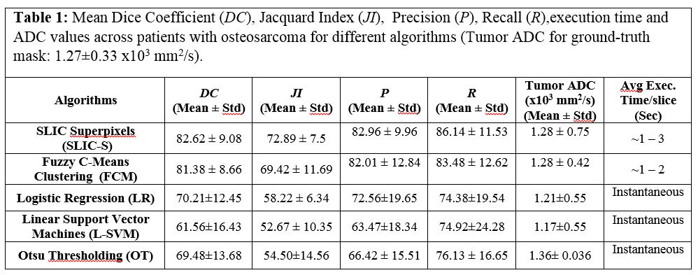
Accuracy-metrics: Ground truth ROIs and segmentation results were compared to evaluate the performance using jacquard index(JI)=│A∩B│/│AUB│×100 , dice-coefficient(DC)=2│A∩B│/(│A│+│B│)×100 , precision(P)=│A∩B│/│B│×100 and recall(R)=│A∩B│/│A│×100 , where A and B are areas demarcated as tumor by radiologist and segmentation algorithm respectively. Segmentation methods and accuracy calculation were implemented in MATLABR2016a and python2.7.
Result:
Figure1 shows an illustrative example of segmented images with all five methods corresponding to the original image (Figure1.a) from one representative patient with OS. Table1 shows the accuracy-metric for segmentation methods across tumor volume for all patients and the execution-time/slice. Clustering based SLIC-S and FCM methods showed quantitatively and qualitatively better segmentation with DC:~81-83%;JI:~69-73%;P:~82-83%;R:~84-86% than the ML based methods LR and L-SVM (DC:~62-70%;JI:~53-58%;P:~64-73%;R: ~74-75%) or thresholding based OT (DC:~70%;JI:~55%;P:~66%;R:~76%). OT, LR, L-SVM methods produces the result almost instantaneously; while SLIC-S, FCM took comparable ~1–3 sec to process one slice image. For SLIC-S and FCM, mean ADC in segmented tumor region were close (1.28x103mm2/s) to the actual ADC value 1.27±0.33x103mm2/s calculated using ground-truth ROI; LR and L-SVM produced comparatively lower (<1.21x103mm2/s) and OT produced comparatively higher (1.36x103mm2/s) value of ADC.Discussion & Conclusion:
A comparative analysis of segmentation techniques using DWI dataset for bone tumor is presented. DWI was used for segmentation as it reflects molecular chages of pathological tissue during treatment more prominently than structural imaging that may help in better delenation of the active tumor and further image analysis.
From our previously reported studies10,11,12, we compare four segmentation algorithms viz semi-automated Otsu threshold-based region-growing(RG) and Active-Contour(AC) requiring manual initial input (seed-points/ROI respectively) and automated energy based graph-cut(GC) and Deep-feed-forward-neural-network(DNN) with the currently implemented methods. AC and OT-RG also produced good segmentation accuracy (DC:~82-83%;JI:~70-71%;P:~77-78%;R: ~87-89%, ~1–6 sec execution-time/slice-image) along with SLIC-S and FCM among all methods. DNN and GC showed comparable accuracy (DC:~72-74%;JI:~58-60%;P:~72-73%;R:~80-84%. while DNN produced results almost instantaneously; GC took comparatively long execution-time ~38-42sec/slice-image. Among supervised ML algorithms, DNN showed highest accuracy (DC:~72%;JI:~58%;P:~73%;R:~80%) than LG or L-SVM. Though ML algorithms produces instantaneous results but it needs considerable time while training. Accuracy parameters might be improved for ML algorithms with a larger dataset. RG and AC methods implementd as semi-automatic methods, need manual intervention for selection of seed-points/ROI. Clustering based SLIC-S and FCM were implemented as fully automatic techniques which produce compratively efficient, accurate, reliable and faster results. FCM method is faster among all methods for segmentation of bone tumor.
Acknowledgements
Authors would like to thank Ministry of Human Resource Development, Government of India for providing funding support as research fellowship to E.B.K. required for the study. Authors would also like to thank and acknowledge the valuable input of the intern team, Abhimanyu Sahai, Rishabh Gupta, Akshay Gupta, Kabir Chhbra in data processing and various stages of implementation.References
1. Geller DS et all. Osteosarcoma : A review of diagnosis , management , and treatment strategies. Clin Adv Hematol Oncol. 2010;8(10):705-718.
2. Mancas M et al. Segmentation Using a Region Growing Thresholding. Proc. SPIE 5672, Image Processing: Algorithms and Systems IV. ; 2005.
3. Achanta R et al. SLIC Superpixels. IEEE Trans Pattern Anal Mach Intell. 2012;34(11):2274-2282.
4. Selvathi D et al. MRI Image Segmentation Using Unsupervised Clustering Techniques. IEEE Proceedings of the Sixth Int.Conf. ICCIMA. ; 2005:5-10.
5. Cox DR. Regression Analysis of Binary Sequences. J R Stat Soc. 2017; 20(2): 215-242.
6. Saitta L. Support-Vector Networks. 1995;297:273-297.
7. Otsu N. A Tlreshold Selection Method from Gray-Level Histograms. IEEE Trans SYSTREMS, MAN, Cybern. 1979;SMC-9(1):62-66.
8. Haralick RM. Statistical and Structural Approaches to Texture. Proc IEEE. 1979;67(5):786-804.
9. Galloway MM. Texture analysis using grey-level run lengths. Comput Graph Image Process. 1975;4:172-179.
10. Mehndiratta et. al. Automated Segmentation of Ewing’s Sarcoma using Diffusion Weighted Imaging, Proc. 24th ISMRM, 2017. https://www.ismrm.org/16/program_files/EP02.htm
11. Mehndiratta et. al. Textural Analysis based Segmentation of Bone Tumors using Diffusion weighted MR image, Proc. 25th ISMRM, 2017. http://archive.ismrm.org/2017/4396.html
12. Mehndiratta et. al. Automatic Segmentation of Bone Tumor with MR imaging, Proc. 26th ISMRM, 2018. http://archive.ismrm.org/2018/2717.html
Figures