4804
Automated Segmentation of Thalamic Nuclei using Convolutional Neural Networks1electrical and computer engineering, university of arizona, tucson, AZ, United States, 2university of arizona, tucson, AZ, United States, 3stanford, stanford, CA, United States, 4medical imaging, university of arizona, tucson, AZ, United States
Synopsis
parcellation of thalamic nuclei is
Introduction
Accurate delineation and volumetry of thalamic nuclei is critical for many applications including targeting for deep brain surgery, longitudinal tracking of diseases such as Alzheimer’s and multiple sclerosis (MS). However, due to poor contrast, thalamic nuclei are not easily distinguishable in conventional T1 or T2 imaging, especially at 3T. Furthermore, due to limitations in spatial resolution as well as reduced fractional anisotropy in the thalamus, diffusion tensor based methods (DTI)1-3 have been successful only in segmenting the larger nuclei. Recently, visualization and manual segmentation of the thalamic nuclei was reported using a white-matter nulled (WMn) magnetization-prepared rapid gradient echo (MP-RAGE) sequence4 at 7T. This was further automated using multi-atlas based segmentation methods5-6. These involve nonlinear registration steps, making it time-consuming. In this study, we investigated the use of convolutional neural networks to achieve fast and robust segmentation of thalamic nuclei at both 3T and 7T.
Methods
MRI acquisition: All images were acquired using a 3T and 7T MRI scanner using the WMn MP-RAGE sequence after informed consent following institutional review board guidelines. The sequence details can be found here 4,7
Preprocessing:
The images were first N4 bias corrected using ANTs and then normalized to the range [0,1] in order to account for variations in image intensity. All 3D images were reformatted to the coronal plane and then split into 2D slices. For 7T, due to the small amount of training data, inputs were warped to a common template space for faster network convergence. For 3T, an automated cropping scheme was developed which intelligently cropped the inputs after rigid registration to a predefined crop in the template space to encompass one of the two thalami for unilateral segmentation.
CNN Architecture:
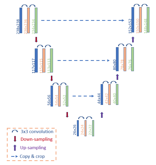
A four-layer U-net architecture was used8 ( Figure 1). The final network consists of two parts; the reducing part consists of a repeated combination of 2 figure maps of size 3x3, a rectified linear unit (ReLu) and a 2x2 max-pooling (with stride/step of 2). This combination is repeated for each layer. A 25% dropout layer was used in training to avoid over fitting. The input images were padded in all directions to achieve a constant input size. For 7T data, the standard U-net was used. For 3T data, a cascaded U-net scheme was used that first segmented the whole thalamus using a U-net and then used the whole thalamus segmentation as an input to the next U-net for segmentation of the thalamic nuclei. .
Model training and validation
For 7T, the network was trained on 19 data sets (a mix of healthy subjects and MS patients) with 32 slices each, using manual labels as ground truth. For 3T, due to lack of manual labels, labels generated using Thalamus Optimized Multi Atlas Segmentation (THOMAS)5-6 were used for training 75 data sets (a mixture of controls and patients with alcohol use syndrome) with 40 slices each. The network was trained using a cross-entropy loss function and an ADAM optimizer, reserving about 10% of the data as validation. Dice coefficients were used as a performance metric. The processing was done on an 8 GPU machine (NVIDIA Tesla P100) with 45 cores and 250 GB RAM.
Results
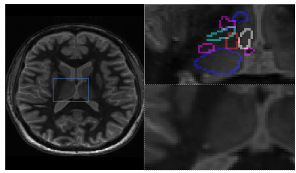
Figure 2 shows a representative WMn MP-RAGE slice, the cropped image that was input to the network and the same input image overlaid with the 12 thalamic nuclei labels generated by the network .
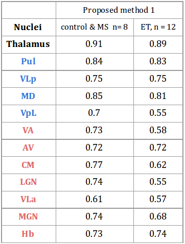
Table 1 shows mean dice (compared against manual segmentation) for the 7T datasets for two populations-8 cases of healthy subjects and patients with MS and 12 cases of patients with ET. Note the high dice achieved with the ET data set even though the network was trained on control and MS data, attesting to the robustness of the network.
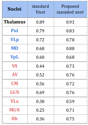
Table 2 compares mean dice for the 3T dataset from the standard U-net and the proposed cascaded U-net. Note the excellent dice achieved on large nuclei and acceptable dice (>0.7) for smaller nuclei. The cascaded U-net method takes about 1.5 min for segmenting all 12 nuclei making it significantly faster than previously reported schemes.
Conclusions
We have presented a CNN based technique for segmentation of the thalamus into 12 nuclei. The cascaded network improves the segmentation performance by utilizing prior information from a first-pass thalamic segmentation. In future, the use of a residual learning network can further improve efficiency.Acknowledgements
No acknowledgement found.References
1. Behrens, Timothy EJ, H. Johansen-Berg, M. W. Woolrich, S. M. Smith, C. A. M. Wheeler-Kingshott, P. A. Boulby, G. J. Barker et al. "Non-invasive mapping of connections between human thalamus and cortex using diffusion imaging." Nature neuroscience 6, no. 7 (2003): 750.
2. Wiegell, Mette R., David S. Tuch, Henrik BW Larsson, and Van J. Wedeen. "Automatic segmentation of thalamic nuclei from diffusion tensor magnetic resonance imaging." NeuroImage 19, no. 2 (2003): 391-401.
3. Battistella, G. et al., 2017. Robust thalamic nuclei segmentation method based on local diffusion magnetic resonance properties. Brain structure & function, 222(5), pp.2203–2216
4. Tourdias, Thomas, Manojkumar Saranathan, Ives R. Levesque, Jason Su, and Brian K. Rutt. "Visualization of intra-thalamic nuclei with optimized white-matter-nulled MPRAGE at 7 T." Neuroimage 84 (2014): 534-545.
5. Jason Su, Thomas Tourdias, Manojkumar Saranathan, Pejman Ghanouni, and Brian Rutt, THOMAS: Thalamus Optimized Multi-Atlas Segmentation at 3T, Proceedings of ISMRM. 2016 May
6. Thomas FT, Su J, Rutt BK, Saranathan M. A method for near realtime automated segmentation of thalamic nuclei. Proc. of the 25th scientific meeting of the ISMRMl Hawaii 2017, p4736
7. Saranathan, Manojkumar, Thomas Tourdias, Ersin Bayram, Pejman Ghanouni, and Brian K. Rutt. "Optimization of white‐matter‐nulled magnetization prepared rapid gradient echo (MP‐RAGE) imaging." Magnetic resonance in medicine 73, no. 5 (2015): 1786-1794.
8. Ronneberger, Olaf, Philipp Fischer, and Thomas Brox. "U-net: Convolutional networks for biomedical image segmentation." In International Conference on Medical image computing and computer-assisted intervention, pp. 234-241. Springer, Cham, 2015..
Figures