4802
Automatic Glioma Segmentation Algorithm Based on Superpixel Features1School of Electronic and Information Engineering, Xi’an Jiaotong University, Xi'an, China, 2Collaborative Innovation Center for Internet Healthcare and School of Software and Applied Technology, Zhengzhou University, Zhengzhou, China, 3Department of Radiology, Henan Provincial People’s Hospital, Zhengzhou, China
Synopsis
This study proposes an algorithm to locate and segment Glioma tumor automatically. The algorithm contains three main steps. Firstly, a self-adaptation simple linear iterative clustering (ASLIC0) algorithm was executed to segment T2 weighted MRI images to superpixels images. Then, 52 features including fractal features, curvature feature and higher order derivative map Haralick texture features was calculated on each superpixel. Finally, a Support Vector Machine was trained as a classifier to select superpixels belong to tumor lesion or not. The Dice overlap measure for the segmented Glioma is 0.87 on the data set from the Henan Provincial People’s Hospital.
Introduction
Glioma is a prevalent fatal brain disease, which accounts for approximately 24.7% of all primary brain and other central nervous system tumors, as well as 74.6% of malignant tumors[1]. With the progress of information technology, especially the development of machine learning, the demand for computer-aided diagnosis, adjuvant surgery, radiotherapy and chemotherapy plan and medical research of Glioma patients is more and more intense. Feature calculation is the first step in applying machine learning which means the quantification of the Region Of Interest (ROI) in Glioma tumor. Therefore, the segmentation of ROI is the basis of all subsequent analysis. In practice, existing Glioma segmentation is hard to be applied in clinical routines because of the heterogeneous nature of Glioma and the image acquisition procedures. At present, it is still a challenge work to make an objective, accurate and repeatable brain tumor segmentation. This study proposes an algorithm to locate and segment Glioma tumor automatically.Methods
The whole process of Glioma segmentation algorithm presented in this paper was shown in Fig. 1. For the training set, an adaptive superpixel segmentation algorithm ASLIC0 was used to calculate the superpixel maps of Glioma MRI images. Then, fractal features, curvature features and Haralick texture features were calculated for each superpixel. Finally, a SVM classifier was trained to classify the superpixel blocks into tumor region or not, and then the ROI of Glioma is segmented.
The superpixel segmentation algorithm (SLIC) [2] uses K-means clustering algorithm to superpixel processing. The selection of parameters has great influence on the segmentation performance. The parameters of the SLIC algorithm include the number of superpixels K and compactness C. The parameter K determines the number of images divided into irregular regions and he parameter C determines the degree of irregularity. A large number of repeated experiments on MRI images of T2-weighted Gliomas found that there is a direct relationship between the parameter K and the complexity of the region of interest on size and edge, as shown in Fig. 2.
In order to quantify the complexity of the tumor boundary, a complexity index TC was proposed. For Glioma images that have not segmented, the tumor area ratio (TAR) are calculated. This paper selected the K = 200, 120, and 40 respectively according to the difference condition of area ratio and boundary complexity. According to experiment, if TAR and TC are greater than the threshold la, lc, then K selects 200, if TAR and TC are less than the threshold la, lc, then K selects 40, and in other cases K selects 120.
Then, 52 features included Haralick texture features[3], curvature features[4] and fractal features[5] were calculated on each superpixel. These features combined with the training label composed a training set, and then, we classify the tumor area and the non-tumor area by an SVM classifier with linear kernel whose optimization routine was Sequential Minimal Optimization (SMO).
Results
In order to verify the accuracy of the segmentation method, Dice[6] was calculated. The closed Dice is to 1, the better segmentation performance is. After experimental comparison, the algorithm is effective for most Glioma images. According to statistics, there are 119 cases’ Dice exceed 0.8 which accounting for 73.92% of all data. For Gliomas with a Dice less than 0.8, the performance of this algorithm is poor and needs to be reviewed by radiologist and then processed by other algorithms.Discussion
After analyzing the segmentation results, the method proposed by this paper has good segmentation results in Glioma images with high contrast between tumor and normal tissues, and can obtain good results on various levels. The specific segmentation result is shown in Fig. 3.Conclusions
The algorithm contains three steps: superpixel segmentation, feature calculation based on superpixel, and classification with an SVM classifier. Experiments shown good performance on most T2 weighted MR images, and the average dice can reach 0.87. The result indicated that this method has good segmentation performance for Gliomas.Acknowledgements
This study was funded by National Natural Science Foundation of China (Grant 81772009), Scientific and Technological Research Project of Henan Province (Grant 182102310162).References
- Q. T. Ostrom et al., "CBTRUS Statistical Report: Primary Brain and Other Central Nervous System Tumors Diagnosed in the United States in 2009–2013," Neuro-Oncology, vol. 18, no. suppl_5, pp. v1-v75, 2016.
- R. Achanta, A. Shaji, K. Smith, A. Lucchi, P. Fua, and S. Süsstrunk, "SLIC superpixels compared to state-of-the-art superpixel methods," IEEE Transactions on Pattern Analysis & Machine Intelligence, vol. 34, no. 11, pp. 2274-2282, 2012.
- R. M. Haralick, K. Shanmugam, and I. H. Dinstein, "Textural Features for Image Classification," Systems Man & Cybernetics IEEE Transactions on, vol. smc-3, no. 6, pp. 610-621, 1973.
- N. Ohtsu, "A Threshold Selection Method from Gray-Level Histograms," IEEE Transactions on Systems Man & Cybernetics, vol. 9, no. 1, pp. 62-66, 1979.
- N. Sarkar and B. B. Chaudhuri, "An efficient differential box-counting approach to compute fractal dimension of image," IEEE Transactions on Systems Man & Cybernetics, vol. 24, no. 1, pp. 115-120, 1994.
- W. R. Crum, O. Camara, and D. L. Hill, "Generalized overlap measures for evaluation and validation in medical image analysis," Medical Imaging IEEE Transactions on, vol. 25, no. 11, pp. 1451-1461, 2006.
Figures
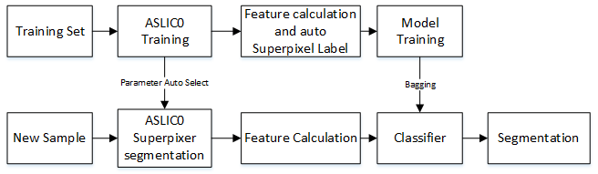
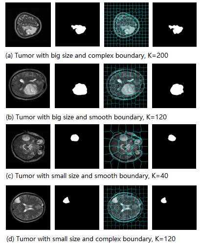
Fig. 2 Influence of tumor area size and boundary complexity on K value.
Note: The first column is the original image, the second column is the manual segmentation, the third column is the superpixel images, the fourth column is the segmentation result by superpixel.
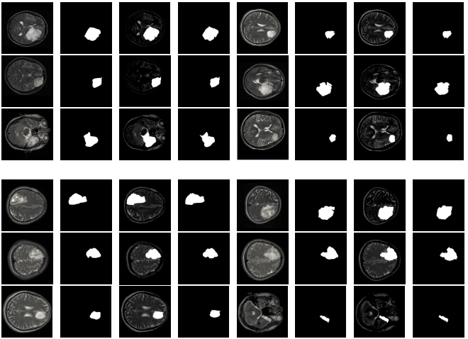
Fig. 3 Glioma image segmentation results.
Note: the first and the fifth columns are the original images, the second and the sixth column are manually defined, the third, the fourth, the seventh, and the eighth columns were segmentation result. The first to third rows were results on low grade gliomas while the fourth to sixth rows were high grade Gliomas.