4787
Accelerated Targeted Coronary MRI Using Sparsity-Regularized SPIRiT-RAKI1Electrical and Computer Engineering, University of Minnesota, Minneapolis, MN, United States, 2Center for Magnetic Resonance Research (CMRR), University of Minnesota, Minneapolis, MN, United States, 3Computer Assisted Clinical Medicine, University Medical Center Mannheim, Heidelberg University, Mannheim, Germany
Synopsis
Long scan duration remains a challenge in coronary MRI. A scan-specific machine learning technique, called Robust Artificial-neural-network for k-space Interpolation (RAKI) has recently shown promising results in accelerating MRI. However, RAKI was originally designed for uniform undersampling patterns. In this study, we propose a technique, called SPIRiT-RAKI that enables RAKI with arbitrary undersampling using scan-specific convolutional neural networks to enforce self-consistency among coils. Regularization terms are also incorporated in the new formulation. Our results indicate that SPIRiT-RAKI can successfully accelerate 3D targeted coronary MRI.
Introduction
Lengthy acquisition times remain a major limitation for coronary MRI. Several acceleration techniques have been proposed for coronary MRI1-6. Recently, a machine learning (ML) technique, called Robust Artificial-neural-network for k-space Interpolation (RAKI), has shown promise in accelerated cardiac MRI7. RAKI uses scan-specific convolutional neural networks (CNN) to nonlinearly interpolate the missing data in undersampled kspace, extending the linear convolutional kernels of GRAPPA8. Scan-specificity of RAKI implies that training the CNN relies on the data from the same scan, which alleviates the need for large datasets used in most ML methods. However, RAKI was originally designed for uniform undersampling patterns. In this study, we utilize the notion of self-consistency of SPIRiT9 to extend RAKI to arbitrary undersampling patterns for accelerating targeted coronary MRI. Similar to SPIRiT, additional priors and regularization terms can also be incorporated to this formulation called SPIRiT-RAKI. We evaluated SPIRIT-RAKI on targeted coronary MRI datasets.Materials and Methods
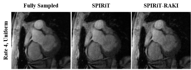
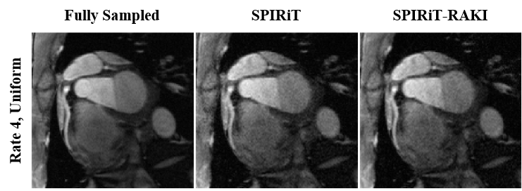
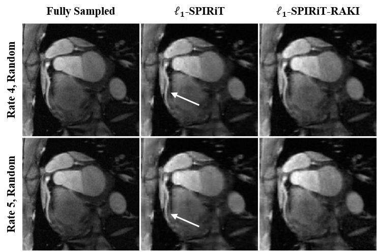
Coronary MRI: Targeted right coronary MRI was acquired on two healthy subjects at 3T with a 30-channel body-coil using a T2-prepared GRE sequence. Imaging parameters were FOV=300x300x60mm3 and resolution=1x1x3mm3. The data were retrospectively undersampled, both uniformly (acceleration rate of 2x2 along ky and kz directions) and randomly (acceleration rates of 4 and 5). For uniform sampling, no regularization term was used for fair comparison to conventional parallel imaging methods. ACS region was selected as the central 45x10 ky-kz lines.
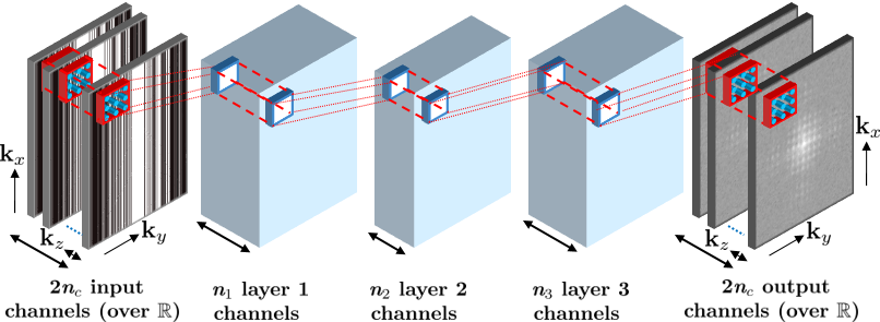
SPIRiT-RAKI: A 4-layer CNN architecture with 3-dimensional kernels was employed to find a nonlinear mapping function from acquired data points to missing data (Fig. 1). In contrast to RAKI, convolutional kernels do not use dilation and the output included the k-space across all coils, which significantly reduced the required time for both calibration and reconstruction. Therefore, the network consisted of input and output channels, where represents the number of coils. The factor of is due to complex k-space being mapped to a real field. All layers except the output layer were followed by rectifier linear units (ReLU). Tikhonov regularization was applied to the weights at each layer to avoid overfitting. The network was trained on the ACS region, with a mean square error objective function and an ADAM optimizer. Following the training of the CNN, reconstruction was performed using the following objective function:
||y - DX||22 + β||x - G(x)||22 + γ||WEx||1
where x is the desired k-space data across all coils, y is the noisy acquired data, D is the under-sampling operator, G(.) represents the CNN nonlinear operations to enforce self-consistency, E is the operator combing coil images into a SENSE Rate-1 image, and W transforms this image into the wavelet domain. β and γ were empirically chosen.
Results
Fig. 2 demonstrates a representative slice from the right coronary MRI of a healthy subject using SPIRiT and SPIRiT-RAKI with uniform ky-kz undersampling rate of 4. SPIRiT amplifies noise slightly more than SPIRiT-RAKI. Fig. 3 depicts a similar trend on another subject. The regularization results from random ky-kz undersampling are shown in Fig. 4 for undersampling rates of 4 (top row) and 5 (bottom row). Both results suffer from blurring artifacts, with more blurring visible for L1-SPIRiT. The white arrows point to patch-like artifacts near the mid portion of the right coronary artery, adversely affecting visualization.Discussion
We proposed SPIRiT-RAKI, which extends the RAKI method to arbitrary undersampling patterns using the self-consistency idea of SPIRiT. This method was evaluated on coronary MRI using both uniform and random undersampling patterns. Our results indicate that SPIRiT-RAKI can improve on SPIRiT, which can potentially facilitate high acceleration rates of 5 for targeted coronary MRI that can reduce the total scan duration to a breath-hold. Wavelet regularization was used in this work. However, previous coronary MRI studies indicate that such regularization may result in blurring artifacts10, which was observed here as well. Hence, more advanced regularizers, which have been used for coronary MRI5,10 may be utilized for higher acceleration rates.Conclusion
SPIRiT-RAKI enables accelerated coronary MRI with arbitrary sampling patterns, by employing CNNs to enforce self-consistency among coils.Acknowledgements
This work was partially supported by NIH R00HL111410, P41EB015894, U01EB025144; NSF CAREER CCF-1651825.References
1. H. Bhat, Q. Yang, S. Zuehlsdorff, K. Li, and D. Li, “Contrast-enhanced whole-heart coronary magnetic resonance angiography at 3T with radial EPI,” Magn Reson Med, vol. 66, pp. 82–91, 2011.
2. P. Hu, J. Chan, et al., “Contrast-enhanced whole-heart coronary MRI with bolus infusion of gadobenate dimeglumine at 1.5 T,” Magn Reson Med, vol. 65, pp. 392–398, 2011.
3. X. Bi, J. C. Carr, and D. Li, “Whole-heart coronary magnetic resonance angiography at 3T in 5 minutes with slow infusion of Gd-BOPTA,” Magn Reson Med, vol. 58, pp. 1–7, 2007.
4. M. Akçakaya, T. A. Basha, et al., “Accelerated contrast-enhanced whole-heart coronary MRI using low-dimensional-structure self-learning and thresholding,” Magn Reson Med, vol. 67, pp. 1434–1443, 2012.
5. M. Akçakaya, T. A. Basha, R. H. Chan, W. J. Manning, and R. Nezafat, “Accelerated isotropic sub millimeter whole-heart coronary MRI: Compressed sensing versus parallel imaging,” Mag Reson Med, vol. 71, pp. 815–822, 2014.
6. D. Piccini, L. Feng, et al., “Four-dimensional respiratory motion-resolved whole heart coronary MR angiography,” Magn Reson Med, vol. 77, pp. 1473–1484, 2017.
7. M. Akçakaya, S. Moeller, S. Weingärtner, and K. Uğurbil, “Scan-specific robust artificial neuralnetworks for k-space interpolation (RAKI) reconstruction: Database-free deep learning for fast imaging,” Magn Reson Med, doi: 10.1002/mrm.27420, 2018.
8. M. A. Griswold, P. M. Jakob, et al., “Generalized autocalibrating partially parallel acquisitions (GRAPPA),” Magn Reson Med, vol. 47, pp. 1202–1210, 2002.
9. M. Lustig and J. M. Pauly, “SPIRiT: Iterative self–consistent parallel imaging reconstruction for arbitrary k-space,” Magn Reson Med, vol. 64, pp. 457–471, 2010.
10. M. Akçakaya, T. A. Basha, et al., “Low-dimensional structure self-learning and thresholding: Regularization beyond compressed sensing for MRI reconstruction,” Magn Reson Med, vol. 66, pp. 756–767, 2011.
Figures