4765
Reconstruction Augmentation by Constraining with Intensity Gradients (RACING)1Computational Radiology Laboratory, Boston Children's Hospital and Harvard Medical School, Boston, MA, United States
Synopsis
Conventional parallel imaging exploits coil sensitivity profiles to enable image reconstruction from undersampled data acquisition. The extent of undersampling that preserves
Introduction
Conventional parallel imaging, such as SENSE [1] and GRAPPA [2], exploit coil sensitivity profiles to enable image reconstruction from undersampled data acquisition. The extent of undersampling that preserves high quality images is limited in part by the total reduction in signal associated with undersampling, and in part by an additional geometry factor that arises from the position of the coil array with respect to the anatomy being imaged. We propose to further constrain the image reconstruction in order to reduce the geometry factor artifact that limits the use of high acceleration factors. We derive equality constraints from the acquisition model that induce a coupling between the signal intensity at a voxel, and the signal intensity at other neighboring voxels. These additional constraints improve the conditioning of the parallel imaging reconstruction by helping to disambiguate aliasing artifact. Consequently, increased undersampling factors have higher image quality. This in turn enables more rapid acquisition of 2D and 3D images. Furthermore, these additional constraints are entirely compatible with alternative sources of information to better condition or regularize the image reconstruction. We propose a SENSE formulation of our augmented image reconstruction equations, derive the equality constraints that reduce the g-factor artifact, and solve for the final image using an Augmented Lagrangian (AL) formulation. We also indicate how our formulation can be extended to incorporate image prior models by adding regularized reconstruction or sparsity constraints.Methods
The discretized version of MR imaging model given by $$d=Ex + n (1)$$
where $$$x$$$ is the unknown MR image, $$$d$$$ is the undersampled $$$k$$$-space data. $$$E=FS$$$ is an encoding matrix, and $$$F$$$ is an undersampled Fourier matrix. $$$S$$$ represents the sensitivity information. Assuming that the interchannel noise covariance has been whitened, the imaging model can be solved and reach the optimal maximum likelihood estimate when $$$E$$$ has full column rank. This can be done by solving the least squares problem
$$\hat{x} =(E^{H}E)^{-1}E^{H}d (2)$$
In a case of undersampled $$$k$$$-space data, Eq.2 is ill-posed. If we consider Cartesian-type sampled $$$k$$$-space, we end up with foldover artifacts in the coil images. The unfolding process in Eq.2 is possible as long as the matrix inversion can be performed. Here, based on the proposed reconstruction framework, we extend the SENSE equations using extra information extracted from first-order derivatives of Eq.1 to improve the condition number of unfolding matrix. Using the additional information, we can extend SENSE equation in the formulation after Fourier transform of the undersampled data: $$\binom{d_{I}}{Dd_{I}} = \binom{S}{DS + SD} \left(x\right) with \ \hat{E} = \binom{E_1}{E_2}$$
where $$$D$$$ represents first-order finite difference operator, and $$$d_{I}=F^H d$$$. Compared with standard SENSE, the number of equations in our reconstruction is doubled. The extended encoding matrices $$$\hat{E}$$$, is too big regarding the size occupied in memory, so instead of solving it through Eq.2, We formulate the problem as an optimization problem T and consider the additional equation, as equality constraint and solve it iteratively through the proposed solution using AL outline [3,4]. $$T: \underset{x,u_0} {minimize} \ \frac{1}{2}\|d-FSx\|_{2}^{2}+\beta\|u_0\|_p \ \ subject \ to\ \ u_0=D d_{I}-DSx-SDx$$
Results
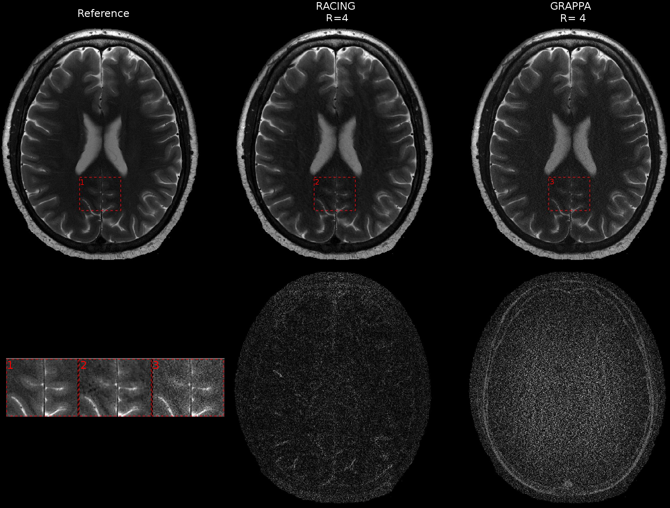
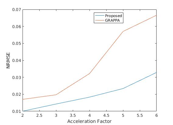
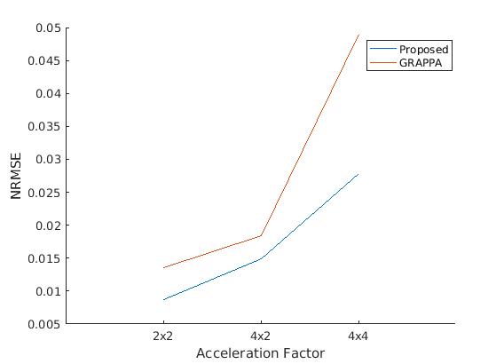
Two data sets have been acquired (2D T2 Fast Spin Echo (FSE), and 3D MPRAGE) on two volunteers. Reconstructions for T2 data using RACING are compared to GRAPPA for undersampling factor (R) of 4 (Figure 1). In Figure 1, GRAPPA shows more severe artifacts and noise amplification compared to the proposed approach. In contrast, the proposed algorithm, which uses sensitivities estimated from exactly the same calibration matrix, allows a better reconstruction accuracy considering the incorporated gradient constraint. Figure 2 provides NRMSE values of the experiments with different R ranging from 2 o 6 for 2D T2 data and as expected the accuracy reduces rapidly as R increases for GRAPPA. Our method improved the results over GRAPPA reconstruction since we took advantage of sensitivity maps unique decay rates and incorporate that additional information in our constraint optimization formulation. Figures 3 and 4 provide the comparison of our method against GRAPPA reconstruction for 3D data. These figures present the results for MPRAGE acquisition with acceleration along both phase encoding dimensions and undersampling factors of 2x2, 4x2 and 4x4. In both figures, RACING consistently produced the best performance at both low and high acceleration factors and outperform reliable GRAPPA reconstruction accuracy.Conclusion
In routine clinical use with multi-channel head coils, the desired image quality arising from parallel imaging in 2D and 3D is limited to a factor of 2-3. Our reconstructions of T2 acquisitions with R=4 have lower reconstruction error than conventional parallel imaging with R=2. For 3D acquisitions, even higher acceleration factors provide satisfactory images. The advantages of spatial gradient constraints for improving the conditioning of parallel imaging reconstruction were confirmed.Acknowledgements
This research was supported in part by NIH grants R01 NS079788, R01 EB019483, R42 MH086984, and by a research grant from the Boston Children's Hospital Translational Research Program.References
[1] Pruessmann KP,Weiger M, Scheidegger MB, Boesiger P, et al. SENSE: sensitivity encoding for fast MRI. Magnetic resonance in medicine 1999;42(5):952–962.
[2] Griswold MA, Jakob PM, Heidemann RM, Nittka M, Jellus V,Wang J, et al. Generalized autocalibrating partially parallel acquisitions (GRAPPA). Magnetic resonance in medicine 2002;47(6):1202–1210.
[3] Ramani S, Fessler JA. Parallel MR image reconstruction using augmented Lagrangian methods. IEEE Transactions on Medical Imaging 2011;30(3):694–706.
[4] Bertsekas DP. Multiplier methods: a survey. Automatica 1976;12(2):133–145.
Figures