4757
Varying Undersampling Dimension for Accelerating Multiple-Acquisition Magnetic Resonance Imaging1Bio and Brain Engineering, Korea Advanced Institute of Science and Technology, Daejeon, Korea, Republic of, 2Bio and Brain Engineering, Korea Advanced Institute of Science and Technology, Dajeon, Korea, Republic of
Synopsis
We proposed a new sampling strategy for efficiently accelerating multiple acquisition MRI. The new sampling strategy is to obtain data along different phase encoding directions across multiple acquisitions. The proposed sampling strategy was evaluated in multi-contrast MR imaging (T1, T2, proton density) and multiple phase cycled (PC) balanced steady-state free precession (bSSFP) imaging by using compressed sensing (CS) algorithms and convolutional neural networks (CNNs) with central and/or random sampling pattern. Sampling along different phase encoding directions across multiple acquisitions was advantageous for accelerating multi-acquisition MRI, irrespective of reconstruction method, sampling pattern or datasets, with further improvement through transfer learning.
Introduction
Routine MRI protocols often consist of multiple data acquisitions for imaging a single anatomical structure. For example, multiple images are independently acquired using multiple MRI sequences for the same field of view (FOV), to obtain a variety of tissue contrasts (e.g., T1, T2, proton density) for more accurate diagnosis. Imaging with balanced steady-state free precession (bSSFP) sequence is often repeated at multiple phase cycling (PC) angles, to suppress banding artifacts1-5. As such, MR imaging often requires imaging multiple times on the same anatomy, which we call multi-acquisition MRI hereafter. Recently, convolutional neural networks (CNNs) have been applied to accelerate MRI scan6-10. Preliminary results of CNNs to share anatomical information from images acquired with different pulse sequences were demonstrated recently11. By sharing information between images acquired with the same or different pulse sequences, CNN can improve the reconstruction of multi-acquisition MRI. In multi-acquisition MRI, acceleration of data acquisition may be enhanced by a sampling strategy that can effectively share information between the multiple images. Since structural information is constant regardless of PC, repetitive sampling of the same high frequency contents can be redundant. The acceleration of multiple PC-bSSFP can be improved by reducing the correlation between the sampling patterns of multiple PC datasets in heuristic12 or automatic manners13. In contrast to the importance of the issue, the sampling strategy for accelerating multi-acquisition MRI has been underexplored. In this study, we propose a new sampling strategy that reduces the overlap of k-spaces across multiple acquisitions in a simple and effective manner. The proposed sampling strategy was tested in multi-contrast MRI and multiple PC-bSSFP imaging by using CS algorithms and CNNs. The aim was to demonstrate that the new strategy is widely applicable for acceleration of multi-acquisition MRI.
Methods
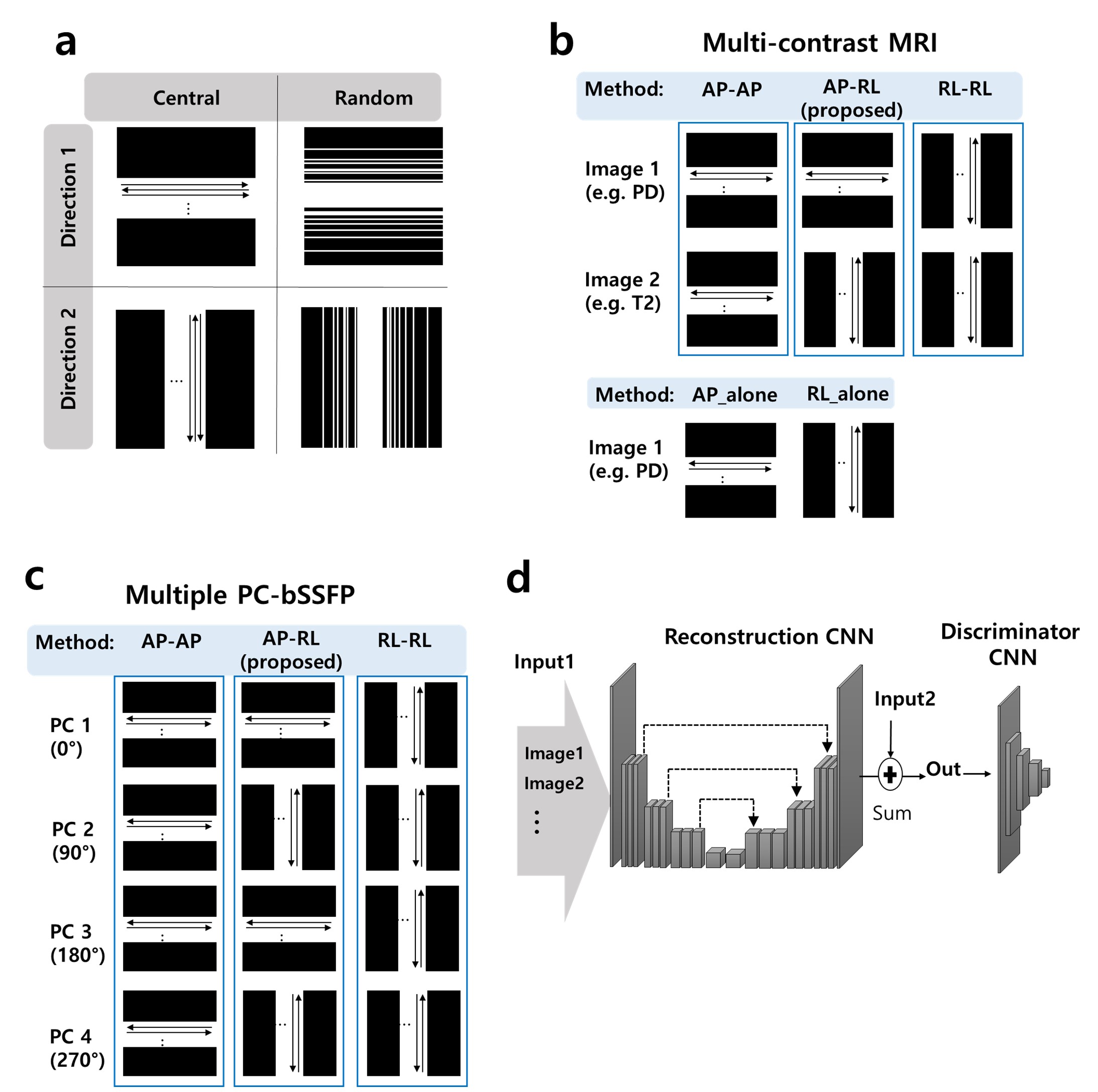
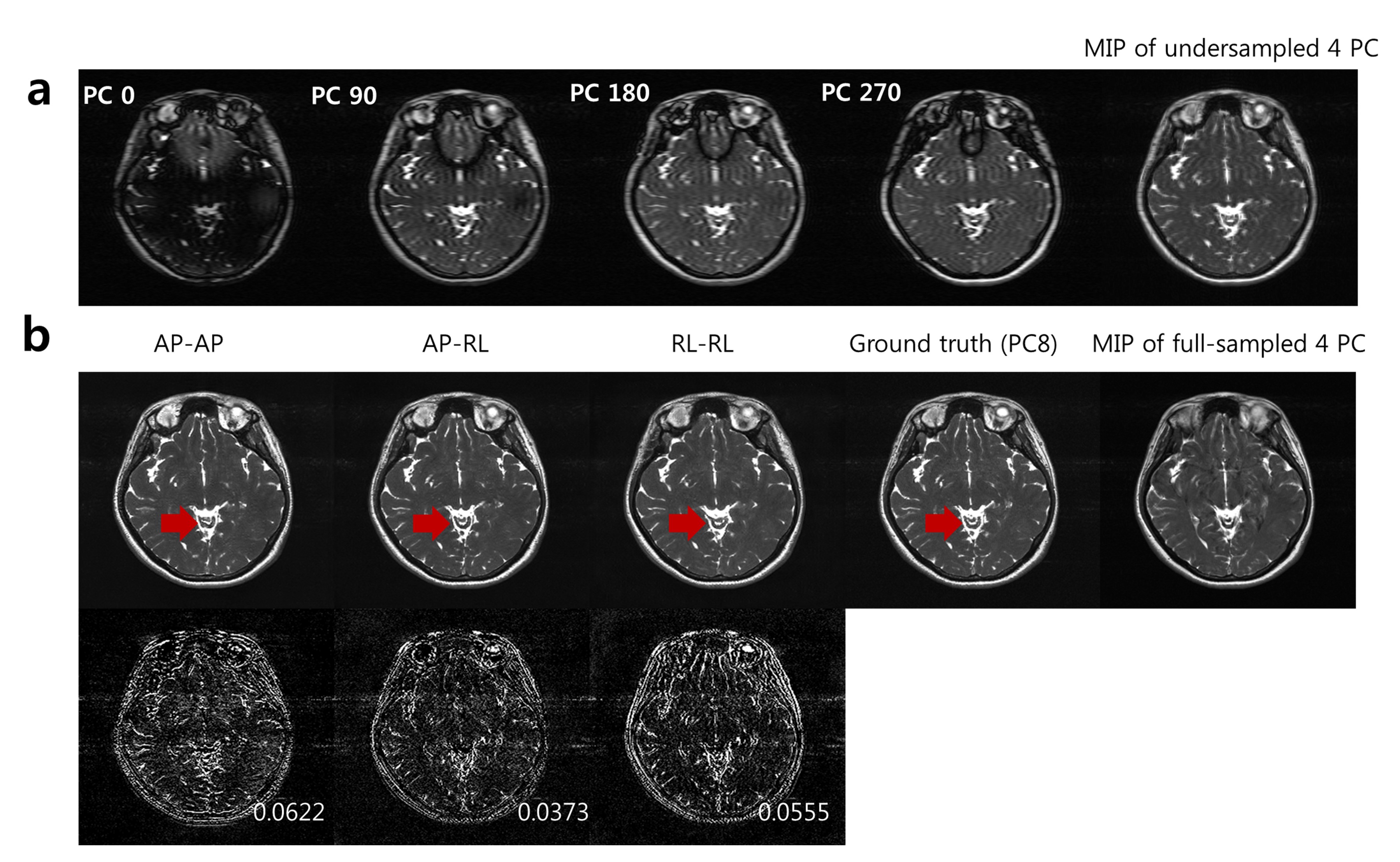
The new sampling strategy is to acquire data along different phase-encoding (PE) directions across multiple acquisitions. This method can reduce the overlap of the sampled k-space data across acquisitions in a simple way (Fig. 1). The proposed sampling strategy with the central sampling pattern (Fig. 1a) applicable to reconstruction using CNNs and the proposed strategy with the random sampling (Fig. 1a) applicable to reconstruction using both CNNs and CS algorithms were used for data acquisition. CNNs were trained to predict the difference between the full-sampled images and the undersampled images in the spatial domain. The proposed sampling strategy with the random sampling pattern was also applied to the reconstruction of multi-contrast MRI using Bayesian CS (BCS)14 and fast multi-contrast CS (FMC-CS)15 that reconstructs two undersampled images simultaneously. Differences between the outputs and the ground truths were evaluated with the metrics of structural similarity (SSIM)16 and normalized root mean square error (NRMSE).Results and Discussion
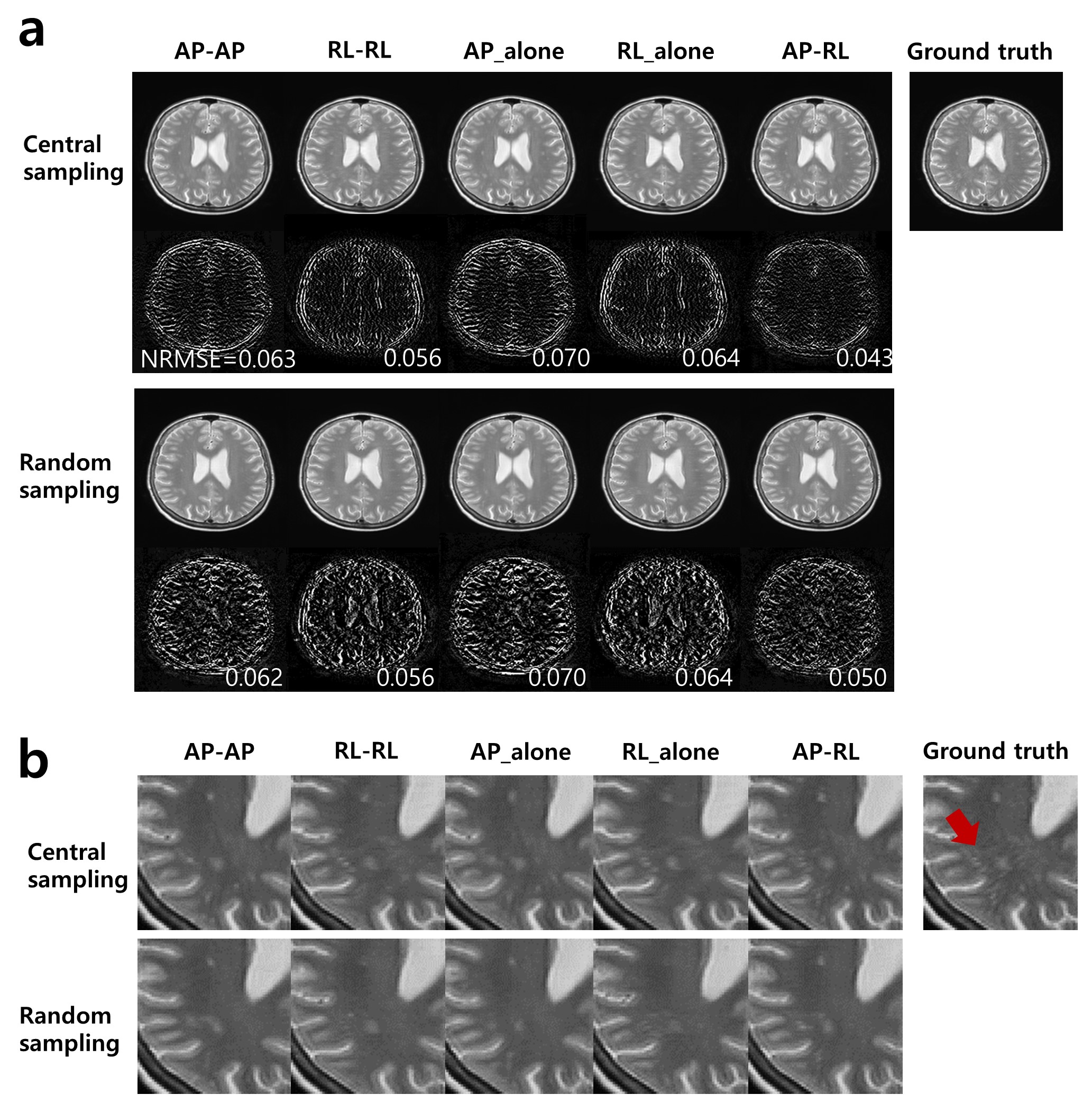
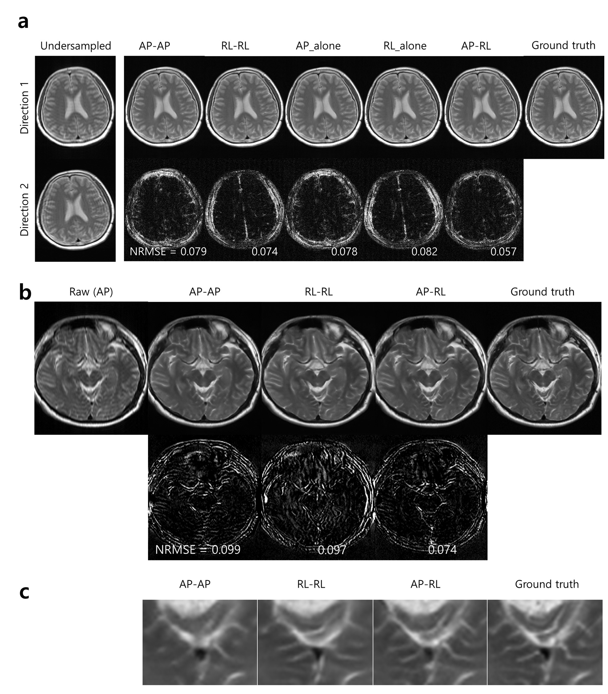
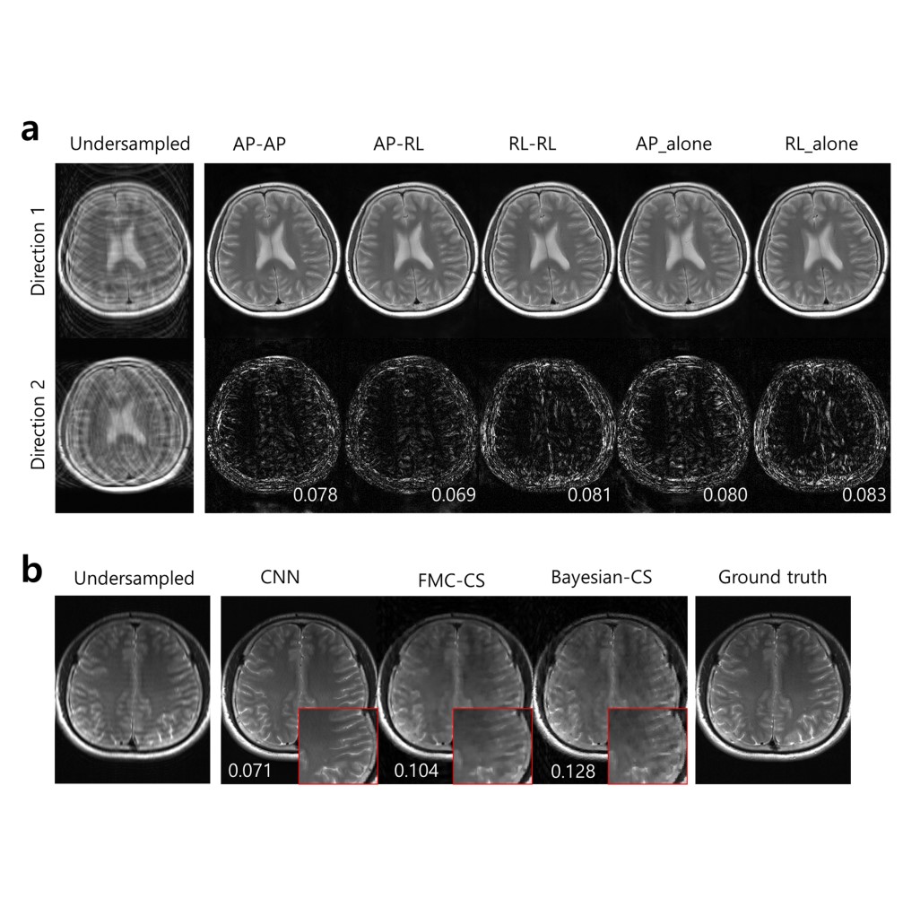
In the public data, the proposed sampling strategy (AP-RL) showed the lowest NRMSE and the highest SSIM values, irrespective of the acceleration factor and the sampling pattern. Visually the proposed strategy provided images closest to the ground truth (Fig. 2a). In magnified view, pathologic high signal intensities in white matter were most clearly detected in the proposed strategy (AP-RL) (Fig. 2b). The results of CNNs in in vivo data were consistent with those of the public data. The proposed strategy (AP-RL) showed the lowest error (Fig. 3 and Fig.4). In addition, the two CS algorithms using the proposed strategy (AP-RL) provided the lowest NRMSE values than those using other sampling strategies. The reconstruction of the undersampled two images with different contrasts took about 30 sec, 5 mins, and 60 ms in FMC-CS, BCS, and CNNs, respectively. Figure 5 demonstrated that the proposed strategy with the central sampling pattern reconstructed details of the cerebellum accurately, while the results of the same PE direction showed distortions in the details. In this study, we have proposed the sampling strategy to efficiently accelerate such multi-acquisition MRI in a simple manner. The advantage of the proposed sampling strategy (i.e., alternating phase-encoding directions across acquisitions) was demonstrated in both multi-contrast MR imaging and multiple PC-bSSFP imaging. We also confirmed that the proposed sampling strategy worked well regardless of the reconstruction method (CNN, CS) or sampling pattern (central, random).Conclusion
The proposed sampling strategy can improve multi-acquisition MRI by integrating anatomical information from other images undersampled along different PE directions. The proposed strategy was applicable to CS algorithms and CNNs using central or random sampling patterns. We confirmed the effects of the proposed strategy in multi-contrast MRI and multiple PC-bSSFP, which supports that the proposed sampling strategy may be useful in various applications where multiple images are acquired along the same scan direction.Acknowledgements
References
[1] S. S. Vasanawala, J. M. Pauly, and D. G. Nishimura, "Linear combination steady-state free precession MRI," Magn Reson Med, vol. 43, no. 1, pp. 82-90, Jan 2000. [2] T. Cukur, N. K. Bangerter, and D. G. Nishimura, "Enhanced spectral shaping in steady-state free precession imaging," Magn Reson Med, vol. 58, no. 6, pp. 1216-23, Dec 2007. [3] K. J. Jung, "Synthesis methods of multiple phase-cycled SSFP images to reduce the band artifact and noise more reliably," (in eng), Magn Reson Imaging, vol. 28, no. 1, pp. 103-18, Jan 2010. [4] N. K. Bangerter, B. A. Hargreaves, S. S. Vasanawala, J. M. Pauly, G. E. Gold, and D. G. Nishimura, "Analysis of multiple-acquisition SSFP," Magn Reson Med, vol. 51, no. 5, pp. 1038-47, May 2004. [5] K. H. Kim and S. H. Park, "Artificial neural network for suppression of banding artifacts in balanced steady-state free precession MRI," Magn Reson Imaging, vol. 37, pp. 139-146, Apr 2017. [6] S. Wang et al., "Accelerating magnetic resonance imaging via deep learning," 2016 IEEE 13th International Symposium on Biomedical Imaging (ISBI), pp. 514-517, 13-16 April 2016 2016. [7] J. Schlemper, J. Caballero, J. V. Hajnal, A. Price, and D. Rueckert, "A Deep Cascade of Convolutional Neural Networks for Dynamic MR Image Reconstruction," arXiv preprint, vol. arXiv:1704.02422, April 1, 2017 2017. [8] D. Lee, J. Yoo, and J. C. Ye, "Deep artifact learning for compressed sensing and parallel MRI," arXiv preprint, vol. arXiv:1703.01120, March 1, 2017 2017. [9] Y. Yang, J. Sun, H. Li, and Z. Xu, "ADMM-Net: A Deep Learning Approach for Compressive Sensing MRI," ArXiv preprints, vol. 1705, May 1, 2017 2017. [10] K. H. Kim, S. H. Choi, and S. H. Park, "Improving Arterial Spin Labeling by Using Deep Learning," Radiology, vol. 287, no. 2, pp. 658-666, May 2018. [22] K. H. Kim, W. J. Do, and S. H. Park, "Improving resolution of MR images with an adversarial network incorporating images with different contrast," Med Phys, May 4 2018. [11] E. Gong, J. Pauly, and G. Zaharchuk, "Accelerating Multi-Contrast Imaging in Neuro-Exam with Sharable Information," in In proceedings of the 25th Annual Meeting of ISMRM, Honolulu, Hawaii, USA, 2017. Abstract No. 0235. [12] T. Cukur, "Accelerated phase-cycled SSFP imaging with compressed sensing," IEEE Trans Med Imaging, vol. 34, no. 1, pp. 107-15, Jan 2015. [13] L. Kerem Senel et al., "Statistically Segregated k-Space Sampling for Accelerating Multiple-Acquisition MRI," ArXiv preprints, vol. 1710, October 1, 2017 2017. [14] B. Bilgic, V. K. Goyal, and E. Adalsteinsson, "Multi-contrast reconstruction with Bayesian compressed sensing," Magn Reson Med, vol. 66, no. 6, pp. 1601-15, Dec 2011. [15] J. Huang, C. Chen, and L. Axel, "Fast multi-contrast MRI reconstruction," Magn Reson Imaging, vol. 32, no. 10, pp. 1344-52, Dec 2014. [16] W. Zhou, A. C. Bovik, H. R. Sheikh, and E. P. Simoncelli, "Image quality assessment: from error visibility to structural similarity," IEEE Transactions on Image Processing, vol. 13, no. 4, pp. 600-612, 2004.Figures