4753
Non-linear Inverse Compressed-Sensing Reconstruction for Self-Gated Multidimensional Cardiac MRI: XD-NLINV1Institute for Diagnostic and Interventional Radiology, University Medical Center Göttingen, Göttingen, Germany, 2partner site Göttingen, German Center for Cardiovascular Research (DZHK), Göttingen, Germany
Synopsis
Motion is a perpetual challenge in cardiac MRI: for comfortable free-breathing exams, both cardiac and breathing motion need to be resolved. Self-gating approaches have been proposed to automatically bin MRI data into appropriate motion states. Here, we propose a new combined parallel imaging/compressed sensing reconstruction for such multi-dimensional datasets. This method, termed XD-NLINV, solves the non-linear parallel imaging problem, simultaneously estimating images and coil sensitivities. This assures efficient use of the available data and removes the need for pre-calculating the coil profiles. We present initial results showing high image quality for self-gated cardiac short-axis data, resolving both breathing and cardiac motion.
Introduction
Cardiac MRI is especially challenging because it necessarily contains motion that needs to be resolved: Cardiac motion cannot (and should not) be stopped. Additionally, while breath-hold exams are possible and are the current standard, they rely on compliant and able patients, which cannot be guaranteed in all cases. Therefore, resolving both cardiac and heart motion is advantageous for robust cardiac MRI exams. Using radial MRI, it was shown that motion states (e.g. breathing and heart phase) can be sorted by self-navigating approaches [1]. This multidimensional dataset can then be reconstructed in a combined parallel imaging/compressed sensing reconstruction [2]. Conventionally, pre-calculated coil sensitivity profiles are used so that the resulting problem is linear. Here, we propose an extension of NLINV [3] for multidimensional reconstruction, termed XD-NLINV, that simultaneously estimates the coil sensitivity profiles and the images. An advantage is that this method uses all available data to estimate the coil sensitivities and that coil profiles which change during the acquisition are taken into account.Theory
Given a vector $$$Y$$$ of measurements from $$$N_C$$$ coils, the image $$$m$$$ and the vector of spatial coil sensitivity profiles $$$C$$$ can be recovered by solving the non-linear problem [3]: \[ \DeclareMathOperator*{\argmin}{arg\,min} \argmin_{m,\,C} \|Y-\mathcal{PF}Cm\|_2^2. \] Here, $$$\mathcal{F}$$$ is the (2D or 3D) Fourier transform and $$$\mathcal{P}$$$ the projection onto the acquired pattern/trajectory in k-space. This problem is bilinear: it is linear in the image $$$m$$$ and in the vector of coil profiles $$$C$$$. Therefore, it can be solved in an alternating minimization scheme. Since both subproblems are generally ill-posed, regularization is added to both. \[ \DeclareMathOperator*{\argmin}{arg\,min} \begin{split} \argmin_{C}\: \| Y - A_C C \|_2^2 &+ \alpha R_1(C)\\ \text{with: } A_C &= \mathcal{PF}m \end{split} \quad\quad\quad \begin{split} \argmin_{m}\: \| Y - A_m m \|_2^2 &+ \beta R_2(m)\\ \text{with: } A_m &= \mathcal{PF}C \end{split} \] with regularization functions $$$R_1$$$ and $$$R_2$$$. These two problems are then optimized in an alternating manner, until some convergence criterion is satisfied. In this work, we updated image and coils 5 times each, decreasing the regularization parameters in each iteration. As the coil profiles are known to be smooth, a weighting matrix $$$W$$$ penalizing high frequencies in k-space is added there: $$$R_1=\|WC\|_2^2$$$ and the resulting problem is solved using the conjugate gradient method. For the image subproblem, ℓ1-total-variation (TV) penalties in both heart-state and breathing-state dimension are added as well as spatial ℓ1-wavelet regularization. As ℓ1-regularization is not differentiable, the subproblem for the images was solved using ADMM [4]. For compressed sensing, sparsity-promoting regularization like ℓ1-wavelets or a total-variation (TV) penalty is added instead of the ℓ2-penalties. This scheme has been implemented in the Berkeley Advanced Reconstruction Toolbox (BART) [5] and applied to a short-axis view of the human heart acquired at 3 T using a 30 channel thorax coil and a golden-angle radial FLASH trajectory. Acquisition parameters were: TR/TE=2.30/1.47 ms, FA= 10°, matrix size: 160 × 160, FOV: 256 × 256 mm², slice thickness: 7 mm, acquisition time: 30 s. This dataset, consisting of 13044 individual TRs, was then binned into 30 cardiac and 12 respiratory states using adapted Singular Spectrum Analysis (SSA-FARI) [6].
Results
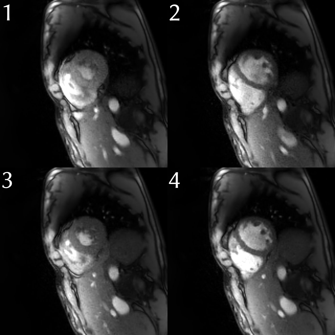
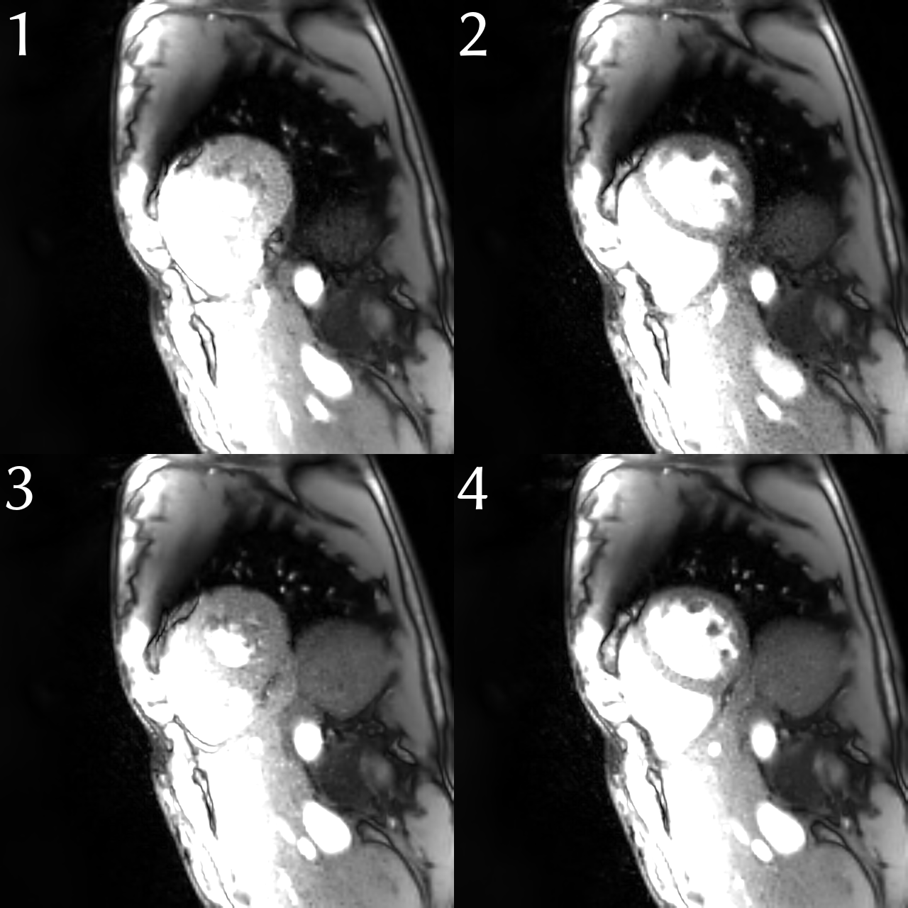
The reconstruction results for end-diastole and end-systole for end-exhalation and end-inhalation are shown in Figs. 1 & 2, where Fig. 2 is windowed such that the breathing state is more visible. As can be seen in Fig. 1, XD-NLINV offers high-quality reconstruction of cardiac data, resolving papillary muscles inside of the left ventricle as well as the heart wall.Conclusion
Extending NLINV to multidimensional data and to compressed sensing enables high-quality reconstruction of non-Cartesian cardiac datasets. Furthermore, no pre-calculation of the coil sensitivity profiles is necessary, as they are estimated simultaneously from the same dataset.Acknowledgements
References
[1]: Feng L et al.: "XD-GRASP: Golden-angle radial MRI with reconstruction of extra motion-state dimensions using compressed sensing", Magn. Reson. Med. 2016; 75:775–788. [2]: Block KT et al.: "Undersampled radial MRI with multiple coils. Iterative image reconstruction using a total variation constraint", Magn. Reson. Med. 2007; 57:1086–1098. [3]: Uecker M et al.: "Image reconstruction by regularized nonlinear inversion — joint estimation of coil sensitivities and image content", Magn. Reson. Med. 2008; 60:674–682. [4]: Afonso MV et al.: "An Augmented Lagrangian Approach to the Constrained Optimization Formulation of Imaging Inverse Problems", IEEE Trans. Img. Proc. 2011; 20:681–695. [5]: Uecker M et al.: "Berkeley advanced reconstruction toolbox", Proc. Intl. Soc. Mag. Reson. Med. 2015; 23:2486. [6]: Rosenzweig S et al.: "Robust Cardiac and Respiratory Self-Gating using an adapted Singular Spectrum Analysis", Proc. 22nd SCMR 2019; (accepted).
Figures