4752
Toward single breath-hold whole-heart coverage compressed sensing MRI using VAriable spatial-temporal LAtin hypercube and echo-Sharing (VALAS)1UIH America Inc., Houston, TX, United States, 2Capital Medical University, Beijing LuHe Hospital, Beijing, China, 3Shanghai United Imaging Healthcare Co., Ltd, Shanghai, China
Synopsis
The main goal is to design and implement a sampling and reconstruction strategy that enables full heart coverage in a single breath-hold, with a relatively high spatial resolution (2.5 × 2.5 mm2) and temporal resolution (40 ms). The challenge in sampling pattern design is how to sample most efficiently. In this work, we present a 10 fold accelerated real‐time cardiac cine MRI pulse sequence using a combination of compressed sensing and parallel imaging.
Introduction
Cardiovascular MRI (CMR) is the recognized gold standard for clinical evaluation of cardiac structure and function. Standard segmented CMR applications rely on ECG1-3 gating coupled with several breath-holds to provide high diagnostic image quality. Yet this protocol may not work for patients with arrhythmias. Whole heart real-time cine imaging using Cartesian sampling trajectories is desirable in that it achieves high efficiency of kt-space coverage, and do not need ECG gating4,5. In this work, the main goal is to design and implement a sampling and reconstruction strategy that enables full heart coverage in a single breath-hold, with a relatively high spatial resolution (2.5 × 2.5 mm2) and temporal resolution (40 ms). The challenge in sampling pattern design is how to sample most efficiently. In MRI, the most important information is around the low frequencies in k-space. Therefore most random sampling methods full samples in the center low frequency region and random samples in the outer region5,6. However, in high spatial-temporal resolution cardiac MRI, images among adjacent phases are highly correlated. As a result, it is desired to decouple the sampling correlations in k-t space. Latin Hypercube Sampling (LHS) is a type of stratified sampling7. It works by controlling the way that random samples are generated for a probability distribution. In this work, we present a 10 fold accelerated real‐time cardiac cine MRI pulse sequence using a combination of compressed sensing and parallel imaging.Method
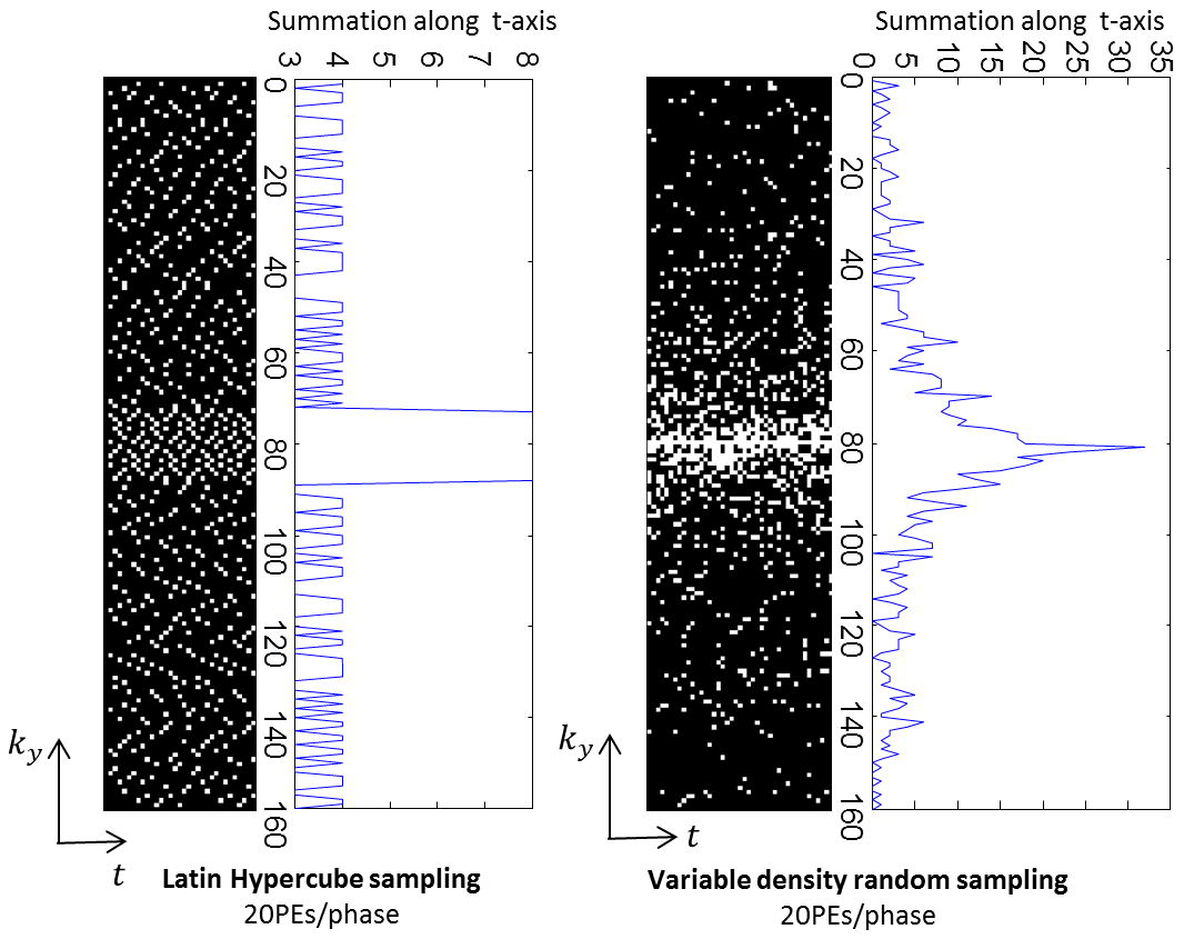
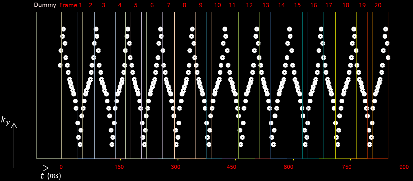
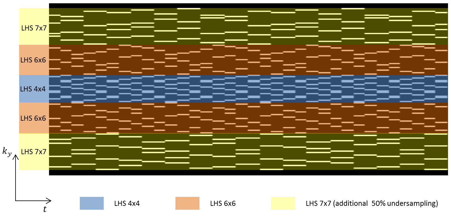
The proposed sampling scheme, called VAriable spatial-temporal LAtin hypercube and echo-Sharing (VALAS), is based on constrained distribution of sample positions on a spatiotemporal grid. Sampling density along time has less statistical fluctuations on LHS (Shown in Fig. 1).In data acquisition, the k-space is continuous prospectively undersampled (Shown in Fig. 2) using a lookup table. The lookup table was generated using variable LHS, with the center k-space 4-fold accelerated, transition region 6-fold accelerated, and outer region 14-fold accelerated (Fig. 3). In reconstruction, adjacent phases share 4 k-space echo data in the outer region, which results in an equivalent 7-fold acceleration in the outer region. We further assume the dynamic CMR series to be sparse in the spatial and temporal total variation (TV) domain. As a result, the image recovery problem is calculated by the following optimization function:
$$$\bf{x} = \arg\min_{\bf{x}} \frac{1}{2} \sum_{j} ||\Omega\bf {F} S_j \bf{x} - d_j|| ^2 + \lambda_1||TV_s(\bf{x})||_1 + \lambda_2||TV_t(\bf{x})||_1 $$$
where the first term is the data consistency term, and the latter two terms are the sparsity constraints; $$$\Omega$$$ represents the undersampling operator, $$$\textbf{F}$$$ is the 2 dimensional fast Fourier transform, $$$S_j$$$ represents the j-th coil sensitivity profile, $$$d_j$$$ the undersampled k-space data from the j-th coil, and j counts all channels.
Results
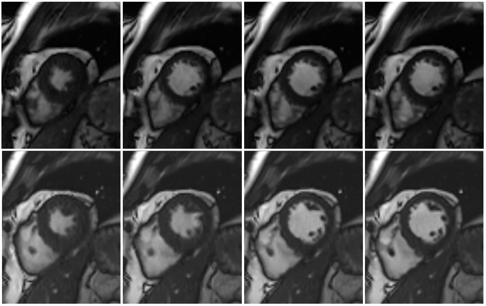
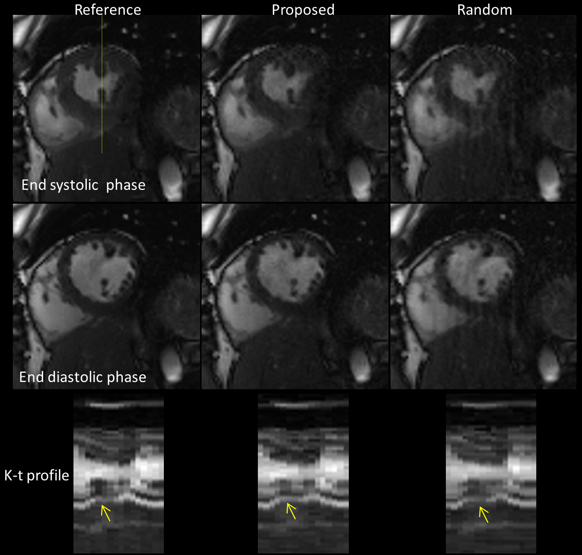
The proposed VALAS technique was implemented with a bSSFP sequence on a clinical 3T scanner (uMR 790 United Imaging Healthcare, Shanghai, China) with the approval of local IRB. Imaging parameters were: imaging matrix: 192 x 165 x 11(slices), TR/TE = 2.7/1.3 ms, FOV = 350 x 300 mm2, slice thickness = 8 mm, flip angle =80°, bandwidth = 1500 Hz, spatial resolution = 1.82x1.82 mm2, 15 Phase encoding lines / phase, reconstructed cardiac phase=20. Cine images of 1 healthy volunteer acquired using VALAS were shown in Fig. 4 (Perspective undersampling, inline reconstruction; top row: Slice 1; bottom row: Slice 2; left-to-right: systolic-to-diastolic). To compare the performance of VALAS over the random sampling pattern, fully sampled data were also acquired and used as golden standard. Fig. 5 shows reconstructions from retrospective undersamplings using VALAS (middle column), variable density random sampling (right column), compared with the reference images (left column). It is seen that VALAS has better image quality and better temporal profile.Conclusion
We have proposed an approach to efficiently undersample the k-t space data for whole heart CMR with high spatial and temporal resolutions. The approach effectively exploits parallel imaging, compressed sensing, and echo sharing techniques. The proposed method has shown to achieve 10-fold acceleration with much reduced artifacts.Acknowledgements
No acknowledgement found.References
[1] Larson A et al., Cardiac and respiratory self-gated projection reconstruction cine imaging; Proceedings of the 89th Scientific Assembly and Annual Meeting, RSNA; Chicago, IL. 2003. p. 591.
[2] Yuan Q, et al., Cardiac-respiratory gating method for magnetic resonance imaging of the heart. Magnetic Resonance in Medicine 43.2 (2000): 314-318.
[3] McLeish K, et al., A study of the motion and deformation of the heart due to respiration, IEEE transactions on medical imaging 21.9 (2002): 1142-1150.
[4] Feng L, et al., Highly accelerated real‐time cardiac cine MRI using k–t SPARSE‐SENSE. Magnetic resonance in medicine 70.1 (2013): 64-74.
[5] Ahmad R, et al., Variable density incoherent spatiotemporal acquisition (VISTA) for highly accelerated cardiac MRI. Magnetic resonance in medicine 74.5 (2015): 1266-1278.
[6] Kim W, et al., Conflict-cost based random sampling design for parallel MRI with low rank constraints. Compressive Sensing IV. Vol. 9484. International Society for Optics and Photonics, 2015.
[7] McKay MD, et al., Comparison of three methods for selecting values of input variables in the analysis of output from a computer code. Technometrics 21, no. 2 (1979): 239-245.
Figures