4743
Automatic Segmentation of Carotid Vessel Wall on GOAL-SNAP Images using SE-UNet1Center for Biomedical Imaging Research, Department of Biomedical Engineering, Tsinghua University, Beijing, China, 2School of Biomedical Engineering and Imaging Sciences, King's College, London, United Kingdom, 3Department of Radiology, University of Washington, Seattle, WA, United States
Synopsis
In this work, we proposed a deep learning structure called SE-UNet for carotid vessel wall segmentation on 3D golden angle radial k-space sampling simultaneous non-contrast angiography and intraplaque hemorrhage (GOAL-SNAP) images. The structure of network consisted of an encoder path for feature extraction and a decoder path for precise localization. The squeeze-and-excitation (SE) module was introduced to the encoder part to learn the context between channels. The proposed SE-UNet achieved high IOU of 0.786, and high pixel-wise sensitivity of 0.976, specificity of 0.850.
Introduction
The carotid atherosclerotic plaque would lead to cardiovascular events. Thus, identification of high risk plaque is of great importance. Traditionally, multi-contrast MRI1-3 was used to image and characterize the carotid vessel wall. However, multi-contrast MRI and the analysis of plaque is time-consuming. Recently, a 3D golden angle radial k-space sampling simultaneous non-contrast angiography and intraplaque hemorrhage (GOAL-SNAP) sequence4 was proposed to acquire multi contrast images using a single sequence with short scan time, providing a fast imaging solution for carotid plaque imaging. Thus, developing automatically analysis method of the GOAL-SNAP images is important for fast carotid plaque evaluation. This study aims to build a deep learning structure for automatic segmentation of carotid vessel wall using GOAL-SNAP images.Methods
Population: 33 patients (18 men and 15 women; median age: 70 years; range: 56-77 years) with carotid atherosclerotic plaque were retrospectively recruited, with institutional review board approval.
Imaging: 3D GOAL-SNAP and 3D MERGE5 images were acquired on Philips 3.0T MR scanner (Achieva; Philips, Best, the Netherlands) with a customer designed carotid coil. The imaging parameters were: GOAL-SNAP: TR/TE=11/4ms, flip angle=8°, field of view=160x160x32mm3, voxel size=0.8x0.8x0.8mm3; MERGE: TR/TE=9/4ms, flip angle=6°, field of view=200x200x32mm3, voxel size=0.8x0.8x0.8mm3.
Image Analysis: For GOAL-SNAP, 9 contrasts at different inversion time (TI) were reconstructed (TI=125, 340, 630, 810, 950, 1090, 1200, 1350, 1600ms) and a T1 mapping image was generated4. Then, each contrast and T1 map of GOAL-SNAP and MERGE images were resliced to generate 48 images centered at the left carotid bifurcation (re-slicing slice thickness: 1mm, in-plane resolution: 0.5mmx0.5mm,). The resliced MERGE and GOAL-SNAP T1 mapping images were reviewed by 2 experienced radiologists in CASCADE6 software using a primary review and peer review strategy. The lumen and outer wall contours were drawn on the MERGE images and mapped to GOAL-SNAP images. The inter-slice and intra-slice registration were done manually.
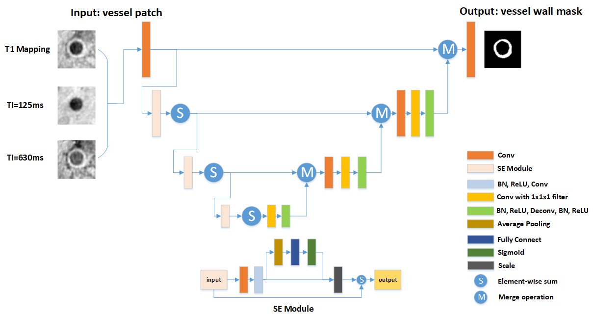
SE-UNet: As shown in Figure 1, the SE-UNet have a similar structure of UNet7, except for using a SE-ResNet8 as the encoder path to extract features and down-sample the images, which can learn the context between different channels without adding too many parameters. In the decoder path, feature maps were then up-sampled and concatenated with previous layers and finally produced a 0-1 mask of segmentation. Among 9 contrasts at different TI, images at TI=125ms and 630ms had the clearest carotid lumen and wall boundaries so we used these 2 contrasts combining T1 mapping contrast as 3 input channels. Of 1584 cross-sectional images, 3/4 images were used as the training dataset, while the rests were test dataset. As a comparison, the traditional UNet was also trained and tested with the same datasets. Data analysis: To evaluate the performance of SE-UNet, the intersection over union (IOU), pixel-wise sensitivity and specificity were calculated with the original UNet as a comparison. Different thresholds at producing 0-1 mask from pixel probability were tested to find an optimal value. The wall thickness and wall area were also calculated for the segmentations of SE-UNet and UNet, and compared with manual segmentation using Spearman correlation.
Results
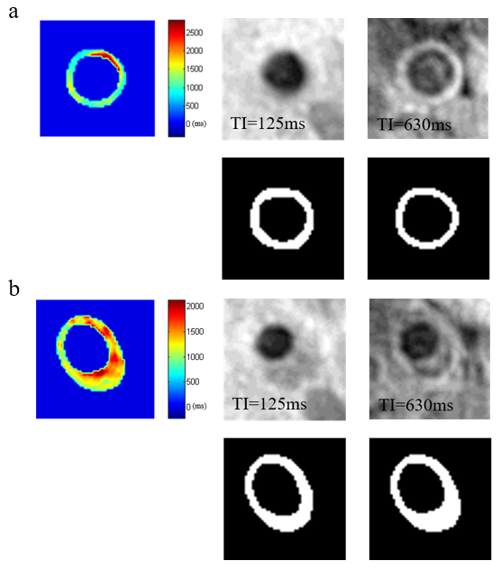
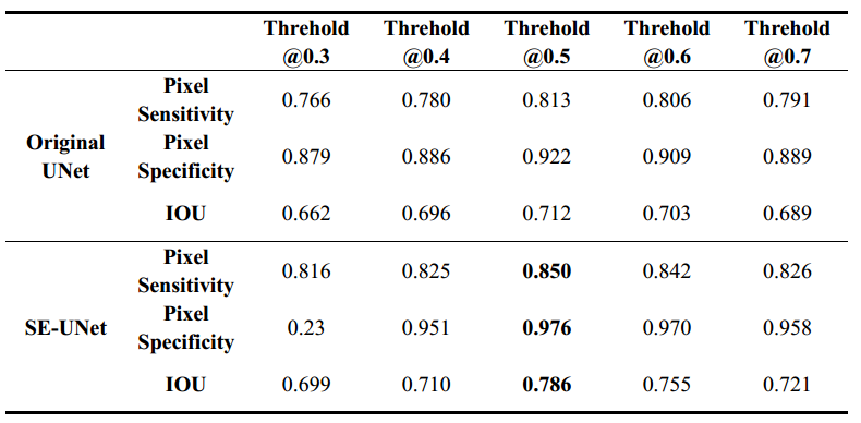
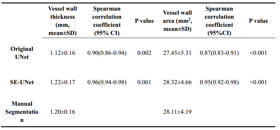
As shown in Table 1, the IOU, pixel-wise sensitivity and specificity of SE-UNet were all higher than the traditional UNet at all threshold. The highest IOU (0.786), pixel wise sensitivity (0.850) and specificity (0.976) can be achieved by SE-UNet at a threshold of 0.5. Table 2 showed that the vessel wall thickness and vessel wall area measured from SE-UNet had higher correlation coefficient than that of the original UNet with the manual segmentation (vessel wall thickness: r=0.96, p=0.001 vs. r=0.90, p=0.002; vessel wall area: r=0.95, p<0.001 vs. r=0.87, p<0.001). Figure 2 showed examples of the segmentation results of SE-UNet at the threshold of 0.5, comparing with the original GOAL-SNAP images and manual segmentation.Discussion and Conclusion
In this study, the feasibility of the proposed SE-UNet in carotid vessel wall segmentation on GOAL-SNAP images was demonstrated. Compared with traditional UNet, the proposed SE-UNet had higher IOU, pixel-wise sensitivity and specificity, indicating adding the SE-ResNet as encoder path can achieve better performance. Besides, GOAL-SNAP sequence can generate different contrast, which allow us to reconstruct best contrasts to facilitate lumen and outer wall segmentation. Although the SE-UNet have higher correlation coefficient with manual segmentation than traditional UNet in vessel wall thickness and area measurement, both methods have high correlation coefficients, suggesting GOAL-SNAP is suitable for lumen and vessel wall delineation.Acknowledgements
None.References
1. Moody AR, Murphy RE, Morgan PS, et al. Characterization of complicated carotid plaque with magnetic resonance direct thrombus imaging in patients with cerebral ischemia. Circulation. 2003;107(24):3047–3052.
2. Zhu DC, Ferguson MS, DeMarco JK. An optimized 3D inversion recovery prepared fast spoiled gradient recalled sequence for carotid plaque hemorrhage imaging at 3.0 T. Magn Reson Imaging. 2008;26(10):1360–1366.
3. McNally JS, Kim SE, Yoon HC, et al. Carotid magnetization-prepared rapid acquisition with gradient-echo signal is associated with acute territorial cerebral ischemic events detected by diffusion-weighted MRI. Circ Cardiovasc Imaging. 2012;5(3):376–382.
4. Qi H, Sun J, Qiao H, et al. Carotid Intraplaque Hemorrhage Imaging with Quantitative Vessel Wall T1 Mapping: Technical Development and Initial Experience. Radiology, 2018;287(1):276.
5. Balu N, Yarnykh VL, Chu B, Wang J, Hatsukami T, Yuan C. Carotid plaque assessment using fast 3D isotropic resolution black blood MRI. Magn Reson Med 2011;65(3):627–637.
6. D Xu, WS Kerwin, T Saam, M Ferguson, and C Yuan. Cascade: Computer aided system for cardiovascular disease evaluation. ISMRM. 2004:1922.
7. Ronneberger O., Fischer P., Brox T. U-Net: Convolutional Networks for Biomedical Image Segmentation. In: Navab N., Hornegger J., Wells W., Frangi A. MICCAI. 2015:9351.
8. Hu J, Shen L, Sun G. Squeeze-and-Excitation Networks. CVPR. 2018:201.
Figures