4729
Propagation Neural Network for cardiac segmentation1Université de Lorraine, Nancy, France, 2U1254, INSERM, Nancy, France, 3Université de Lorraine, Nancy, France, Metropolitan, 4Universität zu Lübeck · Institut für Medizinische Informatik, Lübeck, Germany
Synopsis
To perform a fully-automated segmentation of cardiac volumes, current Convolutional Neural Networks (CNNs) process each slice independently, not taking the depth information into consideration. Networks using 3D convolutions being memory-hungry, we propose a CNN model with a low memory demand and processing the whole volume. The network is based on propagating the redundant depth information from slice to slice. Following a 4-fold cross validation on the MICCAI/ACDC challenge dataset, our network obtained better results than a standard 2D network, improving the average DICE score of 1.7% computed over three cardiac structures (myocardium, left and right ventricle).
Introduction
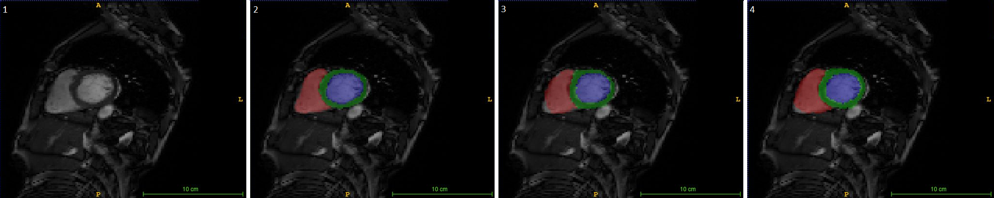
Cardiac Magnetic Resonance Images is a highly effective diagnosis tool, as it offers morphological1 but also functional view of the heart2. To provide a diagnosis radiologists have to manually delineate the different structures (left ventricle, myocardial scar,…), which can be very time-consuming and is prone to variability. There is therefore a growing need for the development of accurate fully-automated cardiac segmentation methods. The most recent techniques relying on Deep Learning and Convolutional Neural Networks (CNN) have shown huge promises for pattern recognition and are now translated into the medical image processing field3.Purpose
2D CNN have shown better segmentation results in the literature4 and require less memory than 3D models. The purpose of this study is to further increase the segmentation accuracy by incorporating the redundant depth information provided by a whole 3D heart volume.Method
We developed a CNN based on the DenseNet structure5. The DenseNet consists in a contracting and an expanding path, a bottleneck, and skip connections. The feature extraction is performed by dense blocks, consisting in a sequence of convolutional layers extracting the same number of feature maps (aka growth rate), batch normalisation, Relu activation, and direct connections between the layers. The exact architecture is depicted in figure 1. The proposed network is divided into 3 sub-networks (all based on the DenseNet structure): an initialization network, a forward and a backward propagation network. The initialization network takes the slice located in the middle of the stack as single input to perform the segmentation and consists in the standard 2D CNN. Once the network initialized, both propagation networks propagate the existing segmentations to their neighboring slices. These sub-networks are fed with the slice to be segmented and the existing neighboring segmentation. Both networks start from the already segmented slice and propagate the segmentation to both ends of the stack, one slice at the time. The training of the initialization network is performed independently first. The forward and backward networks are then jointly trained. The proposed network was then compared to a standard 2D and 3D DenseNet models. The sub-networks of the proposed model were designed so that the number of trainable parameters of the whole network matches the one of the 2D network for a fair comparison (around 8M). It has to be noted that the proposed network required more than 10 times less memory than the 3D network. The 2017 MICCAI/ACDC challenge dataset4 was used to assess our architectures. It contains 3D stacks of short axis images and their segmentation for 100 patients during both the end diastole and the end systole. The images were resized to 128x128 pixels and their intensity normalized. The networks were trained and tested using a 4-fold cross-validation. The DICE score, computed over each of the 3 classes of interest (right ventricle, left ventricle and myocardium), was used to assess the network performance. The training was performed over 100 epochs using ADAM optimizer. The unweighted cross-entropy was chosen as loss function, as it led to better performance than using the DICE score directly.Results
The DICE scores obtained by the different networks are assembled in Table 1. The propagation network showed the best results with an average DICE score of 93.5% computed on the three structure, resulting in an improvement of 1.7% compared to the 2D network. The 3D network could not be tested under optimal conditions due to memory limitations, resulting in poor performance.Discussion
The propagation network showed superior results compared to the 2D one, implying the segmentation process does benefit from the depth information contained in the stack of images. We demonstrated the need to develop light memory demanding networks for volume segmentation. One useful outcome of the propagation network is its flexibility: here trained on propagating the information spatially, it could also be used to propagate the information temporally and catch the heart motion during a cardiac cycle, making it a useful tool for motion correction.Acknowledgements
We gratefully acknowledge the support of NVIDIA Corporation with the donation of the Titan Xp GPU used for this research.
The authors would also like to acknowledge the Region Grand Est and the Doctoral School "IAEM" from the Université de Lorraine for funding Benjamin Roussel's PhD.
References
[1] Rickers, et al. "Utility of cardiac magnetic resonance imaging in the diagnosis of hypertrophic cardiomyopathy." Circulation 112.6 (2005): 855-861.
[2] Gatehouse, et al. “Applications of phase-contrast flow and velocity imaging in cardiovascular MRI.” European radiology. 15.10 (2005):2172-84.
[3] Litjens, et al. "A survey on deep learning in medical image analysis." Medical image analysis 42 (2017): 60-88.
[4] Pop, et al., Statistical Atlases and Computational Models of the Heart. ACDC and MMWHS Challenges: STACOM 2017, Revised Selected Papers. 10663, 2018.
[5] Huang, et al. "Densely Connected Convolutional Networks." CVPR. Vol. 1. No. 2. 2017.
Figures