4717
Accelerating high resolution DWI via deep learning1Neusoft Medical Systems, Shenyang, China
Synopsis
The conventional multi-shot diffusion weighted imaging (DWI) techniques, such as MUSE, have not been widely adopted clinically due to long scan time. In this study, an accelerated multi-shot DWI method based on deep learning is proposed. By learning a fully convolutional neural network to enhance DWI images, more structural details and less noise can be achieved, especially when with fewer shots or NSA (Number of Signal Average), in the meantime the reconstruction time can be reduced by over 200 times. It means the proposed approach reduces the scan and reconstruction time dramatically while keeping high quality of the images, which makes it a potential technique for high resolution multi-shot DWI in routine clinical study.
INTRODUCTION
Diffusion weighted imaging (DWI) is a promising technique for investigating the microscopic properties of tissues. Single-shot DWI is currently the most widely accepted approach mainly due to its insensitiveness to motion and easy implementation. However, it inherently suffers from low resolution, low SNR and geometric distortions. To address these challenges, multi-shot DWI techniques, such as MUSE 1, were employed, but it increases scan and reconstruction time significantly. In this study, a deep-learning-based multi-shot DWI method is proposed, which is able to achieve high image quality with fewer acquisitions and reduce reconstruction time dramatically, thus making fast and high resolution DWI feasible.METHODS
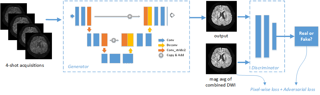
We proposed a fast multi-shot DWI method using the generative adversarial network (GAN), which consisted of a generator and a discriminator (see Fig. 1). The architecture of the generator was based on UNet 2 with skip connections, which learned a non-linear mapping between from respectively reconstructed one-shot DWI images and multi-shot DWI ground truth. The discriminator adopted a CNN-based classification network to differentiate the reconstructed images from the ground truth. Experiments were conducted on a 1.5T MR system (NSM S15P, Neusoft Medical Systems, China). The brain DWI dataset, consisting of 30 subjects, was acquired using 4-shot interleaved EPI sequence with TR/TE=4200/107ms, slice thickness=5mm, slice number=22, matrix size=192*192, FOV=230mm*230mm and 3 diffusion directions at b=1000s/mm2. The coil sensitivity information was obtained by the reference scan using GRE sequence. In the training process, Multi-shot DWI with NSA=4 (Number of Signal Average) was served as ground truth, which was combined with parallel imaging method. Data augmentation was implemented before feeding into the network to improve robustness. The loss function was a combination of the weighted sum of pixel-wise Mean-Square-Error (MSE) loss and the adversarial loss, which preserved texture and sharpness of edges. The Adam optimization algorithm was adopted with momentum of 0.5 and the initial learning rate of 0.0001 was halved every 10 epochs.RESULTS
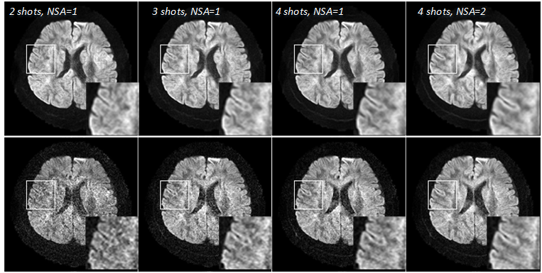
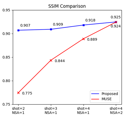
Visual comparison of the results from the proposed method (upper row) and MUSE (lower row) are shown in Fig.2. A series of experiments were conducted with different number of acquisitions, from 2 shots, NSA=1 to 4 shots, NSA=2. As can be clearly seen that, compared to MUSE, the results from the proposed method consistently have more details and less noise, especially when with fewer shots or NSA. Figure 3 shows the quantitative comparison between the two methods in terms of structural similarity index (SSIM), where the result from the proposed method with 4 shots, NSA=1 is numerically close to the result from MUSE with 4 shots, NSA=2 (0.918 vs. 0.924), which means 50% scan time can be reduced with approximately the same image quality. Additionally, the reconstruction time of the proposed method is less than 100ms, which is over 200 times faster than that of 4-shots MUSE.DISCUSSION
Benefited from the powerful image enhancement capability of deep neural network, our proposed method achieves higher image quality level than single shot DWI. In the meantime, the risk of motion artifacts can be reduced due to the shorter scan time, compared with conventional multi-shot DWI methods. Furthermore, the proposed method has the potential to obtain even better results with more sophisticated ground truth, which can be generated by averaging over more acquisitions or using enhanced reconstruction methods 3.CONCLUSION
In this work, an accelerated multi-shot DWI approach using deep learning is proposed, which reduces the scan and reconstruction time dramatically while keeping high quality of the images. Our proposed method can make multi-shot DWI more clinically acceptable.Acknowledgements
No acknowledgement found.References
1. Chen N K, Guidon A, Chang H C, et al. A robust multi-shot scan strategy for high-resolution diffusion weighted MRI enabled by multiplexed sensitivity-encoding (MUSE). Neuroimage, 2013, 72(2):41-47.
2. Ronneberger O, Fischer P, Brox T. U-Net: Convolutional Networks for Biomedical Image Segmentation. 2017, 9351:234-241.
3. Zhang Z, Huang F, Ma X, et al. Self-feeding MUSE: a robust method for high resolution diffusion imaging using interleaved EPI. Neuroimage, 2015, 105(105):552-560.
Figures