4696
A Validation Approach for Imperfect Training Data Fidelity using Signal + Artifact + Noise-based Neural Net (SAN3)-derived Directionalized Streaking Removal1Biomedical Engineering, Illinois Institute of Technology, Chicago, IL, United States, 2Electrical, Electronics, and Information Engineering, Gifu University, Gifu City, Japan, 3Medicine, The University of Chicago, Chicago, IL, United States
Synopsis
While Deep Neural Network (DNN)-based sub-Nyquist reconstruction approaches are well-suited for high-fidelity static imaging targets such as the brain, temporally constrained (i.e. dynamic) sequences may potentially be ill-suited for DNN as these would often embed unresolved MR artifacts into the Training Data. Here, we describe an assessment approach for a generalizable DNN-based dynamic MRI reconstruction method that outputs such artifacts as characterizable and filterable streaks. This work further validates the DNN-model coding process to ensure the desired artifact/noise properties into the DNN output. Using Fourier properties, we demonstrate such validation of streaking directionalization using DNN.
Introduction:
Deep-Neural-Network (DNN)-based MRI reconstructions [1-3] often require both fully sampled and artifact-free reference images for training. Additionally, such DNN models typically require careful tuning, which can be both computationally expensive, and labor-intensive. Accordingly, non-static MR approaches that are more susceptible to image acquisition artifacts, such as cardiac imaging, are not as well-suited for such DNN-based applications. We recently proposed a generalizable DNN-based strategy that requires neither high training data nor optimized DNN modeling that embeds both artifact (A) and noise (N) as quantifiable vectorized projections that may become separable from the invariant signal source (S) [4]. In this work, we exploit fundamental invariance properties in k-space sampling across Fourier and Radon transform domains to establish an appropriate metric to quantitatively assess A+N vector projection using our DNN-based ‘streaking artifact’ direction properties.Generalized DNN Model Assumptions:
Our generalizable DNN-based approach employs the following two assumptions: first, true training data fidelity from MR is limited in non-static acquisitions; and second, the DNN model can be deliberately designed to linearize contributions of the artifact (A) and noise (N) respectively onto a set of projection vectors in mathematically represented domain, while the target signal (S) remains invariant. The goal of this abstract is to develop an approach to characterize this vector projection step derived from the DNN model.Methods:
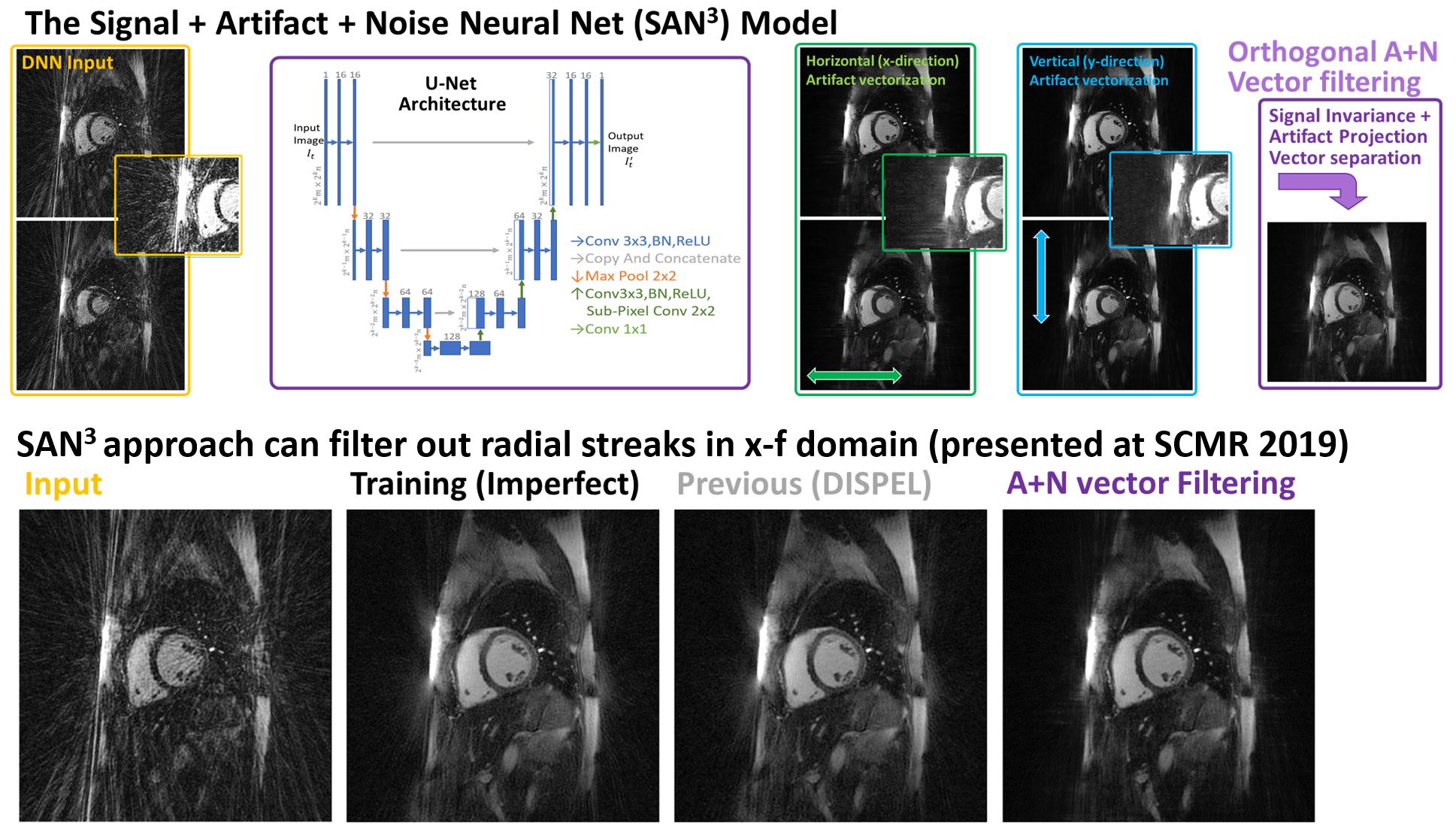
Overview: We outline a generalizable method to exploit the Fourier properties of MRI k-space and how these map to the A+N projection space. In this abstract, we demonstrate this using an illustrative x-t and y-t (i.e. image y-axis and x-axis) projection approach using radial cine MRI approach [5] with R=5 acceleration (i.e. 36 out of 180 Nyquist-satisfied spokes) for visual ease.
DNN Training Description: Figure 1 shows the schematics of the DNN scheme, for which a Nyquist-satisfied radial Cine-MR with weighted [KWIC] temporal window [6] that embeds motion-induced radial streaking [1] is employed as the imperfect training model. This DNN is specifically designed to project the A and N contributions along the image- and time- (i.e. x-t) domain onto the image y-column, and vice-versa onto the image x-column. For this DNN training, an ‘all-but-one’ cyclic training/validation approach was employed via from 10 healthy subjects (for each 15 cine-slice, a set of 400x x-t slices were generated, with 9x15x400 = 54000 total slices per DNN training). This training was re-run using y-t slices (i.e. A+N vector projection onto the x-axis), and thereby ensuring mutual exclusivity of the training data between the DNN models for subsequent validation.
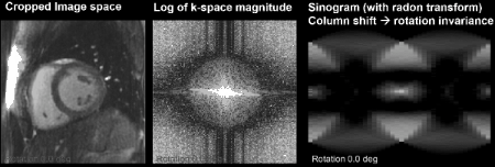
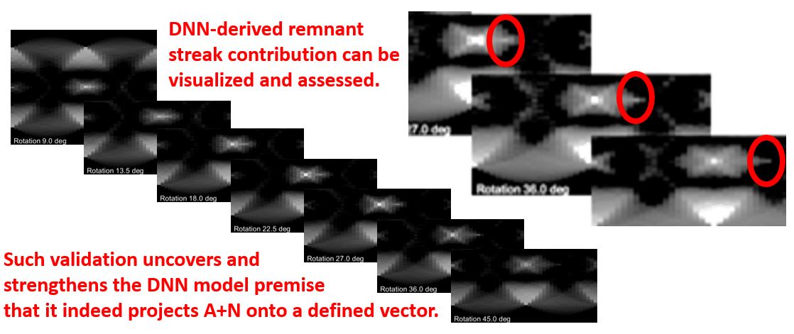
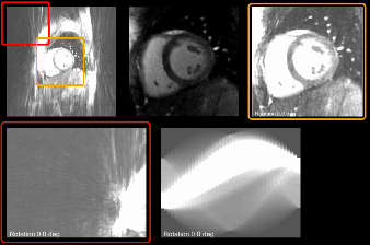
Proposed Assessment: First, we cross-validate our assumption that directionalized A+N components are indeed projected as vectors in known directions by DNN design. For this, we exploit the MRI radial k-space sampling by repeatedly applying a known rotation θ, where this angulation also yields an θ rotation in the image domain by the Fourier Slice Theorem. Upon passage through DNN model, the output embeds invariant signal S, along with A+N in the presumed vector projections. These projections are next examined in the Fourier domain, where a-priori knowledge of the A+N projection streak directions are further exploited; in this case, the Radon transformation at fixed angles θ were performed on the MRI k-space magnitude was performed for subsequent quantification. Further characterization exploits the sinogram’s column shift properties, as further illustrated in the subsequent Results section (Figures 2-4).
Results:
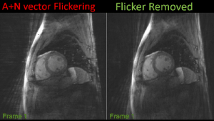
Animated Figure 2 shows the pipeline for DNN-derived output. In this example, θ = [0, 4.5, 9.0, 13.5, … 180] were used. Of note, the DNN-designed A+N projection streaking is clearly observed with respect to the invariant signal S (Figure 3). Visual confirmation of this linearized streak representation peaks as shown here indeed validates our DNN model assumption in forming A+N projection vectors. Accordingly, these directional A+N streaks can be filtered against orthogonal streaks using S invariance; Animated Figure 4 shows the use of DISPEL filter [5] for high-frequency flicker removal.
Finally, animated Figure 5 shows an example of potential pitfall; we caution the use of domain-specific assumptions, such as conducting this assessment in the image domain. Instead of in the x-y space, we note the Fourier-domain magnitude representation of MRI k-space is in fact translation-invariant, and such domain-specific properties must therefore be considered in a careful manner.
Discussions:
We describe a method to systematically assess DNN-based reconstruction algorithms using known properties of embedded artifact and noise. This approach clarifies how the DNN coding step may be validated, such as demonstrating the A+N vector projection directionalization as shown in this work. The combined application and validation of such DNN designs based on Fourier properties will enable further verification/quantitation of presence and extent of such DNN-embedded artifacts.Acknowledgements
The initial two listed authors contributed equally for this work.References
1. Yoon J. et al. NeuroImage (2018) Oct; 179:199-206.
2. Ul Hassan Dar S. et al. Proc. ISMRM (2018).
3. Marandi M. et al. Proc. ISMRM (2018).
4. Suzuki et al. SCMR 2019. In Press.
5. Kawaji K. et al. Med Phys. (2017) Jul; 44(7):3450-3463.
6. Kim KW, Eur Radiol. (2013) May; 23(5):1352-60
Figures