4693
Effects of Image Sharpening on the Accuracy of quantitative-MRI (qMRI) Maps.1Boston University, Boston, MA, United States
Synopsis
Purpose: To study the effects of image sharpening and low spatial frequency removal on the quality of qMRI maps of T1, T2, and proton density (PD). Methods: Previously developed qMRI algorithms, augmented with specialized image filters, were tested with a gel based phantom containing three distinct solutions of variable gadolinium, sucrose, and agarose concentrations. Results: Images were successfully sharpened without significantly effecting pixel values of T1 and T2 weighted maps, while removing PD map spatial artifacts in the gadolinium vials. Conclusion: Unsharp masking and spatial flattening algorithms are effective methods for enhancing qMRI quality toward generating more accurate Synthetic-MRI maps.
Purpose
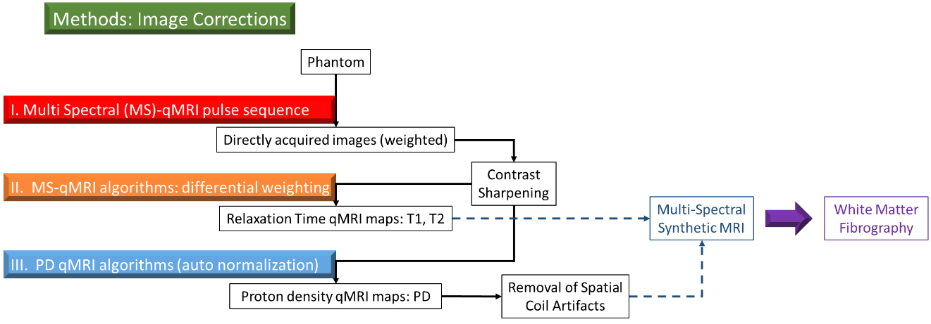
Quantitative MRI (qMRI) algorithms have previously been implemented toward the development of Synthetic-MRI maps using formulated T1, T2, and proton density (PD) weighted maps. Synthetic-MRI maps have further been used to generate three dimensional renderings of human connectomes; however, the resolution in non-axial planes is poor. One method of improving resolution in all imaging planes is unsharp masking, which combats these issues by enhancing the edges of small structures within a medical image, thus sharpening the contrast(1,2). Furthermore, spatial sensitivity intrinsic to the coil used in the MRI scanner creates inhomogeneities in the signal. The purpose of this study is to understand how the process of sharpening and removing the coil profile can impact the quality and accuracy of T1, T2, and PD maps, with implications toward enhancing the resolution of small fibers found in Synthetic-MRI based white matter fibrography (WMF).Methods
The sharpening and spatial coil filtration algorithms programmed for this study were done with Python 3.5 using the Canopy integrated development environment (Enthought, Austin, TX). The sharpening algorithm implements a Gaussian distribution in the spatial domain, and convolves it in the frequency domain with the original image to form a blurred copy. The high frequency components are isolated as weighted edge pixels and added to the original image generating a new, “sharper” representation (Eq. 1).
Eq. 1: $$$UnsharpMask=Original+(Original-Blurred)\times Strength$$$
Spatial coil inhomogeneities were removed through implementation of a two dimensional step function in the frequency domain, filtering out high frequency components of the subject. The remaining low frequency signal represents the spatial profile of the coil and is removed through division into the original image (Eq. 2).
Eq. 2: $$$Flattened=\frac{Original}{Blurred}$$$
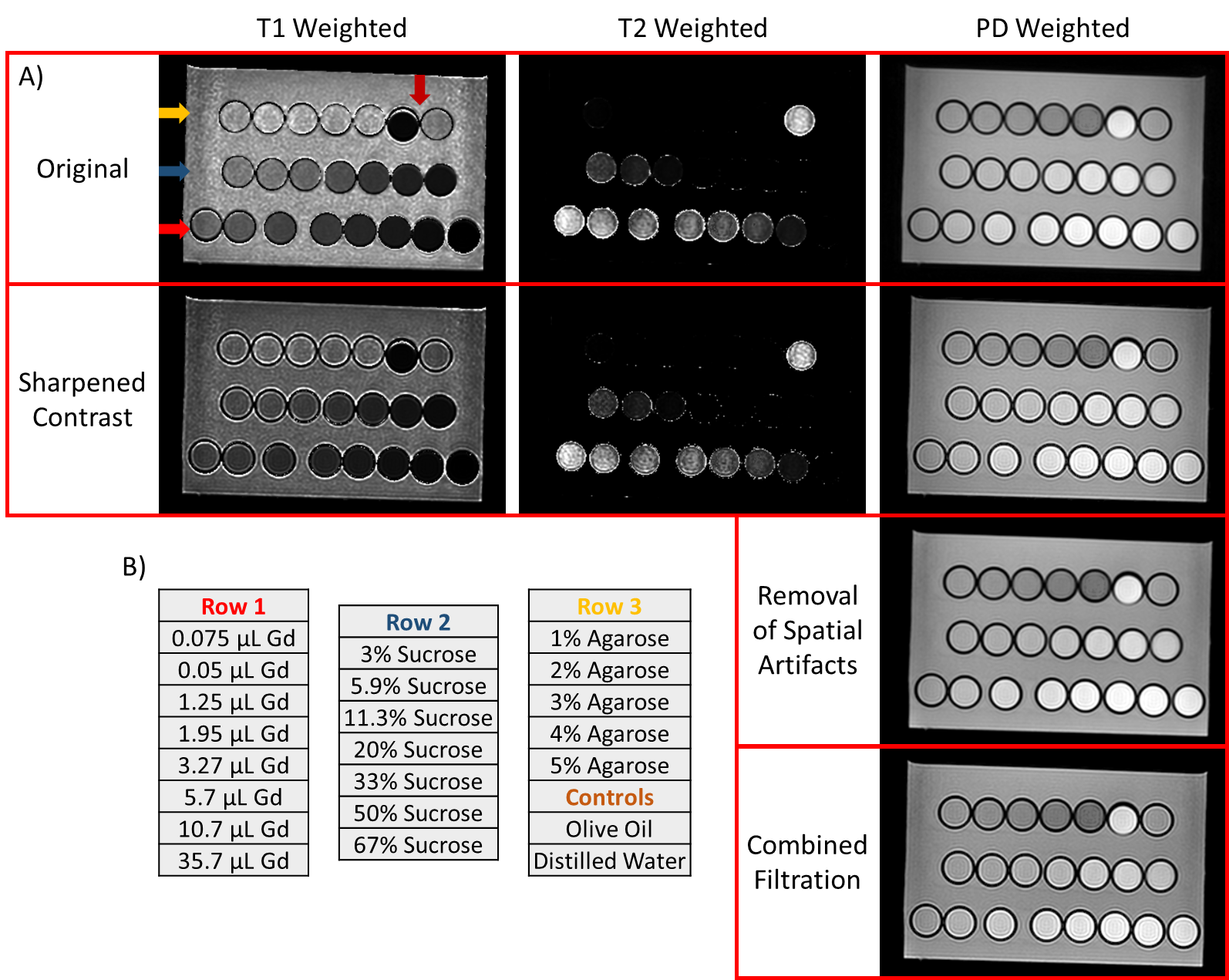
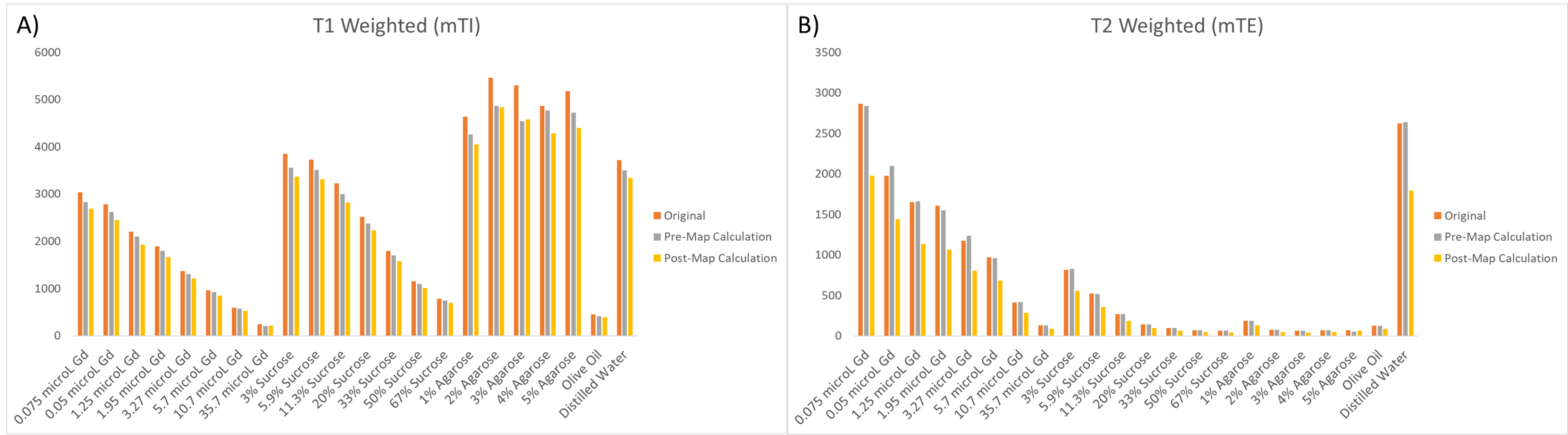
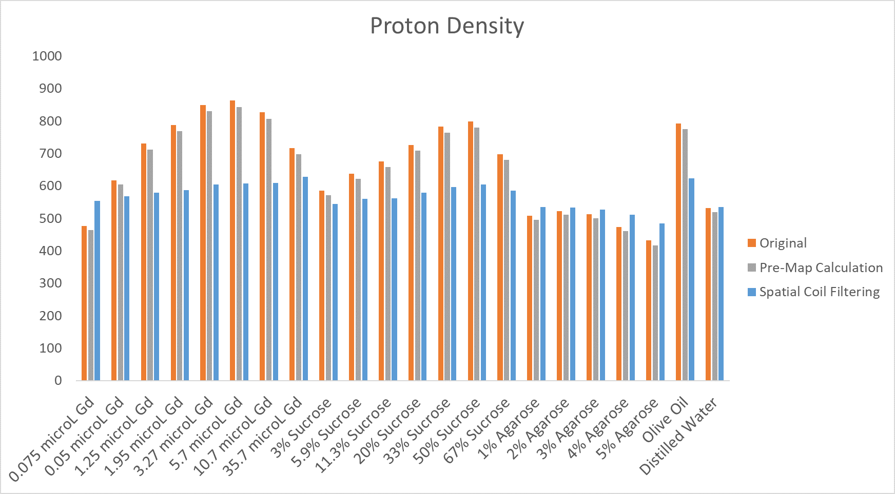
The algorithms were validated by calculating average measurements of a circular region-of-interest (ROI) comprising the majority of each vial within the phantom. Twenty-two vials were embedded in a 4% agarose gel, arranged according to solution type. Gadolinium solutions were prepared by diluting 0.075, 0.5, 1.25, 1.95, 3.27, 5.7, 10.7, 35.7, 73.2, 148.2, and 298.2μL of gadolinium contrast agent in 15mL solutions of distilled water. Sucrose solutions were prepared by dissolving 100g of sucrose in 50mL of distilled water, and further diluting to 67%, 50%, 33%, 20%, 11.3%, 5.9%, and 3% sucrose relative to the original. Finally, gels of 1%, 2%, 3%, 4%, and 5% agarose were prepared along with olive oil and pure distilled water controls.
Results
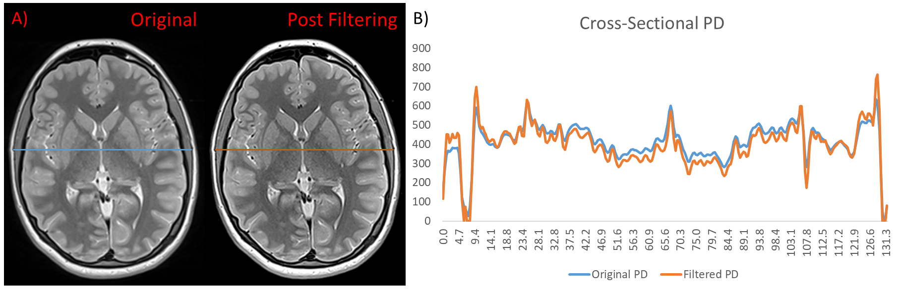
Python algorithms were shown to reliably enhance the resolution and accuracy of MRI phantoms. Increased sharpness and flattening of pixel values (Figure 2) are demonstrated at various stages of the qMRI image processing protocol (Figure 1). Sharpening of the original phantom data or qT1 maps did not significantly decrease the average T1 value for each vial while also maintained the full range of reference qT1. Conversely, sharpening of the qT2 maps drastically decreased the T2 values of each solution, relative to sharpening of the original phantom data (Figure 3). Furthermore, removal of spatial artifacts was shown to flatten the PD values to a constant level for the gadolinium and sucrose vials, demonstrating the filter’s ability to remove the intrinsic profile of the MRI scanner (Figure 4). Finally, the algorithms were implemented on a human brain inducing flattened data at the center of the brain and accentuated the edges of various small neural structures (Figure 5).Discussion and Conclusion
This phantom study demonstrates the ability of python algorithms to improve MRI scans for potential applications in enhancing the resolution of Synthetic-MRI towards superior white matter fibrograms. Furthermore, this work identified pre-map application of the unsharp mask filter as the optimal phase for ensuring accurate differential weighting maps. Phantom data MRI scans containing solutions of variable gadolinium, sucrose, and agarose, were successfully sharpened demonstrating the reliability of computational models to remove MRI scanner artifacts and enhance resolution. As this work is purely experimental in nature, future work is required to ensure these algorithms are optimized for brain tissue, thus the theory behind them must be fully understood. Based on these results, digital image processing has implications toward enhancing the resolution of Synthetic-MRI, thereby allowing the construction of increasingly ultra-high resolution connectomes.Acknowledgements
This work was supported in part by the National Institute of Neurological Disorders and Stroke (5U01NS040069-05 and 2R01NS040069-09), National Institutes of Health Office of the Director (1UG3OD022348-01), and the National Institute of Child Health and Human Development (5P30HD018655-28). We are indebted to Mr. Mitchell Horn for his assistance in preparing and scanning the phantom.References
1. Jabri KN, Wilson DL. Quantitative assessment of image quality enhancement due to unsharp-mask processing in x-ray fluoroscopy. JOSA A. 2002;19(7):1297-307.
2. Panetta K, Zhou Y, Agaian S, Jia H. Nonlinear unsharp masking for mammogram enhancement. IEEE Transactions on Information Technology Inbiomedicine. 2011 Nov 1;15(6):918.
Figures