4684
Simultaneous Functional MRI and Proton Echo-Planar Spectroscopic Imaging in Human Brain (fPEPSI)1Neurology,Physics and Astronomy, University of New Mexico, Albuquerque, NM, United States, 2Neurology, University of New Mexico, Albuquerque, NM, United States, 3Center for Magnetic Resonance Research, Radiology, University of Minnesota, Minneapolis, MN, United States
Synopsis
This study introduces simultaneous fMRI and MRSI integrates multi-slab echo-volumar-imaging (MEVI) into the water suppression module of Proton-Echo-Planar-Spectroscopic-Imaging to simultaneously acquire fMRI, water suppressed and water reference data in a single scan (fPEPSI). FMRI image quality and BOLD sensitivity acquired with 4x4x6 mm3 resolution was comparable to our recently developed MEVI method. Spectral quality and sensitivity of 3D metabolite maps acquired with 4x4x7 mm3 resolution were comparable to conventional PEPSI. This hybrid fMRI/MRSI approach considerably reduces scan times in multi-modal clinical research studies and it is applicable to characterizing neurotransmitter and lactate concentration changes in relation to BOLD signal changes.
INTRODUCTION
Functional MRI (fMRI) and MR Spectroscopic Imaging (MRSI) provide complementary functional and metabolic information for clinical evaluation of patients with neurological disorders1-4,5,6,7. However, the long scan times and considerable differences in spatial resolution of conventional fMRI and MRSI methods have been barriers for clinical integration and for acquiring both modalities in a single scanning session. In the present study, we show proof-of-concept of simultaneous fMRI and MRSI by developing a novel hybrid fMRI/MRSI sequence that integrates multi-slab echo-volumar-imaging (MEVI) into the water suppression module of Proton-Echo-Planar-Spectroscopic-Imaging (PEPSI) to simultaneously acquire fMRI, water suppressed (WS) and water reference (WR) data in a single scan (fPEPSI).
METHODS
Design considerations:
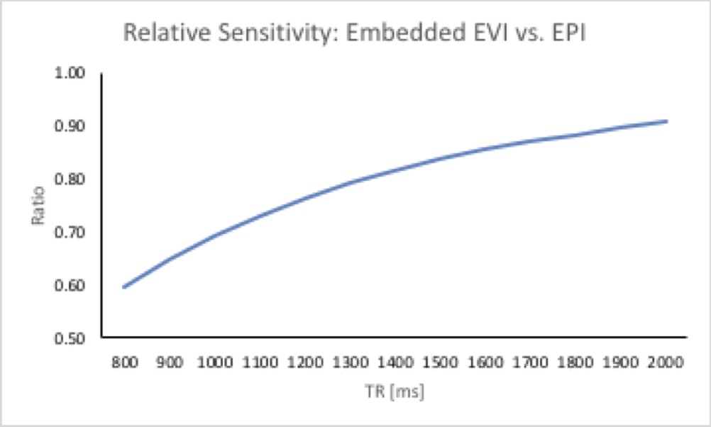
The integration of MEVI into the first water suppression module of PEPSI reduces sensitivity compared with conventional fMRI, since a flip angle close to 90o is required for optimal water suppression and the water saturation recovery period is reduced by the duration of the water suppression sequence. The sensitivity of this hybrid fMRI approach relative to conventional fMRI, which is shown in Fig.1 as a function of TR, is given by:
$$
R_(fPEPSI/fMRI)=((1-e^((-TR)⁄T_1 ))sinα_Ernst (1-cosα_WS
e^((-(TR-T_WS))⁄T_1 )))/((1-cosα_Ernst e^((-TR)⁄T_1 ))(1-e^((-(TR-T_WS))⁄T_1
))sinα_WS )
$$$ Eq.1
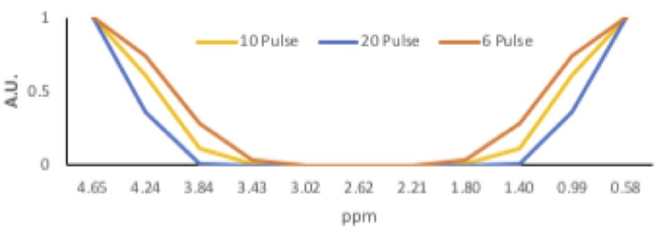
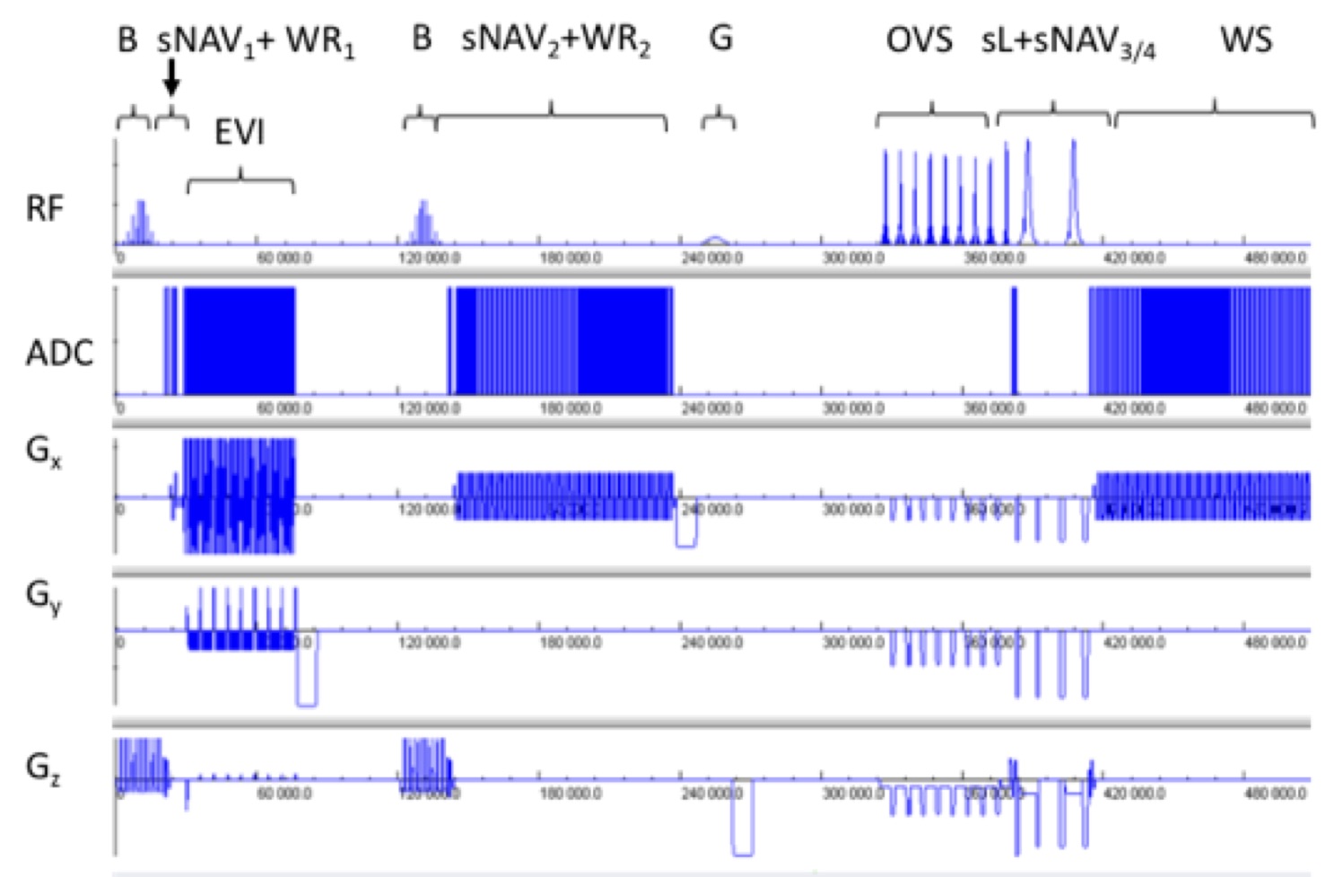
, where aErnst is the Ernst angle, aWS is the flip angle of the first WET water suppression RF pulse and TWS is the duration of the WET sequence. A binomial RF pulse, which offers excellent spectral selectivity performance with tolerance to B1 inhomogeneity, was chosen for slab and spectrally selective water suppression. A simulation of the excitation profile (Fig.2) showed that a 10 RF subpulse design with 2 ms inter-pulse spacing provides an acceptable compromise between the non-excitation spectral range for metabolites of interest (including inositol, lactate and lipids), B0 offset tolerance, water suppression bandwidth and RF pulse duration. Implementation: The multi-slab fPEPSI pulse sequence (Fig.3) acquires the fMRI data in the 1st water suppression module using 4xGRAPPA acceleration and 6/8 partial Fourier acquisition of ky. WR data for metabolite scaling are acquired using a bipolar PEPSI readout before the MEVI module. WR data for eddy current correction are acquired in the 2nd water suppression module using a short PEPSI readout. WS data are acquired using semi-Laser pre-localization and conventional PEPSI readout.
Data acquisition and analysis:
3 healthy adults (1F,2M) were scanned on Siemens 3T scanner with 32-channel head array coil. Institutionally approved informed consent was obtained. A 3 min block-design motor/visual fMRI experiment (finger tapping and eyes open vs. rest and eyes closed) and a 3 min resting state fMRI experiment were concurrently performed during the MRSI acquisition (TR/TE: 1500/30 ms, 64x64x8 spatial matrix, spectral width: 735 Hz, voxel size: 4x4x7 mm3 = 0.11 cc). WR (spectral resolution: 11.5 Hz) and WS data (spectral resolution: 1.4 Hz) were reconstructed online as described previously9. Spectral quantification was performed using LCModel fitting10. FMRI data were acquired using 64x64x8 spatial matrix with 4x4x6 mm3 voxel size and reconstructed offline using custom MATLAB tools. Model-based and seed-based fMRI analysis of the reconstructed EVI data was performed using the TurboFIRE fMRI analysis software11.
RESULTS
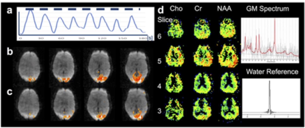
FMRI image quality (uniformity, distortion, ghosting) was comparable to our recently developed MEVI method. The fMRI time course from the visual cortex measured with fPEPSI shows a 4 % signal change with contrast-to-noise ratio > 10 (Fig.4a) and average correlation coefficient of 0.63 with spatial extent of 71.1cc in the activation map (Fig.4b). The average correlation coefficient in the visual resting-state network was 0.62 with spatial extent of 74.4 cc (Fig.4c). The high spatial resolution metabolite maps of Cho, Cr and NAA clearly delineate the ventricles. Spectral quality and sensitivity of fPEPSI was comparable to conventional PEPSI (Fig.4d). The SNR of NAA in cortical gray matter was 3-4, which is consistent our previous studies using conventional PEPSI.
DISCUSSION
This study demonstrates the feasibility of simultaneous fMRI and short TE MRSI using a novel hybrid fMRI/PEPSI sequence with MRSI sensitivity comparable to conventional PEPSI. While this approach slightly reduces the SNR of fMRI and limits the minimum TR compared with conventional fMRI, task-based activation and resting-state connectivity maps were comparable to conventional fMRI. We have previously extended EVI to simultaneous multi-slab, and a similar approach can be used to increase the currently limited volume coverage and implement slab-specific shimming to mitigate the B0 offset sensitivity of water-excitation based fMRI. Averaging of MRSI data across multiple fMRI scans can be used to increase sensitivity.
CONCLUSIONS
This hybrid fMRI/MRSI approach considerably reduces scan times in multi-modal clinical research studies while maintaining MRSI sensitivity with minor decreases in fMRI sensitivity. This approach is applicable to characterizing neurotransmitter and lactate concentration changes in relation to BOLD signal changes.
Acknowledgements
1R21EB018494-01A1, 2P20GM103472-06, 1P30GM122734-01, P41 EB015894References
[1] G. Oz, J. R. Alger, P. B. Barker, R. Bartha, A. Bizzi, C. Boesch, P. J. Bolan, K. M. Brindle, C. Cudalbu, A. Dincer, U. Dydak, U. E. Emir, J. Frahm, R. G. Gonzalez, S. Gruber, R. Gruetter, R. K. Gupta, A. Heerschap, A. Henning, H. P. Hetherington, F. A. Howe, P. S. Huppi, R. E. Hurd, K. Kantarci, D. W. Klomp, R. Kreis, M. J. Kruiskamp, M. O. Leach, A. P. Lin, P. R. Luijten, M. Marjanska, A. A. Maudsley, D. J. Meyerhoff, C. E. Mountford, S. J. Nelson, M. N. Pamir, J. W. Pan, A. C. Peet, H. Poptani, S. Posse, P. J. Pouwels, E. M. Ratai, B. D. Ross, T. W. Scheenen, C. Schuster, I. C. Smith, B. J. Soher, I. Tkac, D. B. Vigneron, R. A. Kauppinen, and M. R. S. C. Group, "Clinical proton MR spectroscopy in central nervous system disorders," Radiology, vol. 270, pp. 658-679, Mar 2014.
[2] A. Bizzi, V. Blasi, A. Falini, P. Ferroli, M. Cadioli, U. Danesi, D. Aquino, C. Marras, D. Caldiroli, and G. Broggi, "Presurgical functional MR imaging of language and motor functions: validation with intraoperative electrocortical mapping," Radiology, vol. 248, pp. 579-589, Aug 2008.
[3] C. Giussani, F. E. Roux, J. Ojemann, E. P. Sganzerla, D. Pirillo, and C. Papagno, "Is preoperative functional magnetic resonance imaging reliable for language areas mapping in brain tumor surgery? Review of language functional magnetic resonance imaging and direct cortical stimulation correlation studies," Neurosurgery, vol. 66, pp. 113-120, Jan 2010.
[4] D. Zhang, J. M. Johnston, M. D. Fox, E. C. Leuthardt, R. L. Grubb, M. R. Chicoine, M. D. Smyth, A. Z. Snyder, M. E. Raichle, and J. S. Shimony, "Preoperative sensorimotor mapping in brain tumor patients using spontaneous fluctuations in neuronal activity imaged with functional magnetic resonance imaging: initial experience," Neurosurgery, vol. 65, pp. 226-236, Dec 2009, 2796594.
[5] R. Hourani, L. J. Brant, T. Rizk, J. D. Weingart, P. B. Barker, and A. Horska, "Can proton MR spectroscopic and perfusion imaging differentiate between neoplastic and nonneoplastic brain lesions in adults?," AJNR Am J Neuroradiol, vol. 29, pp. 366-372, Feb 2008, 2946840.
[6] M. Law, S. Yang, H. Wang, J. S. Babb, G. Johnson, S. Cha, E. A. Knopp, and D. Zagzag, "Glioma grading: sensitivity, specificity, and predictive values of perfusion MR imaging and proton MR spectroscopic imaging compared with conventional MR imaging," AJNR Am J Neuroradiol, vol. 24, pp. 1989-1998, Nov-Dec 2003.
[7] W. Wang, Y. Hu, P. Lu, Y. Li, Y. Chen, M. Tian, and L. Yu, "Evaluation of the diagnostic performance of magnetic resonance spectroscopy in brain tumors: a systematic review and meta-analysis," PLoS One, vol. 9, p. e112577, 2014, PMC4231038.
[8] S. Posse, E. Ackley, R. Mutihac, J. Rick, M. Shane, C. Murray-Krezan, M. Zaitsev, and O. Speck, "Enhancement of temporal resolution and BOLD sensitivity in real-time fMRI using multi-slab echo-volumar imaging," Neuroimage, vol. 61, pp. 115-130, May 15 2012, 3342442.
[9] A. E. Amin AE, Fotso K, Dager SR, Posse, S, "Integration of Water Referencing with Water Suppression for Absolute Quantification of High-Speed MR Spectroscopic Imaging.," in Proc. International Society for Magnetic Resonance in Medicine (ISMRM) Honolulu, HI, 2017, p. 5503.
[10] S. W. Provencher, "Estimation of Metabolite Concentrations from Localized in-Vivo Proton Nmr-Spectra," Magnetic Resonance in Medicine, vol. 30, pp. 672-679, Dec 1993.
[11] S. Posse, F. Binkofski, F. Schneider, D. Gembris, W. Frings, U. Habel, J. B. Salloum, K. Mathiak, S. Wiese, V. Kiselev, T. Graf, B. Elghahwagi, M. L. Grosse-Ruyken, and T. Eickermann, "A new approach to measure single-event related brain activity using real-time fMRI: Feasibility of sensory, motor, and higher cognitive tasks," Human Brain Mapping, vol. 12, pp. 25-41, Jan 2001.
Figures