4683
3D Cartesian Fast-interrupted Steady-state Sequence (FISS) with Intrinsic Fat Suppression1School of Biomedical Engineering & Imaging Sciences, King's College London, London, United Kingdom, 2Siemens Healthineers, Erlangen, Germany
Synopsis
Fat-suppressed balanced steady-state free precession (bSSFP) is a rapid imaging sequence often used in cardiovascular MRI. Recently a fast interrupted steady-state (FISS) sequence was proposed which periodically interrupts the steady-state of the bSSFP. The resulting frequency response modulation can be leveraged for suppression of the off-resonant fat signal without the need of fat preparation pulses. FISS was demonstrated for 2D radial acquisitions, however challenges to apply this approach to 3D Cartesian have been reported. Here we propose to extend FISS to 3D Cartesian imaging and investigate its behavior by extended phase graph simulations and in-vivo leg, abdominal measurements.
Introduction
Balanced steady-state free precession (bSSFP) is a rapid imaging sequence often used in cardiovascular MRI. bSSFP relies on adequate fat suppression to depict vessels and to avoid fat-related artifacts. Usually spectrally selective fat suppression pulses are applied which may however be sensitive to B0/B1 field inhomogeneities with fat suppression only fulfilled around TI and may increase scan time. While bSSFP has the advantage of high signal-to-noise ratio and good intrinsic contrast, it is susceptible to off-resonance effects which lead to a periodic amplitude modulation of the transverse steady-state magnetization resulting in the so called banding artifact1. Recently a fast interrupted steady-state (FISS) sequence was proposed which periodically interrupts the steady-state of bSSFP2,3. FISS modulates the frequency response of the off-resonant signal in terms of the offset, width and shape of the banding artifacts to improve robustness against off-resonance. Moreover, this modulation can be exploited to suppress fat at 3.5ppm without the need of additional fat preparation pulses. As reported in2, the application of FISS to Cartesian imaging poses several challenges and can lead to serious artifacts. Here we propose a 3D Cartesian FISS sequence which solves the observed artifacts and illustrates its potential for upper thigh angiography and fat-suppressed abdominal MRI.Methods
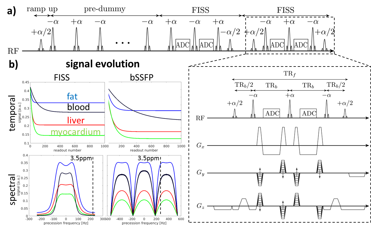
The pulse sequence diagram of FISS2 is depicted in Fig.1. A FISS module is composed of a [+α/2,(-α,readout,+α)n,-α/2] train where α denotes the RF excitation and n is the number of bSSFP readouts surrounded by the ±α/2 ramp-up/down pulses which cause the steady-state interruption. bSSFP readouts are repeated with a repetition time of TRb. In order to extend FISS to 3D Cartesian imaging here we propose to also consider: a) RF phase cycling, b) gradient spoiling and c) dummy preparation pulses. Between FISS modules the RF phase is incremented according to
$$\varphi_j = \varphi_{j-1}+j\cdot \varphi_0$$
yielding the phase of the j-th FISS module with a golden angle $$$\varphi_0=111.3^\circ$$$ initialization. Together with phase-encoding gradient spoiling, i.e. unbalanced gradients, this ensures sufficient spoiling of residual transverse magnetization before the next excitation. Dummy preparation pulses at the beginning of the sequence bring the magnetization to bSSFP steady-state before it is interrupted. Otherwise, it can occur that off-resonant transient magnetization is excited and even amplified instead of suppressed. This effect is in the Cartesian case more pronounced in the final image, because of the different phase-encoding steps per readout.
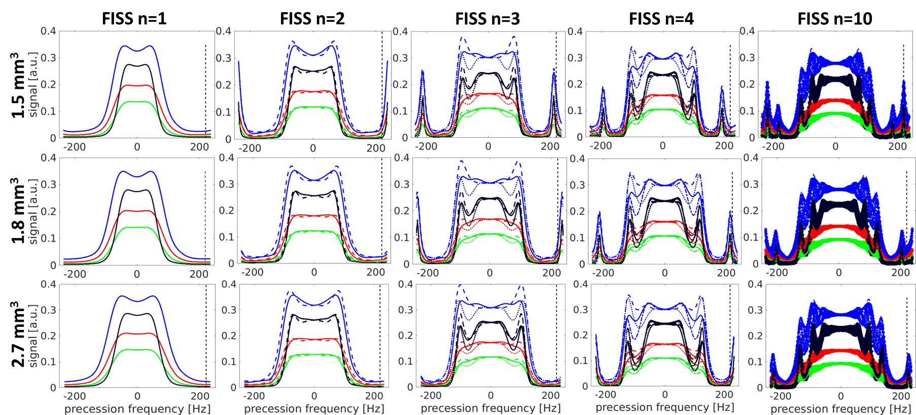
FISS signal evolution and frequency behavior were simulated using extended phase graphs4 (EPG) for fat (250/70 ms), liver (500/40 ms), myocardium (1000/45 ms) and blood (1600/250 ms) with respective T1/T2 values.
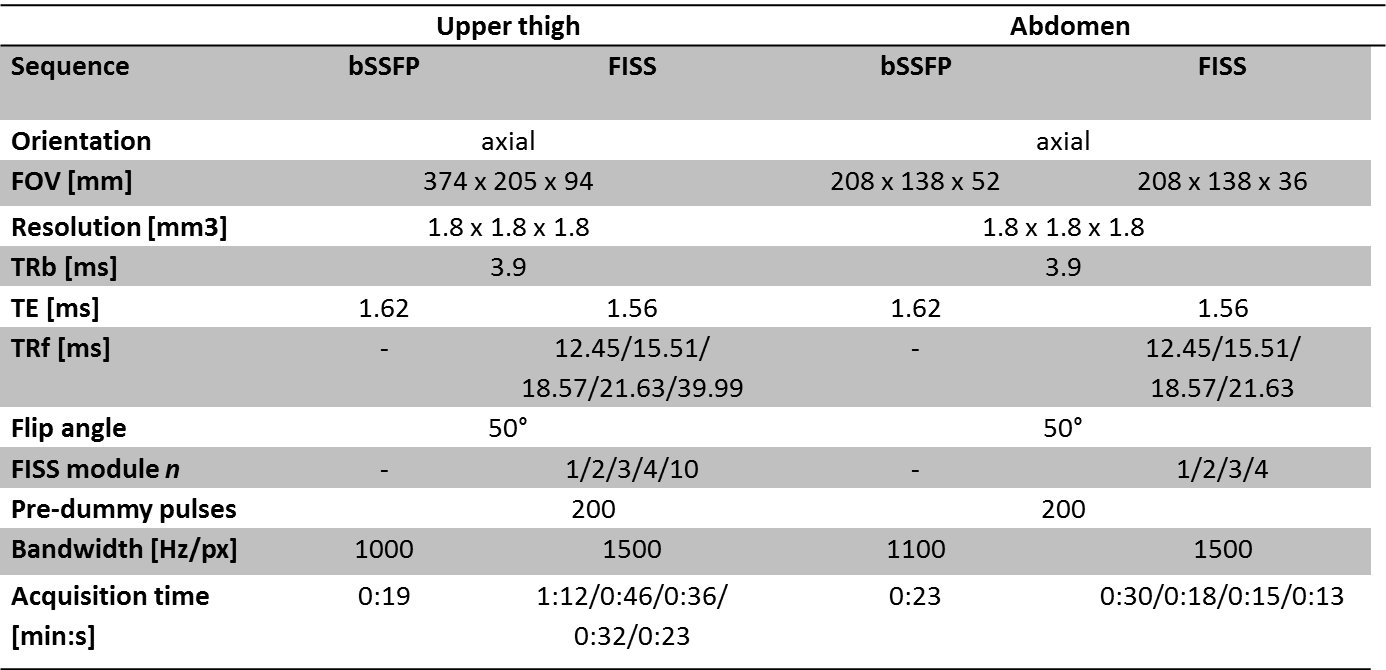
In-vivo data was acquired on a 1.5T MR scanner (Aera, Siemens Healthineers) with imaging parameters as defined in Tab. 1. Images were obtained in 5 healthy subjects (2 female, age = 29±3 years) for upper thigh and abdomen.
Results and Discussion
An exemplary comparison of FISS versus bSSFP is depicted in Fig. 1b) where the spectral frequency response indicates the fat suppression capability. At around 220 Hz (3.5ppm at 1.5T) a broad stop band provides the desired signal suppression for different tissues. The frequency response pattern changes for varying FISS interruption rates which is mainly determined by the timing parameters TRb and n. For fast acquisitions a minimal TRb is desired with maximal bandwidth, hence, the timing is primarily determined by the imaging resolution and therefore FISS behavior changes with imaging resolution and the number of bSSFP readouts n. These dependencies are investigated theoretically via EPG simulations in Fig. 2 and substantiated by measurements (Fig.3 and 4). The reliable operating range of FISS is determined by n and imaging resolution as depicted in Fig. 2 and tends to smaller values n for higher resolutions.
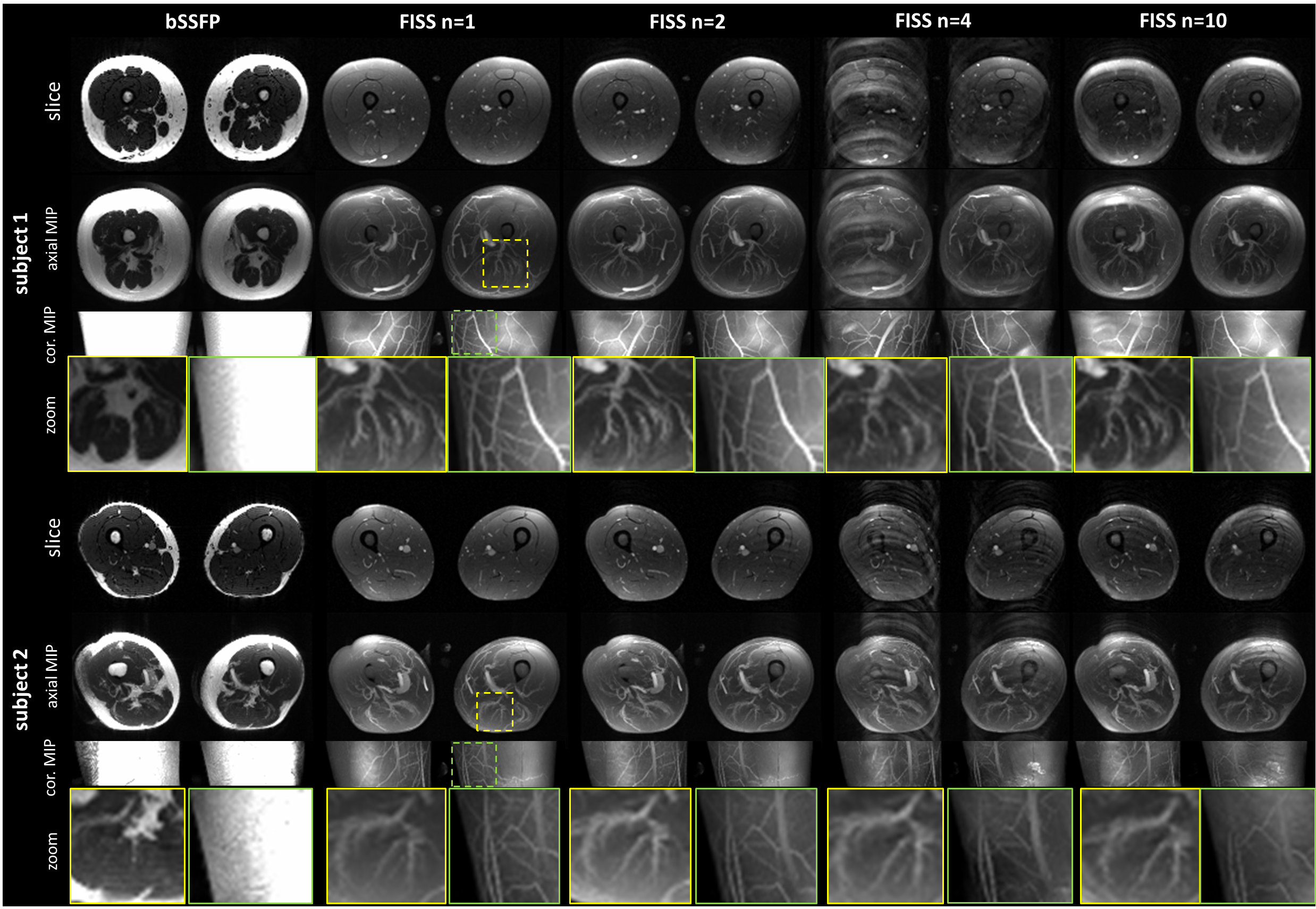
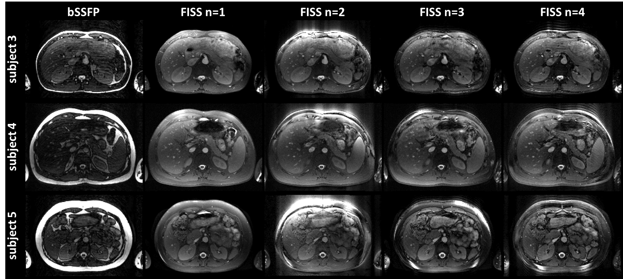
Images
for different length n of FISS in
comparison to bSSFP are shown for upper thigh in Fig. 3 and for abdomen in Fig.
4. Fat suppression is excellent and consistent over the whole volume and amongst subjects with varying body mass index with FISS
while absent in bSSFP (without fat-saturation pulses). Contrast between muscle
and vessel is improved (see MIP). Small vessel structures can be observed in
FISS and are lost in bSSFP. FISS fat suppression is susceptible to B0 inhomogeneities as observed in the abdominal imaging (Fig.4), but is still able to provide good fat suppression.
Conclusion
3D Cartesian FISS is feasible and offers homogeneous intrinsic fat suppression without the need for dedicated pre-pulses which makes it an ideal candidate for fast fat-suppressed imaging acquisitions such as 3D peripheral angiography or 3D abdominal imaging. Extensions of this approach to free-running 3D whole-heart MRI are currently under investigation.Acknowledgements
This work was supported by EPSRC (EP/P007619) and Wellcome EPSRC Centre for Medical Engineering (NS/A000049/1).References
1. Bieri, O. and K. Scheffler, Fundamentals of balanced steady state free precession MRI. Journal of Magnetic Resonance Imaging, 2013. 38(1): p. 2-11.2. Koktzoglou, I. and R.R. Edelman, Radial fast interrupted steady-state (FISS) magnetic resonance imaging. Magnetic Resonance in Medicine, 2018. 79(4): p. 2077-2086.
3. Edelman, R.R., et al., Cardiovascular cine imaging and flow evaluation using Fast Interrupted Steady-State (FISS) magnetic resonance. Journal of Cardiovascular Magnetic Resonance, 2018. 20(1): p. 12.
4. Weigel, M., Extended phase graphs: dephasing, RF pulses, and echoes - pure and simple. J Magn Reson Imaging, 2015. 41(2): p. 266-95.
Figures