4680
Radial Single-Shot Fast Spin Echo: Toward Fast, Motion-Robust, Multi-Contrast Imaging1Global MR Applications and Workflow, GE Healthcare, New York, NY, United States, 2Global MR Applications and Workflow, GE Healthcare, Madison, WI, United States, 3Global MR Applications and Workflow, GE Healthcare, Menlo Park, CA, United States, 4GE Healthcare, Waukesha, WI, United States, 5Global MR Applications and Workflow, GE Healthcare, Houston, TX, United States
Synopsis
We present a radial Single-Shot Fast Spin Echo pulse sequence that is capable of generating multiple contrasts from a single spin echo train via temporal filtering. Undersampling artifacts are minimized by using a long echo train and an aggressive variable refocusing flip angle scheme. In vivo feasibility is demonstrated in the brain.
Introduction
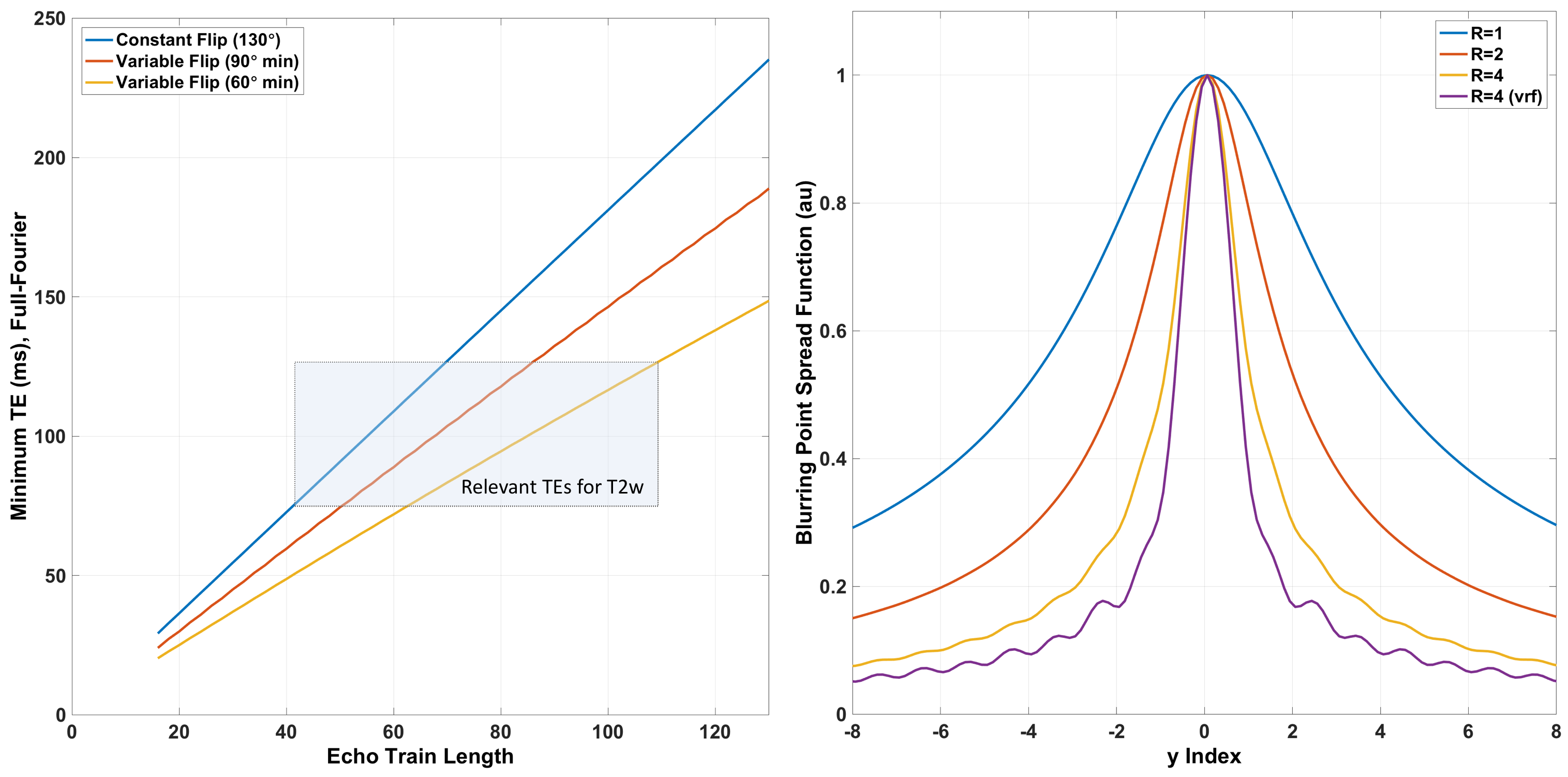
Though multi-shot fast spin echo (FSE, or TSE) remains the de facto gold standard in clinical T2-weighted (T2w) imaging, single-shot fast spin echo (SSFSE, or HASTE, SSH-TSE) often performs well in situations where speed and/or motion-robustness are critical, such as abdominal, pediatric and fetal imaging, at the expense of modest reductions in resolution, contrast, and signal-to-noise ratio (SNR). The advent of parallel imaging has been extremely beneficial for the clinical utility of SSFSE, and variable refocusing flip angle with full-Fourier encoding1 have enabled further gains in image quality. In spite of these improvements, standard Cartesian SSFSE with linear phase encoding is still constrained in several important ways, especially for full-Fourier encoding (Figure 1), making alternative sampling schemes worth considering. In addition, the growing interest in quantitative MRI, and efficient multi-contrast imaging2-5 make non-Cartesian k-space trajectories an interesting possibility for extending the utility of this clinical workhorse. Here, we present the results of our initial experience with a radial SSFSE pulse sequence.Methods
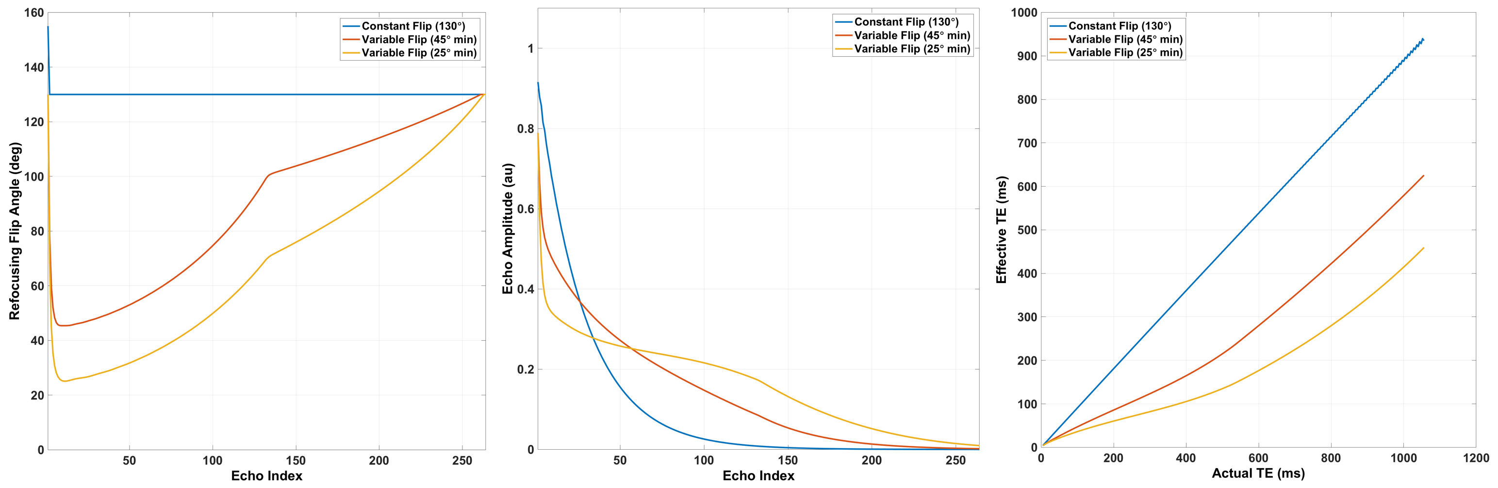
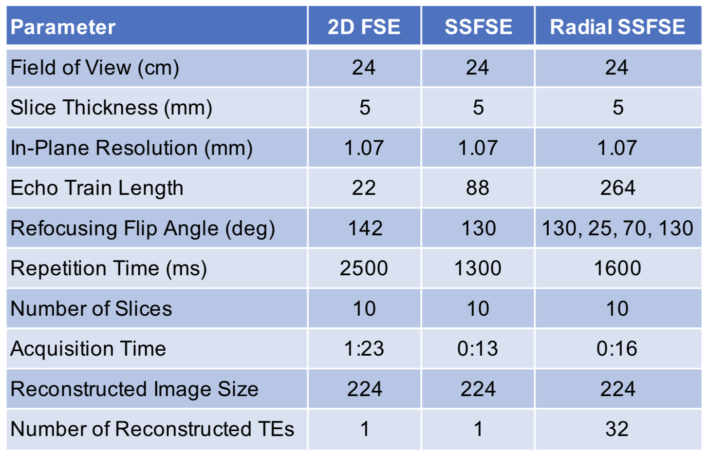
A conventional, Cartesian SSFSE pulse sequence was modified to acquire k-space data with a radial golden angle trajectory. An aggressive variable refocusing flip angle scheme6, with flip angle targets of 130, 25, 70 and 130 degrees, was utilized to prolong T2 decay (Figure 2), and a long echo train length (of 264) was used to minimize aliasing artifacts due to undersampling at the edge of k-space. Under IRB control, several volunteers were imaged on a 3T MRI scanner (Discovery MR750, GE Healthcare) with an 8-channel head coil. The radial SSFSE images were subsequently reconstructed with standard gridding, and an arbitrary number of T2 weightings were generated by repeatedly applying and shifting in the temporal (echo) direction a symmetric "tornado" (or hourglass) filter7. Conventional Cartesian multi-shot FSE and SSFSE images were collected for comparison at a single TE of 100ms and at the same spatial resolution, with parameters further summarized in Table 1.Results & Discussion
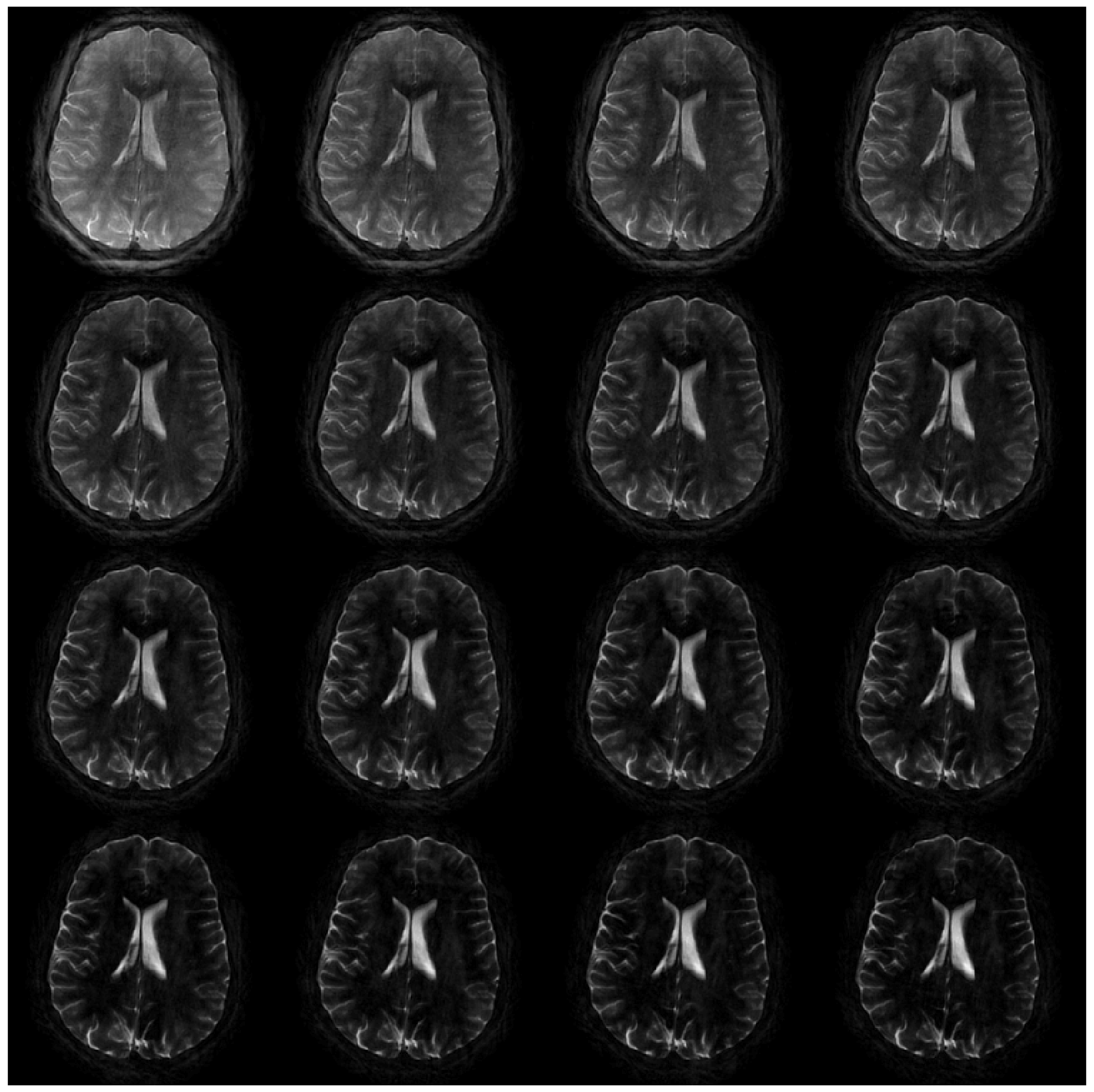
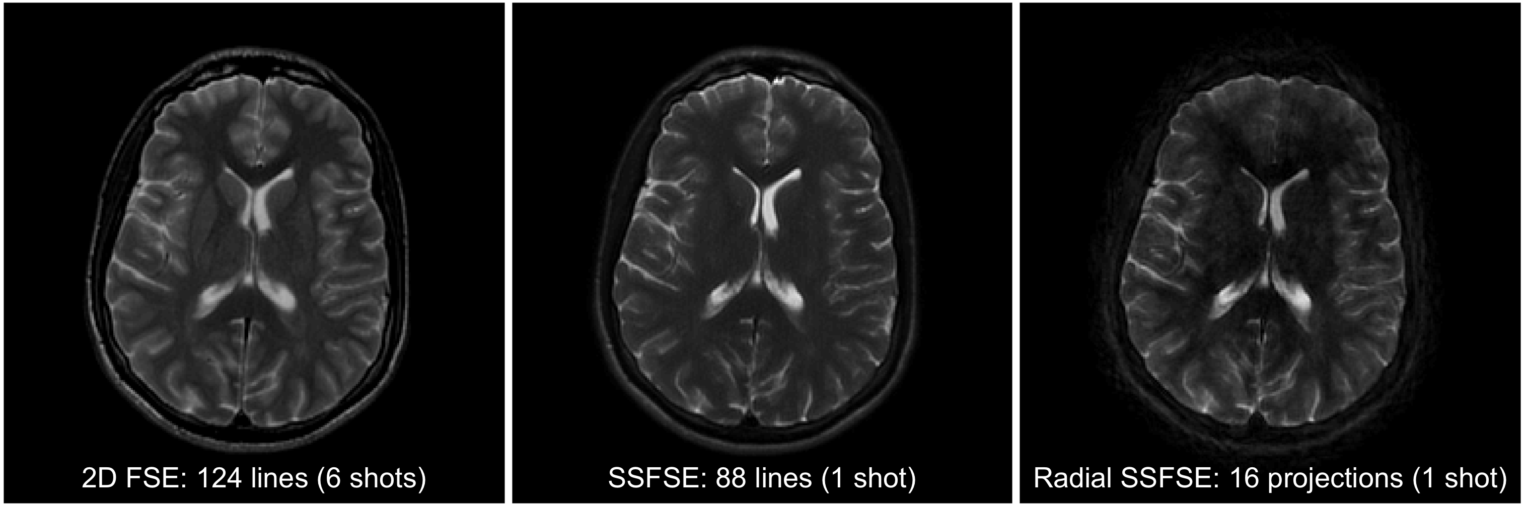
Preliminary in vivo imaging results from the radial SSFSE pulse sequence are shown in Figures 3 and 4. Figure 3 contains the first 16 reconstructed T2 weightings from one imaging shot of the radial SSFSE acquisition, captured in less than two seconds, and arbitrarily reconstructed at a temporal resolution of 32 phases with 50% overlap (i.e. 16 full projections per phase with high-frequency view-sharing via tornado filtering). Early gray/white matter signal decay is clearly visible, while CSF signal persists due to its relatively long T2. Figure 4 contains the conventional, Cartesian multi-shot FSE and SSFSE comparison images acquired at a single TE of 100 ms, versus a single reconstructed temporal phase from the radial SSFSE acquisition, demonstrating comparable T2 contrast and reasonable image quality given the relative reduction in high-frequency k-space sampling density.
These preliminary results demonstrate the feasibility of radial SSFSE imaging, its ability to generate multiple T2 contrasts in a single imaging shot, and its ability to circumvent some of the limitations imposed by standard linear phase encoding, namely the reliance on partial Fourier reconstruction techniques, the proportional relationship between TE and ETL, and the necessary tradeoff between ETL (SNR) and directional T2-blur. The costs of this flexibility and additional contrast information, however, are the image reconstruction challenges common to all non-Cartesian imaging, generally, including blurring and streaking artifacts, and increased computational complexity.
Conclusion
We have presented a novel radial SSFSE pulse sequence that enables the reconstruction of multiple contrasts in a single imaging shot. Undersampling artifacts are minimized in this example with variable refocusing flip angle and a long echo train. Greater undersampling, along with an exploration of advanced image reconstruction techniques, and a methodical evaluation of the motion-robustness of this pulse sequence, remain as future work.Acknowledgements
The authors would like to acknowledge helpful discussions with Andre Fischer and Anne Menini of GE Healthcare.References
- Loening AM, Litwiller DV, Saranathan M, Vasanawala SS. Increased Speed and Image Quality for Pelvic Single-Shot Fast Spin-Echo Imaging with Variable Refocusing Flip Angles and Full-Fourier Acquisition. Radiology. 2017 Feb;282(2):561-568.
- Tanenbaum LN, Tsiouris AJ, Johnson AN, Naidich TP, DeLano MC, Melhem ER, Quarterman P, Parameswaran SX, Shankaranarayanan A, Goyen M, Field AS. Synthetic MRI for Clinical Neuroimaging: Results of the Magnetic Resonance Image Compilation (MAGiC) Prospective, Multicenter, Multireader Trial. AJNR Am J Neuroradiol. 2017 Jun;38(6):1103-1110.
- Tamir JI, Uecker M, Chen W, Lai P, Alley MT, Vasanawala SS, Lustig M. T2 shuffling: Sharp, multicontrast, volumetric fast spin-echo imaging. Magn Reson Med. 2017 Jan;77(1):180-195.
- Keerthivasan, M Saranathan, A Bilgin, DR Martin, MI Altbach, High-resolution 3D T2 mapping using a stack-of-stars radial FSE pulse sequence. Proc. ISMRM 2017.
- Litwiller D, Zhang J, Estkowksi L, Rettmann D, Bayram E. Fast, Motion-Robust, T1-Weighted Neuro Imaging with Single-Shot Fast Spin Echo. Proceedings of the American Society of Neuroradiology (ASNR), 2018, Vancouver, BC, Canada.
- Busse RF, Brau AC, Vu A, Michelich CR, Bayram E, Kijowski R, Reeder SB, Rowley HA. Effects of refocusing flip angle modulation and view ordering in 3D fast spin echo. Magn Reson Med. 2008 Sep;60(3):640-9.
- Barger AV, Block WF, Toropov Y, Grist TM, Mistretta CA. Time-resolved contrast-enhanced imaging with isotropic resolution and broad coverage using an undersampled 3D projection trajectory. Magn Reson Med. 2002 Aug;48(2):297-305.
Figures