4671
Visualizing the Myocardium in-vivo with a 3D uTE Acquisition1Canon Medical Research USA, Mayfield Village, OH, United States, 2Johns Hopkins University School of Medicine, Baltimore, MD, United States, 3Canon Medical Systems Corporation, Otawara-shi, Japan
Synopsis
A dark blood 3D uTE acquisition scheme with an MSDE pre-pulse is shown to provide suppression of flowing blood, while maintaining good definition of the myocardium.
Introduction
The presence of myocardial fibrosis has been studied with MRI in various cardiomyopathies and other heart diseases.[1-4] The methods to characterize the extent of fibrosis have relied on Gadolinium-based contrast agents to highlight the areas of fibrosis. Additionally, a more quantitative approach can be taken with myocardial T1 mapping using variations of the Look-Locker approach such as MOLLI, ShMOLLI, etc... [5]
Recently, ultra-short echo time acquisitions (uTE) have been shown to be effective in characterizing both fibrosis in the heart muscle and plaques in the vasculature [6-9], but remain limited due to cardiac motion and confounding signal from neighboring blood and/or fat. In this study, we aim to implement and optimize a cardiac gated uTE acquisition which nulls both blood and fat signal to allow visualization of myocardial fibrosis.
Methods
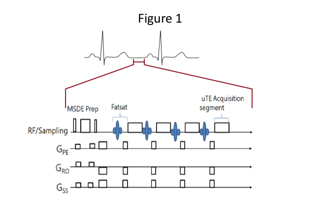
We have created a fat-saturated cardiac gated, uTE
acquisition intended to visualize the myocardium without interference from the
blood pool. The acquisition is a three
dimensional radial acquisition with TE 0.98/TR 4, Flip angle 10, collected with
1.3 x 1.3 x 1.5 mm resolution. In the
images presented, 25000 trajectories were acquired in groups of 50 per
heartbeat to keep the acquisition time during any heartbeat to 200 msec. The total acquisition time was approximately 500
heartbeats (8 minutes). Depending on heart
rate, the acquisition window was delayed approximately 600 msec after the
R-wave to acquire the data in diastole.
Since each trajectory acquires the center of k-space, a Motion Sensitive Driven Equilibrium (MSDE) pre-pulse was used to suppress the blood pool signal over a wide variety of velocities and directions. This pre-pulse was applied once per heartbeat, immediately prior to the imaging segment. To evaluate blood signal nulling, imaging was performed on a healthy female volunteer in accordance with a protocol approved by the local institutional review board.
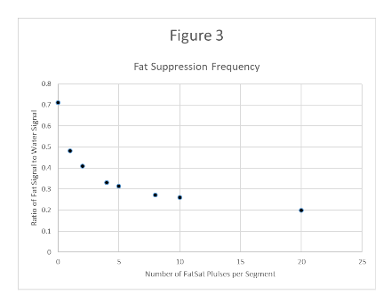
Fat saturation was achieved by broadcasting multiple spatial spectral saturation pulses evenly spaced across the segments. To investigate the effect of fat saturation and the number of pulses, a phantom study was performed on a two-compartment phantom with a water/CuSO4 solution in one compartment and vegetable oil in the other and the ratio of fat:water signal plotted vs number of pulses from 1 to 20.
The pulse sequence diagram in Figure 1 illustrates the final acquisition scheme with 5 fat suppression pulses.
Results
A fat-suppressed, blood nulled, cardiac gated uTE sequence was successfully implemented and gave the following results.
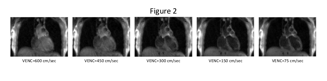
The images in Figure 2 demonstrate the effect of changing the velocity encoding (VENC) from 600 cm/sec to 75 cm/sec, where signal nulling improves with stronger gradient weighting.
Similarly, demonstrating the effect of number of fat presaturation pulses, the ratio of the fat signal to the water signal is plotted in Figure 3 showing diminishing returns employing more than 5 pulses.
Discussion
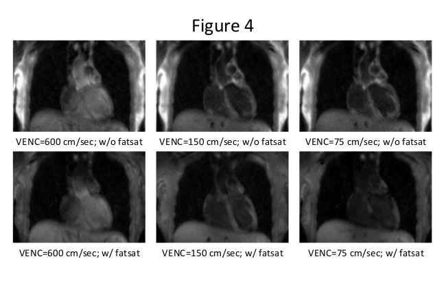
Maintaining a short (200-300 msec) imaging window is essential for 3D uTE imaging of the beating heart. A single application of the MSDE prepulse will eliminate both coherent and incoherent blood signal that would otherwise obscure the myocardium. As shown in Figure 2, for this subject, a VENC of 150 cm/sec is sufficient to remove blood signal, and still maintain good definition of the myocardium, however at 75 cm/sec the myocardial signal begins to disappear. Increasing the frequency of fat suppression within the acquisition segments improves the suppression at the expense of lengthening the data collection window. The addition of fat suppression causes additional attenuation of the myocardial signal, as shown in Figure 4, demonstrating that these two spin preparation events are not independent. In conclusion, the authors have shown a practical uTE acquisition scheme which may be utilized to evaluate myocardial signal without confounding signal from blood or fat.
Acknowledgements
No acknowledgement found.References
1. Mewton N, Liu CY, Croisille P, Bluemke D, Lima JA (2011) Assessment of myocardial fibrosis with cardiovascular magnetic resonance. J Am Coll Cardiol 57: 891-903.
2. Weber KT, Brilla CG (1991) Pathological hypertrophy and cardiac interstitium. Fibrosis and renin-angiotensin-aldosterone system. Circulation 83: 1849-1865.
3. Marijianowski MM, Teeling P, Mann J, Becker AE (1995) Dilated cardiomyopathy is associated with an increase in the type I/type III collagen ratio: a quantitative assessment. J Am Coll Cardiol 25: 1263-1272.
4. Brooks A, Schinde V, Bateman AC, Gallagher PJ (2003) Interstitial fibrosis in the dilated non-ischaemic myocardium. Heart 89: 1255-1256.
5. Donekal S, Lima JAC (2013) Diffuse Interstitial Myocardial Fibrosis by T1 Myocardial Mapping: Review, Transl Med, 3:1
6. Chan CF, Keenan NG, Nielles-Vallespin S, Gatehouse P, Sheppard MN, Boyle JJ, Pennell DJ, Firmin DN (2010) Ultra-short echo time cardio vascular magnetic resonance of atherosclerotic carotid plaque, Journal of Cardiovascular Magnetic Resonance, 12:17
7. Karolyi, et al (2013) Classification of Human Coronary Atherosclerotic Plaques Ex-vivo with T1, T2 and Ultra-short TE MRI, JACC Cardiovasc Imaging. 2013 April ; 6(4): 466–474.
8. Crowe LA, Petramaggiori G, Nielles-Vallespin S, Speier P, Vigato E, Majd H, Vallée J-P (2010) 3D radial UTE MRI for serial assessment of fibrosis development and silicone implant distortion in rat, Proc. Intl. Soc. Mag. Reson. Med. 18.
9. deJong S, et al (2011) Direct detection of myocardial fibrosis by MRI, Journal of Molecular and Cellular Cardiology 51: 974–979.
10. Carl M, Bydder GM, Du J (2016) UTE Imaging with Simultaneous Water and Fat Signal Suppression Using a Time-Efficient Multispoke Inversion Recovery Pulse Sequence, Magn Reson Med 76(2): 577–582
Figures