4660
Parallel Imaging based on k-x Domain Interpolation using Deep Neural Networks1Electrical and Electronic Engineering, Yonsei University, Seoul, Korea, Republic of
Synopsis
In this study, we compare two deep learning approaches to reconstruct multi-channel magnetic resonance (MR) images subsampled along phase-encoding (PE) direction. They are both based on the Fully-Connected (FC) layers but are performed in two different domains : Image domain, and k-x domain which is 1D inverse Fourier transformed (IFT) k-space. We demonstrate that the latter method shows superior performance to the former one in terms of removing the aliasing artifacts and recovering the details of MR images. The performance of the proposed method to the conventional MR reconstruction on image domain was qualitatively and quantitatively evaluated.
Introduction
MR reconstruction has been a very actively studied research area and parallel imaging (PI) has been proposed as a technique to accelerate the MR acquisition time since it utilizes the spatial sensitivity of each multi receiver coil 1,2. Today, deep learning is replacing the conventional MR reconstruction methods and this is also the case in the fields of PI 3. Most of the deep learning-based MR reconstruction methods are performed on either image domain or k-space domain. Recently, a manifold learning framework which directly reconstructs the MR images from the raw data has also emerged 4.
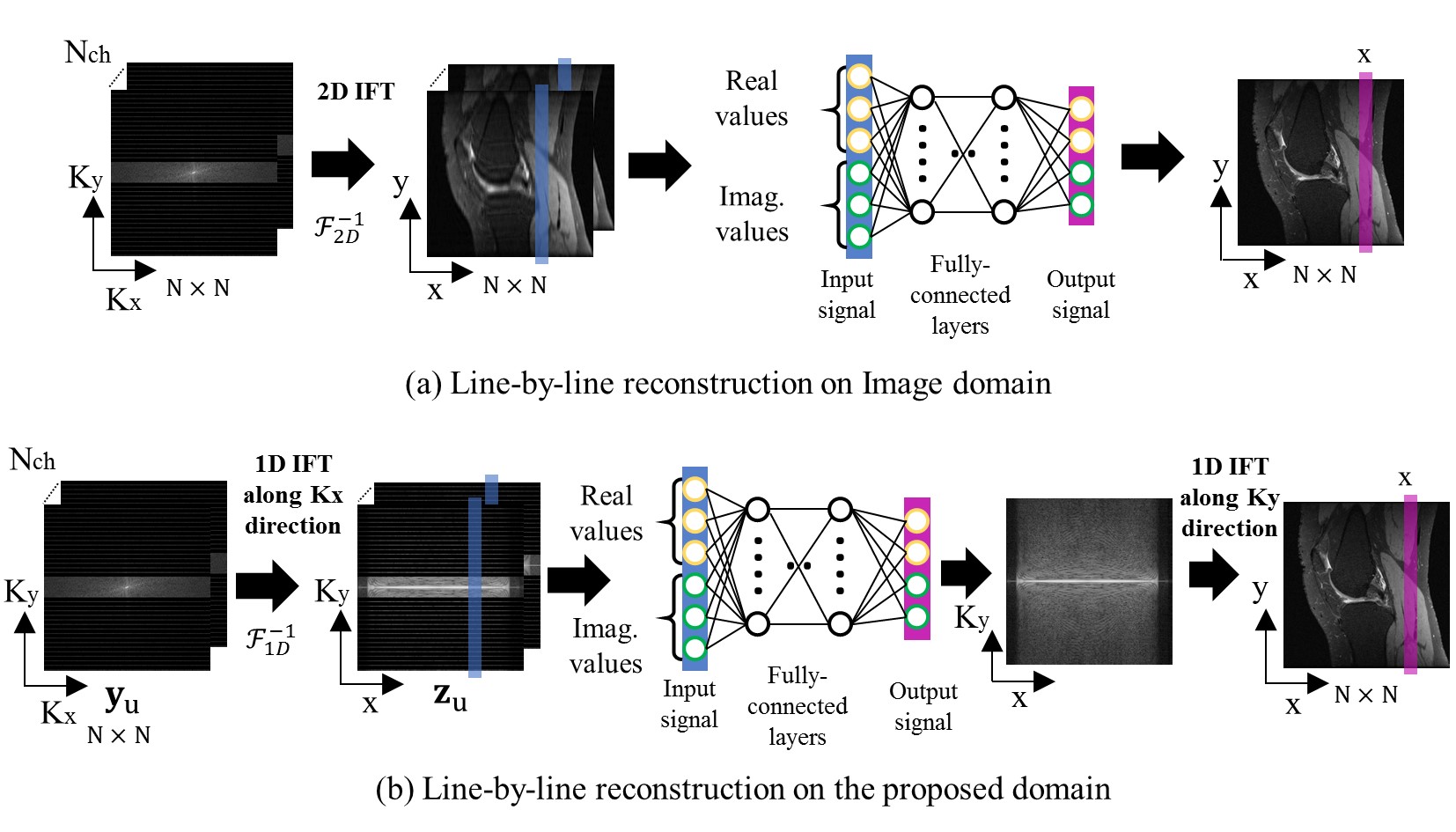
In this work, we propose a deep learning architecture based on FC layers to specifically reconstruct multi-channel MR data under-sampled along PE direction using cartesian sampling pattern. The input to the proposed network is multi-channel k-space data that is initially 1D inverse Fourier transformed along frequency-encoding (FE) direction. This causes no aliasing since the subsampling was performed in only PE direction. Then, the FC network processes the multi-channel column of the input line-by-line by interpolating the missing data using the true-sampled points. The FC network outputs the root-sum-of-squares line but with filled points that were initially missed from under-sampling. Lastly, 1D IFT along PE direction was performed to form the reconstructed images.
The advantage of the proposed method is that it utilizes the true-sampled data which is free of aliasing artifacts induced by inverse transforming the under-sampled data. The initial 1D FT along the FE direction enables line-by-line processing which dramatically reduces the number of parameters required for FC network from N2 to N. In addition, the complexity of the learning is much less compared to recently emerging domain transform learning4 since there is no need for the network to learn domain transform.
Methods
Standford Fullysampled 3D FSE Knee datasets 5 from 10 subjects were used for this study. The imaging parameters were as follows : FOV = 160mm x 160mm x 153.6mm, Matrix size = 320 x 320 x 1, Number of channels = 8, Number of slices = 256, TR/TE = 1550ms/25ms, Flip angle = 90’. Matrix size of each slice was resized to 256 x 256.
Figure.1. shows the structures of the two networks designed for this study. Figure.1-(a) shows the deep learning-based MR reconstruction performed on image domain, which maps multi-channel aliased input images into alias-free root-sum-of-squares images through line-by-line processing. This can be done since the subsampling was performed only in one direction. Figure.1-(b) shows the proposed method. First, the under-sampled input k-space is initially 1D inverse Fourier transformed along FE direction since this causes no aliasing due to the characteristic of the cartesian sampling pattern. Second, each multi-channel column line under-sampled along PE direction passes the FC network which is identically designed as the one in Figure.1-(a) for fair comparison. The FC network predicts the missing points by interpolating the true-sampled points and outputs the root-sum-of-squares line. Then, the output lines of the FC network are 1D inverse Fourier transformed along PE direction to form knee MR images. The mean square error was computed between the reconstructed MR images and full-sampled Ground Truth (GT) MR images.
The input and label data to the networks in Fig.1 were divided into real and imaginary components since the deep learning library used cannot handle complex data. The FC network comprises 4 layers. The first 3 layers contain 1280 neurons each, followed by Rectified Linear Unit (ReLU). The final layer contains 512 neurons, followed by linear activation to predict the desired output.
Result
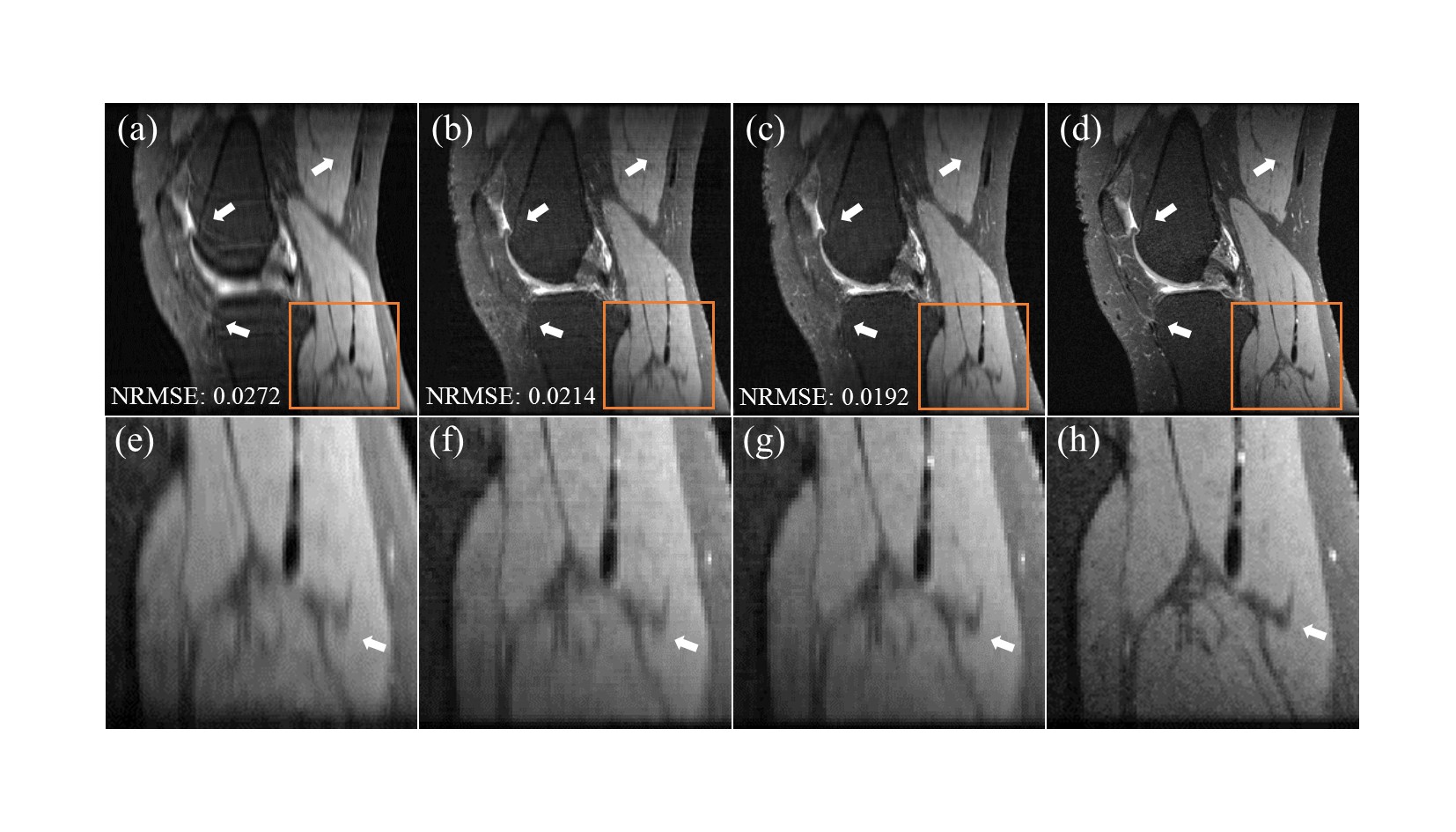
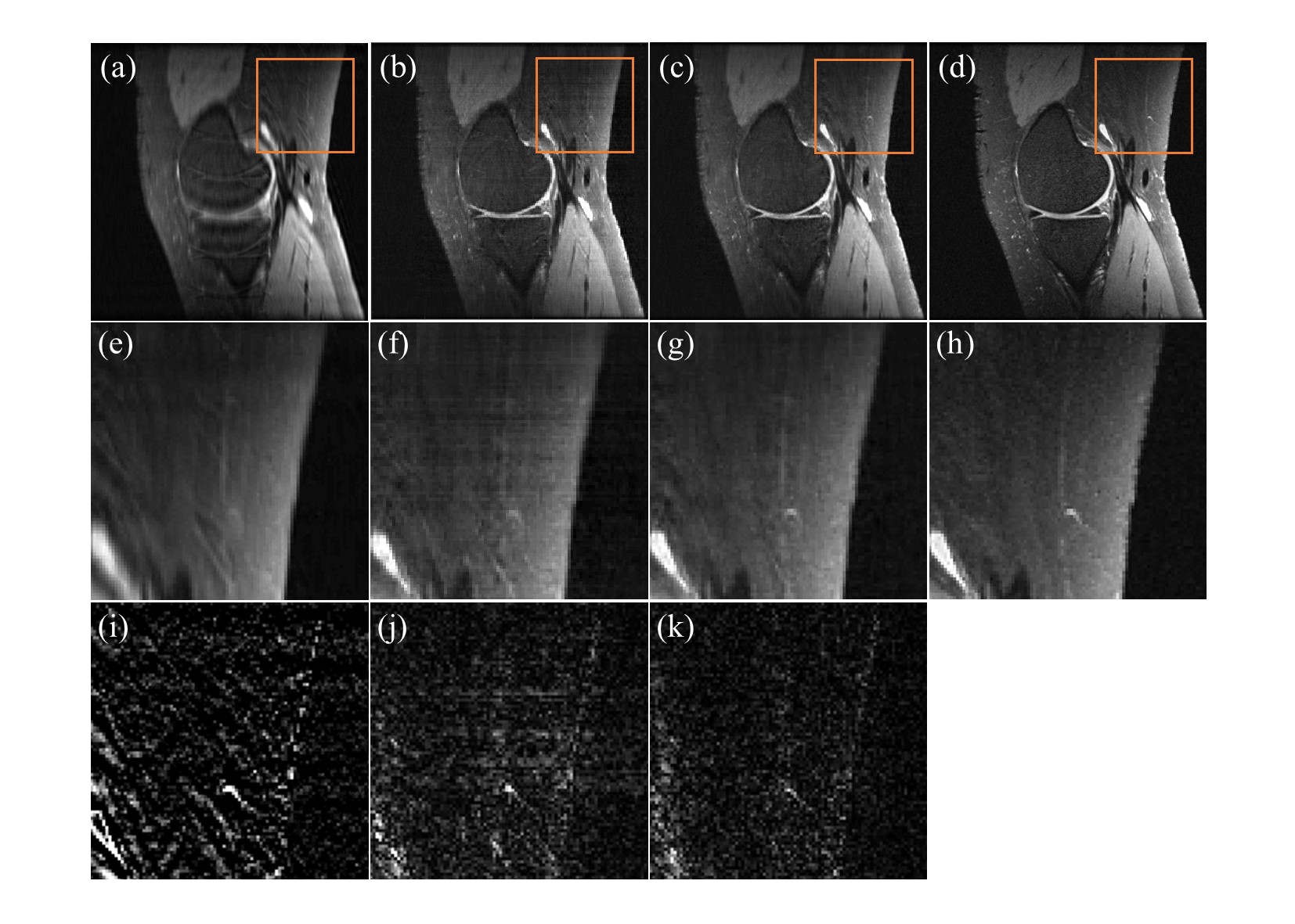
As shown in Figure.2, the proposed method (Figure.1-(b)) showed better reconstruction performance than that of MR reconstruction performed on image domain (Figure.1-(a)). Qualitatively, it showed improvements in reducing the aliasing artifacts and recovering the details of MR images. It was also free of horizontal artifact occurred from line-by-line processing on image domain as denoted in Figure.3. Quantitatively, the proposed method also showed better NRMSE result.Conclusion
In this study, we compare the line-by-line reconstruction performance of the two deep learning frameworks which are based on the same FC architecture but run in two different domains. The proposed method shows better result than MR reconstruction performed on image domain since it utilizes the raw information (i.e., true-sampled points) which is free of distortion occurred by 2D IFT on under-sampled data. This can reduce the complexity of the deep neural network and the number of required parameters.Acknowledgements
This research was supported by the National Research Foundation of Korea (NRF) grant funded by the Korean Government (MSIP) (2016R1A2B4015016) and National Research Foundation of Korea (NRF) grant funded by the Korean government (MSIT) (2018M3C7A1024734).References
1. Pruessmann, Klaas P., et al. "SENSE: sensitivity encoding for fast MRI." Magnetic resonance in medicine 42.5 (1999): 952-962.
2. Griswold, Mark A., et al. "Generalized autocalibrating partially parallel acquisitions (GRAPPA)." Magnetic Resonance in Medicine: An Official Journal of the International Society for Magnetic Resonance in Medicine 47.6 (2002): 1202-1210.
3. Kwon, Kinam, Dongchan Kim, and HyunWook Park. "A parallel MR imaging method using multilayer perceptron." Medical physics 44.12 (2017): 6209-6224.
4. Zhu, Bo, et al. "Image reconstruction by domain-transform manifold learning." Nature 555.7697 (2018): 487.
5. Epperson K, Sawyer AM, Lustig M, Alley M, Uecker M. Creation Of Fully Sampled MR Data Repository For Compressed Sensing Of The Knee. In: Proceedings of the 22nd Annual Meeting for Section for Magnetic Resonance Technologists, Salt Lake City, Utah, USA, 2013.
Figures