4652
Deep Scaled Domain Learning for Compressed MRI using Optional Scaling Transform1Utsunomiya University, Utsunomiya, Japan
Synopsis
Image domain learning designed for image denoiser has superior performance when aliasing artifacts are incoherent; however, its performances will be degraded if the artifacts show small incoherency. In this work, a novel image domain learning CNN is proposed in which images are transformed to scaled space to improve the incoherency of artifacts. Simulation and experiments showed that the quality of obtained image was fairly improved especially for lower sampling rate and the quality was further improved by cascaded network. It was also shown that the resultant PSNR exceeded one of the transform learning method.
Introduction
Recently, deep learning algorithms using convolutional neural network (CNN) show successful results in under-sampled signal reconstruction problem. There are many approaches such as Image domain learning [1,2], Transform learning [3], and k-space learning [4] and so on. Image domain learning can use deep residual learning CNN which is known as powerful image denoiser. However, the performance is not fully demonstrated if aliasing artifacts are not incoherent enough, e.g. 1-dimensional under-sampling in 2-D Cartesian signal acquisition. In this work, a novel image domain learning CNN method is proposed in which images are transformed to scaled space and artifacts are removed in that domain. In addition, cascaded CNN is applied to improve the image quality.Method
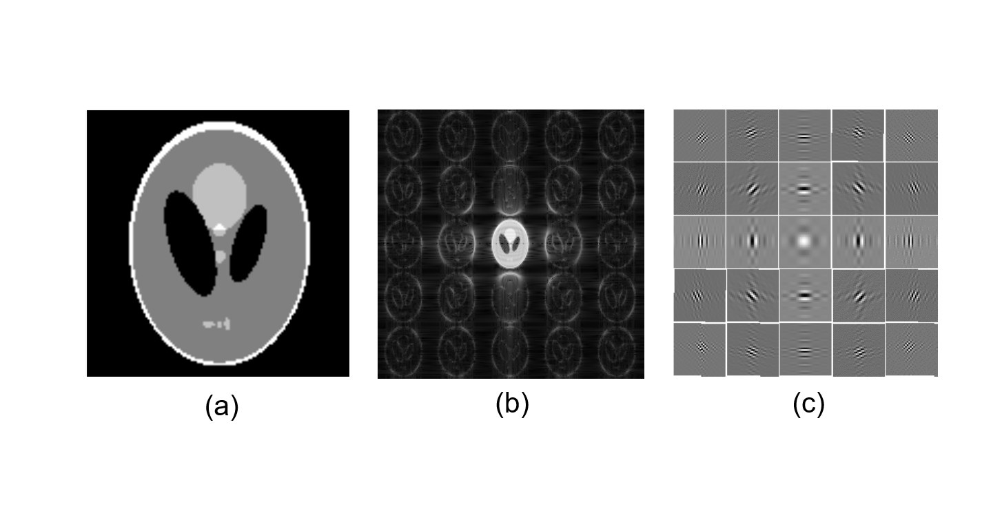
Fresnel transform based multi-resolution analysis (FREBAS) [5,6] is used to down-scale images. Considering one-dimensional signal, a decomposed sub image of $$$m$$$-th index $$$\rho(m,x) $$$ in FREBAS domain can be described equivalently as a convolution integral with the kernel of a band-pass filter function. where $$$\rho (x)$$$ is an image data, $$$\Delta x$$$ is the pixel width, N is number of data and $$$D$$$ is a scaling parameter,
$$ \rho (m,x)= \rho (x-mDN \Delta x) \ast {\rm sinc} \left(\frac{2 \pi x}{D \Delta x} \right) \exp\left( -j \frac{2 \pi m x }{ D \Delta x } \right) …(1)$$
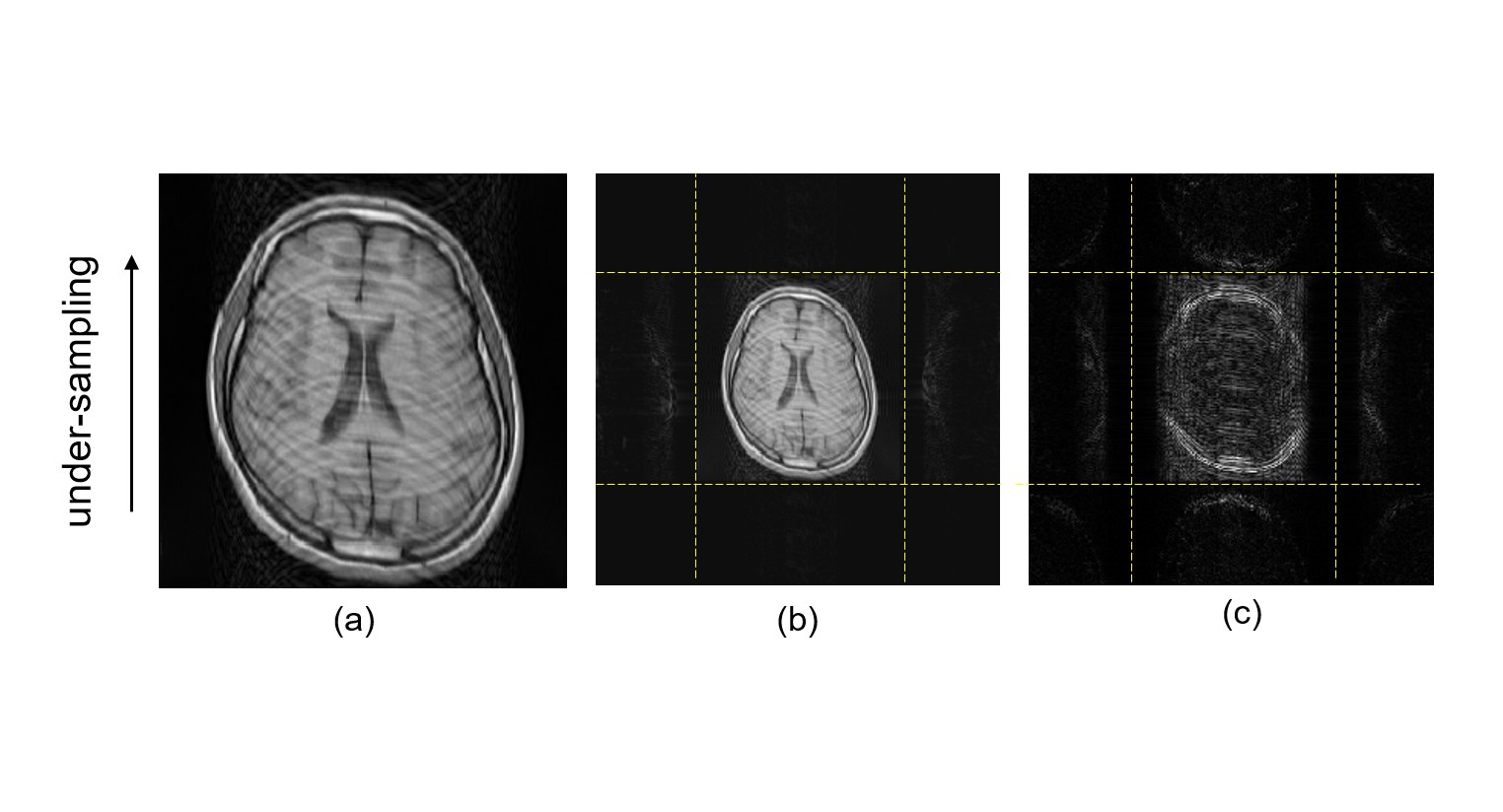
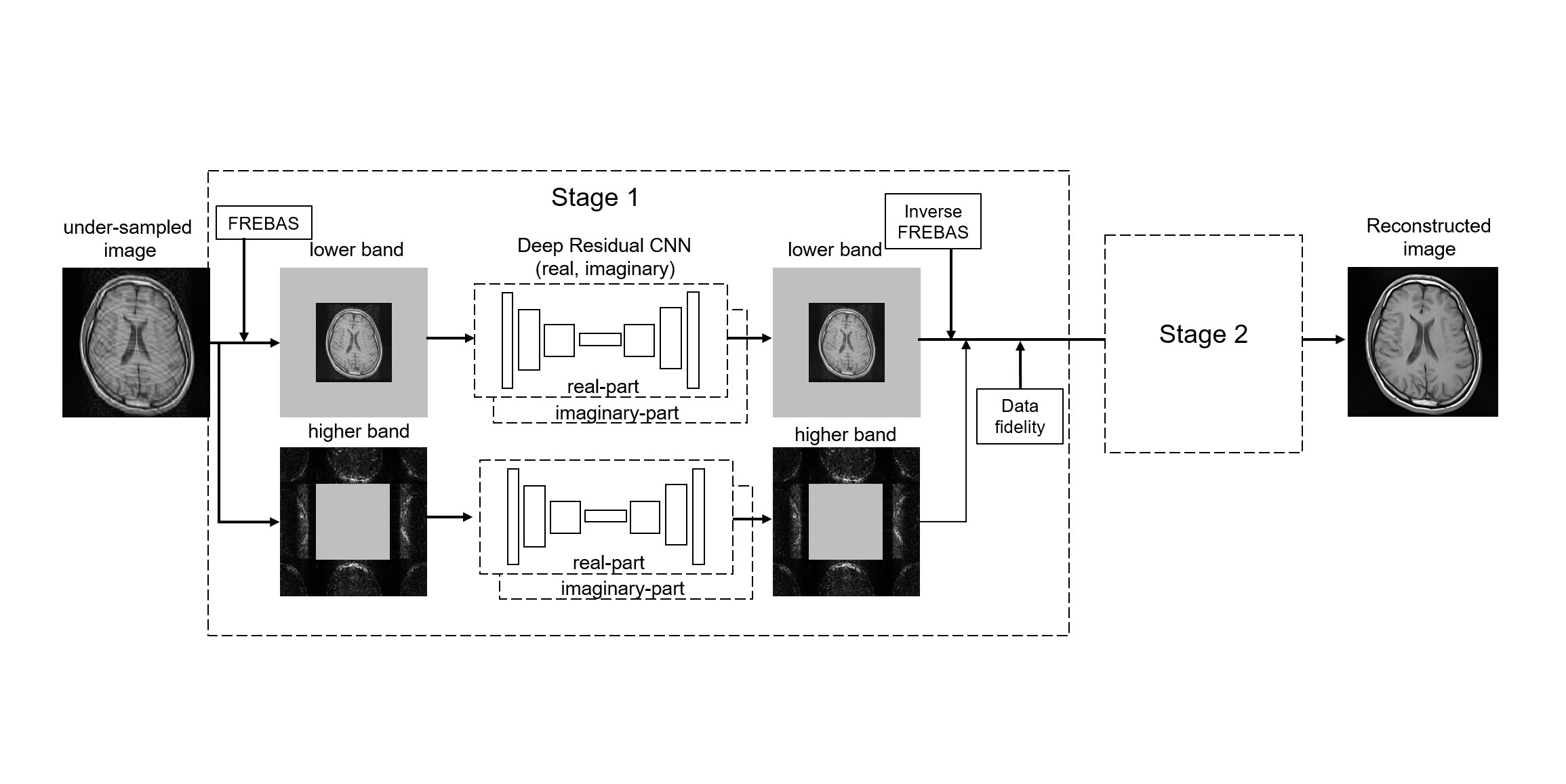
Even though Eq. (1) is described as convolution integral, FREBAS can be calculated using several FFTs and IFFTs. Since FREBAS is complex transform, it can be applied to phase varied images straightforwardly. Figure 1 shows the example of FREBAS transform. Figure 2 shows the FREBAS transform under-sampled image. Figure (a) shows a 1-dimensionally under-sampled image, figs. (b), (c) show the FREBAS transform of (a) using D=1.5 and error image in that domain, respectively. Figure 3 shows the proposed CNN network. As shown in Fig.2, where the magnitude of aliasing artifacts is not distributed uniformly in the FREBAS space, residual learning was performed separately for central lower-band image and higher-band images. Each CNN has two-channel, since FREBAS is a complex transform. Deep CNN based on residual learning and batch normalization [7,8] was used for learning the distribution of aliasing artifacts in the scaled domain. To improve the obtained image quality cascaded 2-stage network was also examined. The depth 17 of CNN was set 17 and corresponding receptive field size was 35x35. Three types of layers were used, (1) Conv+ReLU: for the first layer, 64 filters of size 3 x 3, 2) Conv+BN+ReLU: for layers 2 ~ 16, 64 filters of size 3 x 3 x 64, 3) Conv: for the last layer, 3x3x64 filter were used to reconstruct the output.
Results & Discussions
Simulation
experiments of 2-dimensional Cartesian data acquisition were performed using volunteer images. Under-sampling for phase encoding direction was executed numericaly in the computer. 100 image were used for
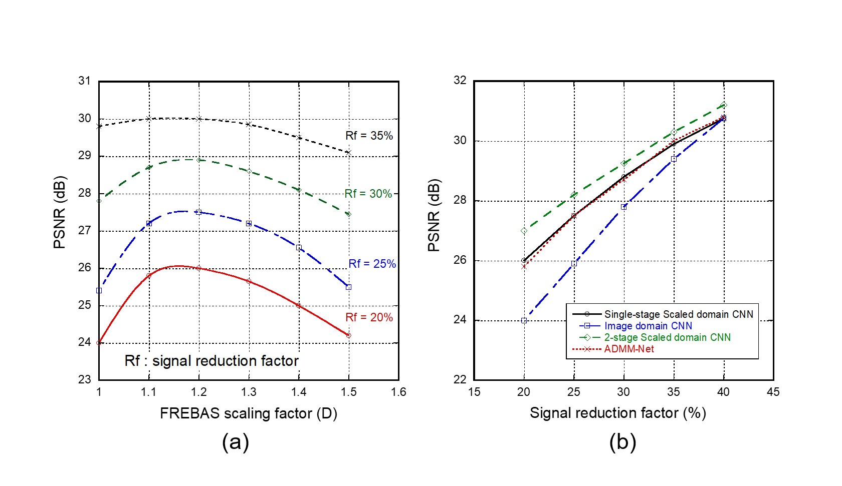
training CNN and 20 images were used for evaluation. Figure 4(a) shows the
relation of PSNR with reference to D. Highest PSNR is obtained when D takes the
value around 1.2 and the improvement of PSNR increases as the signal sampling
rate decreases. Incoherent artifacts will be transformed into coherent-like
artifacts by FREBAS scaling, however since most artifacts exist in lower bands,
reducing the area of the lower band will degrade the amount of removed
artifacts. Therefore, the highest PSNR is obtained when the scaling factor D
takes small value as 1.2. Figure 4(b) shows the results of PSNR evaluation with
reference to signal reduction factor. Proposed method is compared to 5-stage ADMM-Net
[9] in which the
coefficient of ADMM reconstruction network between k-space signal and
reconstructed images is learned.
PSNR is fairly improved to
be equivalent to ADMM-net by applying the scaled domain learning using FREBAS
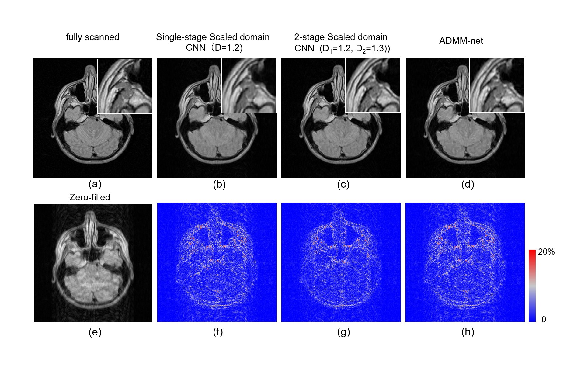
and the PSNR is further improved by using 2-stage CNN learning. Figure 5 shows
comparison of obtained images when the reduction factor Rf is 25%. Figure 5(a)
shows the fully scanned image and (e) shows the zero-filled image using
under-sampled signal. Proposed 2-stage CNN shown in Fig.5(c) used two diferent scaling parameter D1=1.2 and D2=1.3. Comparison of reconstructed images (b), (c), (d) shows that
the structure of images is much more remained and the reconstruction error
become smaller by applying 2-stage CNN learning. These results indicate
that scaled domain learning is effective when the down-scaling factor takes rather
small number such as 1.2 which scale is feasible by FREBAS but is difficult by
Wavelet or other dyadic transforms.Conclusion
Aother image domain learning for CS reconstruction method is proposed and demonstrated. The performances of Image domain learning can be improved by applying it in scaled domain and resultant PSNR exceeds that of ADMM-Net.Acknowledgements
This study was supported in part by JSPS KAKENHI(16K06379). We would like to thank Canon Medical Systems.References
- Kwon K, Kim D and Park H, A parallel MR imaging method using multilayer perceptron. Med. Phys 2017; 44 6209–6224
- Deep Residual Learning for Accelerated MRI using Magnitude and Phase Networks, arXiv:1804.00432
- Zhu B, Liu JZ, Cauley SF: Image Reconstruction by Domain-transform Manifold Learning, Nature, 555: 487-492, 2018
- Akçakaya M, Moeller S, Weingärtner S et al.: Scan-specific Robust Artificial-neural-networks for k-space Interpolation (RAKI): Database-free Deep Learning Reconstruction for Fast Imaging, in: Proc Intl Soc Mag Reson Med, 0576, 2018
- Ito S, Yamada Y, Multiresolution Image analysis using dual Fresnel transform Pairs and Application to Medical Image Denoising. IEEE International Conference on Image Processing 2003, Barcelona, Spain, Map8.7
- Ito S, Yamada Y, FREBAS Domain Super-Resolution Reconstruction of MR Images. ISMRM2010, 2936, Stockholm, Sweden
- He K,Zhang X,Ren S et al. Deep residual learning for image recognition. Conf IEEE CVPR: Las Vegas, 2016, 770-77
- Zhang K, Zuo W, Chen Y et al. Beyond a Gaussian Denoiser: Residual Learning of Deep CNN for Image Denoising. IEEE Tran Image Proc 2017:26, 3142-3155
- Yan Y, Jian S, Hbin L and Zongben Xu, ADMM-Net: A Deep Learning Approach for Compressive Sensing MRI. arxiv.org/abs/1705.06869
Figures