4645
Unsupervised Learning for Improved Fidelity Multi-contrast MRI1Electrical Engineering and Computer Sciences, University of California, Berkeley, Berkeley, CA, United States
Synopsis
Multi-contrast MRI acquisitions from a single scan have tremendous potential to streamline exams and reduce imaging time. However, maintaining clinically feasible scan times necessitates significant undersampling, pushing the limits on compressed sensing and other low-dimensional techniques. While learning methods have been proposed to overcome this limitation, they rely on fully sampled data for training, which are difficult to obtain for multi-dimensional imaging. Here, we present an unsupervised learning approach based on convolutional sparse coding, which learns a structured convolutional dictionary directly from undersampled k-space datasets. We apply the proposed method to T2 Shuffling knee datasets and demonstrate improvements to image sharpness and relaxation dynamics compared to the locally low-rank reconstruction.
Introduction
Multi-contrast MRI acquisitions from a single scan have the potential to shorten and streamline exams. These methods collect data at multiple measurement times and jointly reconstruct multiple images$$$^{1-3}$$$ or quantitative parameter maps$$$^{4-7}$$$. These high-dimensional acquisitions are typically sampling-limited, as many different contrast points must be collected to faithfully recover the underlying dynamics$$$^{2-4}$$$. Hence, reconstructions often rely on sparsity and low rank to take advantage of spatiotemporal redundancy$$$^{2,4-8}$$$, with their associated limitations: bias in the reconstruction, residual artifacts, and blurring. Multi-contrast reconstructions could potentially benefit from data-driven techniques that learn from a large number of existing datasets. Most existing machine learning methods are supervised and require fully sampled reference data. But in many clinical applications, high-quality, fully-sampled k-space datasets are scarce and hard to obtain.
Here, we propose a learning-based method using convolutional sparse coding (CSC)$$$^{9}$$$ to learn a structured convolutional dictionary directly from undersampled k-space$$$^{10}$$$. We apply CSC to T2 Shuffling (T2Sh)$$$^{2}$$$, a recently proposed multi-contrast acquisition that aims to mitigate blurring in conventional 3D fast spin-echo acquisitions by exploiting global and local low-rank information. We compare the image quality of the reconstructed multi-contrast images and demonstrate improved fidelity and reduced bias in the relaxation dynamics.Theory
T2Sh Model: Multi-contrast Representation Using Principle Component Basis
We consider the multi-echo forward model $$$y = \mathbf{E}x$$$, where $$$y$$$ is the under-sampled k-space data at different echo-times (TE), $$$\mathbf{E}$$$ is the forward operator incorporating ESPIRiT-based$$$^{11}$$$ coil sensitivities and sampling, and $$$x$$$ is the time series of images at different TEs. We approximate the time-series through a global subspace $$$\Phi$$$, where $$$\Phi$$$ is a predetermined basis based on anatomy and acquisition parameters$$$^{2,5-7}$$$.
Even with the global subspace, the reconstruction is highly under-determined, as acceleration factors are often greater than the number of coils. Hence, T2sh further constrains the reconstruction through locally low-rank (LLR) regularization$$$^{2,12}$$$. However, LLR does not leverage spatial structure and additionally leads to biases the relaxation dynamics.
Instead, we propose to leverage many under-sampled T2Sh scans previously acquired and replace LLR with a learning-based CSC that adapts to the data. To maintain computational efficiency, we maintain the global subspace constraint to compactly represent the relaxation direction.
Convolutional Sparse Coding for Multi-contrast Image Reconstruction
CSC$$$^{9}$$$ is a machine learning method for learning sparse representations. Here, we combine CSC with an explicit forward model$$$^{10}$$$ to directly learn convolutional kernels from T2Sh undersampled datasets $$$\{y_i\}$$$:$$\mathop{\min}_{\alpha_{i,j},d_j:\|d_j\|^2\leq1}\|\mathbf{E}\Phi(\sum_{j}d_j*\alpha_{ij}-y_i)\|_2^2+\|\alpha_{ij}\|_1,$$where $$$d_j$$$ are the 3D convolutional dictionary kernels and with $$$\alpha_i$$$’s being their the corresponding sparse coefficients, $$$y_i$$$ are the acquired k-space data and $$$\mathbf{E}\Phi$$$ is the encoding system matrix.
During training, the convolutional dictionary kernels $$$d_j$$$ and sparse coefficients $$$\alpha_i$$$ are jointly solved through alternating minimization using mini-batches of k-space data. In inference, the reconstruction uses the trained dictionary to find the sparse coefficients that best fit the acquired k-space. The reconstruction is implemented with the Python package SigPy using GPUs.
Method
T2Sh extended the CUBE pulse sequence (GE Healthcare) to randomly shuffle phase encode orderings (Figure 1). With IRB approval and informed consent/assent, five pediatric patients were scanned with T2Sh at 3T (GE MR750) using a 16-channel flex-coil array. We used 16 slices from the first four scans for training and two slices from the fifth scan for testing. A temporal subspace of size four was pre-computed from simulated signal evolutions. The convolutional dictionary consisted of 128 kernels, sized 11x11x4. We compared our CSC reconstruction with l1-wavelet and LLR reconstructions$$$^2$$$ with the same subspace.
Results
Figure 2 shows the reconstructed multi-contrast time series and subspace coefficients, as well as the magnitude component of the 3D convolutional dictionary kernels (11x11x4), learned directly from the under-sampled T2Sh data. Figure 3 compares the T2Sh reconstructions regularized with l1-wavelets, LLR, and the proposed CSC method. Overall, our method shows similar quality, with some marginal improvement to image sharpness. Figure 4 shows the T2 relaxation curve in trabecular bone and fluid for the three reconstructions. Compared to l1-wavelet and LLR regularizations, which have an artifact-related zero crossing, our learning-based reconstruction better resolves the temporal dynamics, which better explain the physical relaxation behavior. Figure 5 shows an animation of the reconstructed global subspace coefficients and the subsequent multi-contrast images, reconstructed by T2Sh with LLR regularization and by our proposed CSC method. As shown in the dynamics, our method reduces noise/artifact amplification in the third and fourth subspace coefficients, but show some square-like artifacts related to the convolutions, which may improve when scaling to hundreds-to-thousands of training datasets.
Conclusion
A learning-based reconstruction using convolutional sparse coding enables higher fidelity reconstructions of multi-contrast acquisitions, which may be suitable for Synthetic MRI and quantitative parameter mapping.Acknowledgements
We thank Xucheng Zhu for helpful discussion and advice. We also thank the following funding sources: National Institutes of Health (NIH) grants R01EB009690, P41RR09784; Sloan Research Fellowship; Bakar Fellowship; GE Healthcare.References
1. Riederer, S. J., Suddarth, S. A., Bobman, S. A., Lee, J. N., Wang, H. Z., & MacFall, J. R. (1984). Automated MR image synthesis: feasibility studies. Radiology, 153(1), 203-206.
2. Tamir, J. I., Uecker, M., Chen, W., Lai, P., Alley, M. T., Vasanawala, S. S., & Lustig, M. (2017). T2 shuffling: Sharp, multicontrast, volumetric fast spin‐echo imaging. Magnetic resonance in medicine, 77(1), 180-195.
3. Gómez, P. A., Sperl, J. I., Sprenger, T., Metzler-Baddeley, C., Jones, D. K., & Saemann, P. (2015). Joint Reconstruction of Multi-Contrast MRI for Multiple Sclerosis Lesion Segmentation. In Bildverarbeitung für die Medizin 2015 (pp. 155-160). Springer Vieweg, Berlin, Heidelberg.
4. Ma, D., Gulani, V., Seiberlich, N., Liu, K., Sunshine, J. L., Duerk, J. L., & Griswold, M. A. (2013). Magnetic resonance fingerprinting. Nature, 495(7440), 187.
5. Huang, C., Graff, C. G., Clarkson, E. W., Bilgin, A., & Altbach, M. I. (2012). T2 mapping from highly undersampled data by reconstruction of principal component coefficient maps using compressed sensing. Magnetic resonance in medicine, 67(5), 1355-1366.
6. Zhao, B., Setsompop, K., Adalsteinsson, E., Gagoski, B., Ye, H., Ma, D., ... & Wald, L. L. (2018). Improved magnetic resonance fingerprinting reconstruction with low‐rank and subspace modeling. Magnetic resonance in medicine, 79(2), 933-942.
7. Assländer, J., Cloos, M. A., Knoll, F., Sodickson, D. K., Hennig, J., & Lattanzi, R. (2018). Low rank alternating direction method of multipliers reconstruction for MR fingerprinting. Magnetic resonance in medicine, 79(1), 83-96.
8. Doneva, M., Börnert, P., Eggers, H., Stehning, C., Sénégas, J., & Mertins, A. (2010). Compressed sensing reconstruction for magnetic resonance parameter mapping. Magnetic Resonance in Medicine, 64(4), 1114-1120.
9. Zeiler, M. D., Krishnan, D., Taylor, G. W., & Fergus, R. (2010). Deconvolutional networks.
10. Ong, F., Lustig, M. (2018). k-space Aware Convolutional Sparse Coding: Learning from Undersampled k-space Datasets for Reconstruction. In Proc. Intl. Soc. Mag. Reson. Med (Vol. 26, p. 3378).
11. Uecker, M., Lai, P., Murphy, M. J., Virtue, P., Elad, M., Pauly, J. M., ... & Lustig, M. (2014). ESPIRiT—an eigenvalue approach to autocalibrating parallel MRI: where SENSE meets GRAPPA. Magnetic resonance in medicine, 71(3), 990-1001.
12. Trzasko, J. D. (2013, September). Exploiting local low-rank structure in higher-dimensional MRI applications. In Wavelets and Sparsity XV (Vol. 8858, p. 885821). International Society for Optics and Photonics.
Figures
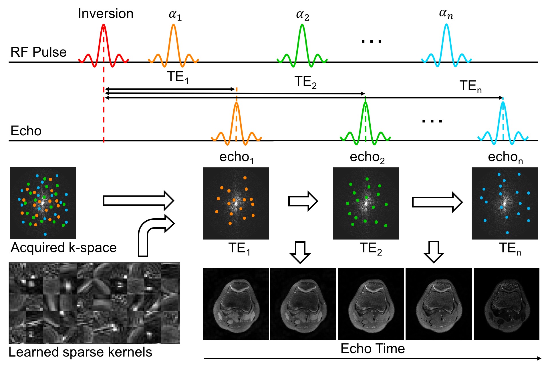


Figure 4. T2 relaxation curve in trabecular bone and fluid. The comparisons between different methods in bone and fluid are shown on the right side respectively. Note, that locally low rank based T2 shuffling suffers from bias and exhibits a non-physical zero crossing, unlike our proposed unsupervised learning-based technique.
