4644
An Unsupervised Deep Learning Approach for Reconstructing Arterial Spin Labeling Images from Noisy Data1Gordon Center for Medical Imaging, Massachusetts General Hospital and Harvard Medical School, Boston, MA, United States
Synopsis
Recently convolutional neural networks (CNNs) have been successfully applied to computer vision tasks and attracted growing interests in medical imaging. One barrier for the application of deep neural networks is the need of large amounts of training pairs, which are not always available in clinical practice. Inspired by the deep image prior method, this work presents a new image reconstruction framework based on CNN representation where no training pairs and pre-training are needed. We demonstrate the effectiveness of the proposed method by performing denoising and image reconstruction using noisy arterial spin labeling (ASL) data with and without undersampling.
Introduction
Convolutional neural networks (CNNs) have achieved great success in computer vision, and currently been applied to medical image denoising and reconstruction tasks [1-11]. One requirement for the successful application of CNN is large amount of training pairs. In applications such as 3D cardiac magnetic resonance (MR) imaging, labels reconstructed from free-breathing fully-sampled data is difficult to get. For image reconstruction tasks, raw k-space data is needed but not easy to collect retrospectively.
Recently, it is shown in the deep image prior framework that CNN can learn intrinsic structures from corrupted images and is possible to restore the clean image from its corrupted version by only employing random noise as network input [12]. Different from natural images, for MR scans, prior images of the same subject, instead of random noise, can be employed as network input, which should further improve the results. Furthermore, instead of using the corrupted image as training labels, k-space data can be utilized as training labels and the training objective function can be formulated based on maximum likelihood.
Inspired by these two observations, we propose a new image reconstruction framework using CNN as image representation. The CNN is trained from scratch along with the reconstruction and no prior training data are needed. The proposed framework can be advantageous in applications such as arterial spin labeling (ASL) imaging [13,14], where the perfusion signal suffers from inherently low signal-to-noise ratio (SNR) [15,16], while the control image has high SNR and can be used as network input. Performance of the proposed method is tested using denoising and undersampled reconstruction cases.
Methods
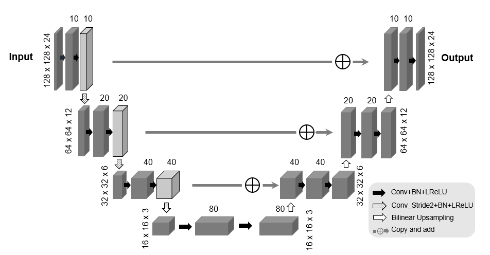
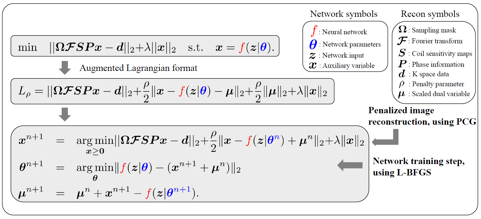
The unknown magnitude image $$$x$$$ is represented by a CNN output: $$x = f(\theta|z),$$ where $$$f:\mathbb{R}\rightarrow\mathbb{R}, \theta, z$$$ indicate the CNN, network trainable parameters and network input, respectively. A modified 3D U-net [17] shown in Fig. 1 is used as the network structure. As proof-of-concept, we choose the control image of ASL as the network input $$$z$$$. For denoising applications, the training function is L2 norm of the difference between the network output and the noisy image: $$\hat{\theta} = \underset{\theta}{\operatorname{argmin}} ||f(\theta | z) - b||_2, $$ where $$$b$$$ denotes the noisy magnitude image. For image reconstruction from undersampled k-space data using the SENSE framework [18], the training function is constructed as $$\hat{\theta} =\underset{\theta}{\operatorname{argmin}} ||\Omega FSPf(\theta | z) - d||_2 + \lambda ||f(\theta | z)||_2,$$ where $$$\Omega, F, P, d$$$ denotes down-sampling mask, Fourier transform, coil sensitivity maps, phase information matrix and k-space data, respectively. As the system matrix is coupled with the neural network, alternating direction method of multipliers (ADMM) [19] (details shown in Fig. 2) is used to separate the reconstruction and network training steps as the network training need more updates. In addition, ADMM allows the direct use of penalized image reconstruction methods at the image reconstruction step, which have been extensively studied with many existing packages and toolboxes. L-BFGS [20] is chosen as the network training algorithm due to its monotonic property. PCG algorithm [21] is used to solve the penalized reconstruction. For each loop the network training step was run 20 epochs and in total 100 outer-iterations were run.
All experiments were performed on a healthy subject using a 3T whole-body MR scanner approved by our local IRB. All ASL images were acquired using pseudo-continuous ASL (pCASL) [22,23] with balanced steady-state free precession readout [24,25]. The pCASL labeling parameters were as follows: flip angle=25°; RF duration/space =0.5/0.92 ms; total tagging duration=1500 ms, PLD time=1.2 s. Imaging parameters were as follows: FOV=240x180x120 mm3; encoding matrix size = 128x96x24, TR/TE=3.96/1.74 ms; total acquisition time=~5.3 min. For the undersampling case, 25% of each repetition was used (4x down-sampling). Signal-to-noise ratio (SNR) was calculated as the mean of the perfusion subtraction signal in the gray matter region divided by the standard deviation of noise.
Results
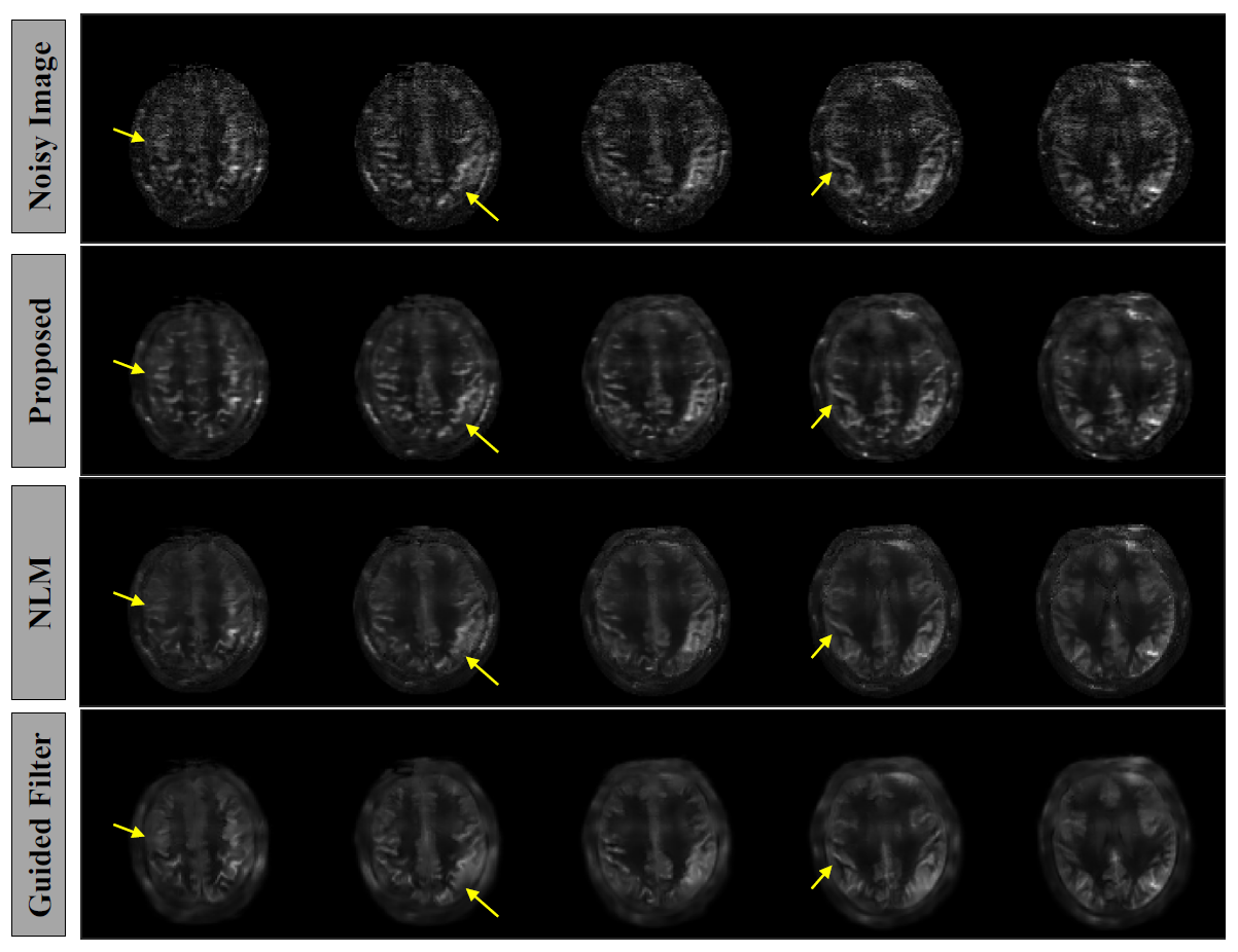
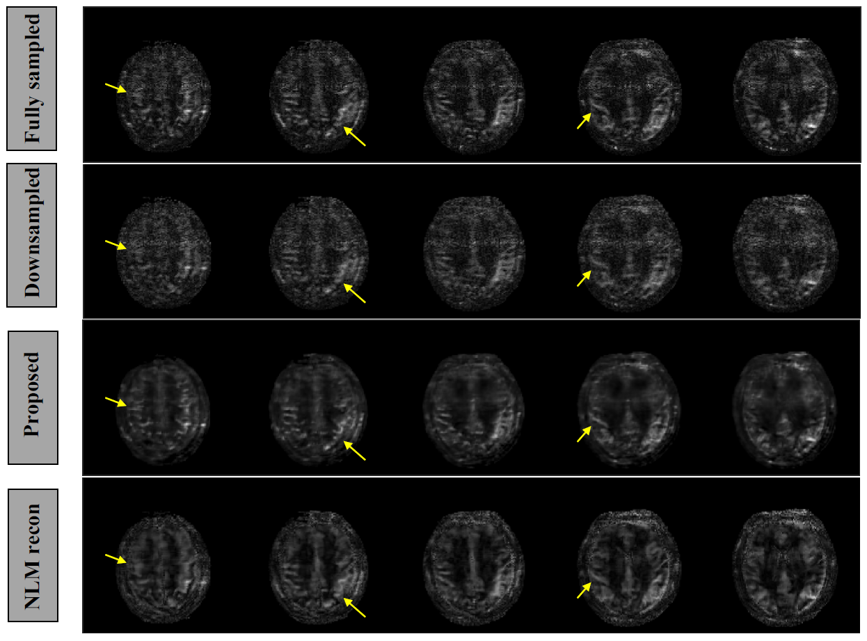
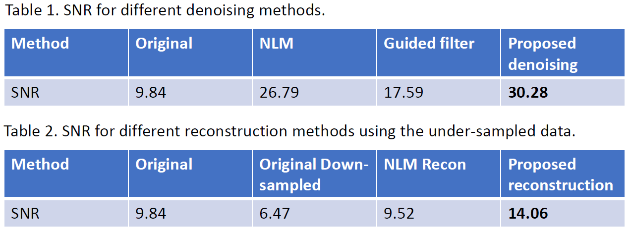
Fig 3. Shows the image denoising results obtained by the proposed method, which were also compared with nonlocal-mean (NLM) [26] and guided filter methods [27]. We can see that the proposed method can reveal more cortex details than the compared methods. SNR values shown in Table 1 of Fig. 5 confirms this observation. Fig 4 shows the reconstructed images using 25% of k-space data for each repetition. Compared with the method including NLM in the reconstruction framework [28], the proposed method produced images with superior quality. Table 2 of Fig.5 confirms this observation.Conclusions
We propose a new reconstruction framework by representing the unknown image as CNN output. Prior training pairs are not needed, but only the subject’s own images from other sequences. Results of both image denoising and reconstruction from undersampled data demonstrate the superior performance of the proposed framework.Acknowledgements
This work was supported by NIH grants R01 AG052653 and P41 EB022544.References
[1] Wang S, Su Z, Ying L, Peng X, Zhu S, Liang F, Feng D, Liang D. Accelerating magnetic resonance imaging via deep learning. InBiomedical Imaging (ISBI), 2016 IEEE 13th International Symposium on 2016 Apr 13 (pp. 514-517).
[2] Sun J, Li H, Xu Z. Deep ADMM-Net for compressive sensing MRI. In Advances in Neural Information Processing Systems 2016 (pp. 10-18).
[3] Schlemper J, Caballero J, Hajnal JV, Price AN, Rueckert D. A deep cascade of convolutional neural networks for dynamic MR image reconstruction. IEEE transactions on Medical Imaging. 2018 Feb;37(2):491-503.
[4] Hammernik K, Klatzer T, Kobler E, Recht MP, Sodickson DK, Pock T, Knoll F. Learning a variational network for reconstruction of accelerated MRI data. Magnetic resonance in medicine. 2018 Jun;79(6):3055-71.
[5] Zhu B, Liu JZ, Cauley SF, Rosen BR, Rosen MS. Image reconstruction by domain-transform manifold learning. Nature. 2018 Mar;555(7697):487.
[6] Mardani M, Gong E, Cheng JY, Vasanawala S, Zaharchuk G, Alley M, Thakur N, Han S, Dally W, Pauly JM, Xing L. Deep generative adversarial networks for compressed sensing automates MRI. arXiv preprint arXiv:1706.00051. 2017
[7] Kim KH, Choi SH, Park SH. Improving arterial spin labeling by using deep learning. Radiology. 2017 Dec 21;287(2):658-66.
[8] Aggarwal HK, Mani MP, Jacob M. MoDL: Model Based Deep Learning Architecture for Inverse Problems. IEEE transactions on medical imaging. 2018 Aug 13.
[9] Han Y, Yoo J, Kim HH, Shin HJ, Sung K, Ye JC. Deep learning with domain adaptation for accelerated projection‐reconstruction MR. Magnetic resonance in medicine. 2018 Sep;80(3):1189-205.
[10] Yang G, Yu S, Dong H, Slabaugh G, Dragotti PL, Ye X, Liu F, Arridge S, Keegan J, Guo Y, Firmin D. Dagan: Deep de-aliasing generative adversarial networks for fast compressed sensing mri reconstruction. IEEE transactions on medical imaging. 2018 Jun;37(6):1310-21.
[11] Qin C, Hajnal JV, Rueckert D, Schlemper J, Caballero J, Price AN. Convolutional recurrent neural networks for dynamic MR image reconstruction. IEEE transactions on medical imaging. 2018 Aug 6.
[12] Ulyanov D, Vedaldi A, Lempitsky V. Deep image prior. arXiv preprint arXiv:1711.10925. 2017 Nov 29.
[13] Detre JA, Leigh JS, Williams DS, Koretsky AP. Perfusion imaging. Magn Reson Med 1992;23(1):37-45.
[14] Williams DS, Detre JA, Leigh JS, Koretsky AP. Magnetic resonance imaging of perfusion using spin inversion of arterial water. Proc Natl Acad Sci U S A 1992;89(1):212-216.
[15] Alsop DC, Detre JA, Golay X, Gunther M, Hendrikse J, Hernandez-Garcia L, Lu H, Macintosh BJ, Parkes LM, Smits M, van Osch MJ, Wang DJ, Wong EC, Zaharchuk G. Recommended implementation of arterial spin-labeled perfusion MRI for clinical applications: A consensus of the ISMRM perfusion study group and the European consortium for ASL in dementia. Magn Reson Med 2015;73(1):102-116.
[16] Borogovac A, Asllani I, Arterial spin labeling (ASL) fMRI: advantages, theoretical constrains and experimental challenges in neurosciences. International journal of biomedical imaging, 2012.
[17] Ronneberger O, Fischer P, Brox T. U-net: Convolutional networks for biomedical image segmentation. InInternational Conference on Medical image computing and computer-assisted intervention 2015 Oct 5 (pp. 234-241). Springer, Cham.
[18] Pruessmann KP, Weiger M, Scheidegger MB, Boesiger P. SENSE: sensitivity encoding for fast MRI. Magnetic resonance in medicine. 1999 Nov 1;42(5):952-62.
[19] Boyd S, Parikh N, Chu E, Peleato B, Eckstein J. Distributed optimization and statistical learning via the alternating direction method of multipliers. Foundations and Trends® in Machine learning. 2011 Jul 26;3(1):1-22.
[20] Zhu C, Byrd RH, Lu P, Nocedal J. Algorithm 778: L-BFGS-B: Fortran subroutines for large-scale bound-constrained optimization. ACM Transactions on Mathematical Software (TOMS). 1997 Dec 1;23(4):550-60.
[21] Barrett R, Berry MW, Chan TF, Demmel J, Donato J, Dongarra J, Eijkhout V, Pozo R, Romine C, Van der Vorst H. Templates for the solution of linear systems: building blocks for iterative methods. Siam; 1994.
[22] Dai W, Garcia D, de Bazelaire C, Alsop DC. Continuous flow-driven inversion for arterial spin labeling using pulsed radio frequency and gradient fields. Magn Reson Med 2008;60(6):1488-1497.
[23] Wu WC, Fernandez-Seara M, Detre JA, Wehrli FW, Wang J. A theoretical and experimental investigation of the tagging efficiency of pseudocontinuous arterial spin labeling. Magn Reson Med 2007;58(5):1020-1027.
[24] Park SH, Wang DJ, Duong TQ. Balanced steady state free precession for arterial spin labeling MRI: Initial experience for blood flow mapping in human brain, retina, and kidney. Magn Reson Imaging 2013;31(7):1044-1050.
[25] Han PK, Ye JC, Kim EY, Choi SH, Park SH, 2016. Whole‐brain perfusion imaging with balanced steady‐state free precession arterial spin labeling. NMR in biomedicine, 29(3), pp.264-274.
[26] Buades A, Coll B, Morel JM. A non-local algorithm for image denoising. InComputer Vision and Pattern Recognition, 2005. CVPR 2005. IEEE Computer Society Conference on 2005 Jun 20 (Vol. 2, pp. 60-65). IEEE.
[27] He K, Sun J, Tang X. Guided image filtering. IEEE transactions on pattern analysis & machine intelligence. 2013 Jun 1(6):1397-409.
[28] Adluru G, Tasdizen T, Schabel MC, DiBella EV. Reconstruction of 3D dynamic contrast‐enhanced magnetic resonance imaging using nonlocal means. Journal of Magnetic Resonance Imaging. 2010 Nov;32(5):1217-27.
Figures