4638
Quiet Dixon Imaging with Looping Star Sequence1Philips Research, Hamburg, Germany, 2Philips Healthcare, Best, Netherlands
Synopsis
Dixon imaging with a conventional bipolar multi-gradient-echo sequence is often loud, mainly because of rapid switching of the strong readout gradient. In this work, the feasibility of using the Looping Star sequence instead is explored, which was recently introduced for quiet radial multi-gradient-echo imaging. Different variants of a dual-acquisition Looping Star sequence are proposed and demonstrated to allow a robust water-fat separation in phantom and volunteer experiments.
Introduction
The zero echo time (ZTE) sequence relies exclusively on frequency encoding and thus dispenses with slice selection and phase encoding gradients. Moreover, it keeps the readout gradient (GR) on during excitation and changes GR only gradually each repetition time (TR).1 In this way, it enables gradient switching and associated acoustic noise to be reduced to a minimum. However, it is commonly restricted to short, non-selective radiofrequency (RF) pulses with small flip angles for excitation and to sampling of free induction decays (FIDs) on center-out k-space trajectories for acquisition. Recently, self-refocusing k-space trajectories were introduced into the ZTE sequence to additionally allow sampling of gradient echoes. The resulting sequence, termed Looping Star, has since been demonstrated in effective transverse relaxation time constant (T2*)-weighted imaging as well as T2* and susceptibility mapping.2 In the present work, its application in chemical shift encoding-based water-fat separation is explored with the aim to quiet Dixon imaging.
Methods
As in the ZTE sequence, the amplitude of GR is kept constant throughout a shot in the Looping Star sequence, but the direction of GR is essentially rotated multiples of 360° to generate a train of gradient echoes from a train of FIDs. This is schematically illustrated in Fig. 1. To avoid high slew rates, the spacing between corresponding FIDs and gradient echoes must be fairly long, which hampers a reliable water-fat separation. Hence it is suggested to pursue a dual-acquisition approach instead. Two variants of it have been considered in the present work. In the first variant, TR is increased for the second acquisition, as shown in Fig. 1. This is achieved by altering the gradient spoiling while preserving the amplitude of GR and the receive bandwidth, or vice versa. The difference in TR is then given by the desired difference in echo time ΔTE = TE2 - TE1, divided by the number of excitations per shot (NE). In the second variant, TR is fixed, but NE is incremented for the second acquisition, as shown in Fig. 2. This involves rotating the direction of GR by a smaller angle per TR. ΔTE is in this case equal to TR.
Experiments were performed on a 1.5 T Ingenia scanner (Philips Healthcare, Best, Netherlands) using different coil arrays. A phantom consisting of a small, oil-filled cylinder inside a larger, water-filled cylinder and selected extremities of volunteers were imaged with the proposed dual-acquisition Looping Star sequence. Typical sequence parameters included an amplitude of GR of 6 – 9 mT/m and a flip angle of 3 – 4°. To suppress contamination of the signal from one gradient echo by the signal from the FIDs or other gradient echoes, a combination of RF and gradient spoiling or phase cycling was applied. Single-acquisition images were reconstructed from the collected data and processed with a two-point Dixon method using a multi-peak spectral model of human fat.3
Results
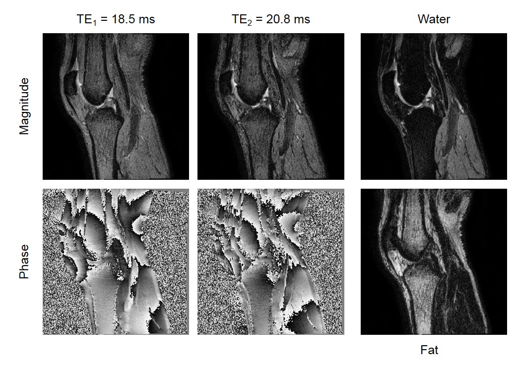
The feasibility of water-fat separation from two acquisitions with the Looping Star sequence is demonstrated in Figs. 3 and 4. The phantom was scanned with an isotropic resolution of 2 mm and a TR of 2.3 ms in the shown example. Using 8 and 9 excitations per shot for the two acquisitions led to an in-phase TE1 and opposed-phase TE2 of 18.4 ms and 20.7 ms, respectively. While the magnitude of the single-acquisition images is similar, the evolution of the phase reflects main field inhomogeneity (ΔB0) and chemical shift. Water and oil signals are well separated and correctly assigned to water and fat images. The knee was scanned with an isotropic resolution of 1 mm and 6 excitations per shot in the shown case. Choosing a TR of 3.1 ms and 3.5 ms for the two acquisitions led to a TE1 and TE2 of 18.5 ms and 20.8 ms, respectively. Water and fat are consistently separated, despite the many phase wraps in the single-acquisition images.
Discussion
Dixon imaging with the ZTE sequence was attempted previously, but struggled with the inherently ill-conditioned water-fat separation in the central k-space covered at acquisition times close to zero and with ΔB0 mapping.4 The Looping Star sequence provides images at non-zero echo times and, using a dual-acquisition approach, with echo spacings favorable for Dixon methods. While requiring higher slew rates than the ZTE sequence, it still promises to reduce acoustic noise substantially compared to a conventional bipolar multi-gradient-echo sequence. A thorough quantitative analysis remains to be carried out. Moreover, scan acceleration needs to be applied to shorten scan times, and sampling of the FIDs needs to be exploited to enhance image quality and contrast.Acknowledgements
No acknowledgement found.References
1. Madio DP, et al. Magn Reson Med 1995; 34:525-529. 2. Wiesinger F, et al. Magn Reson Med 2018; Early view. 3. Eggers H, et al. Magn Reson Med 2011; 65:96-107. 4. Froidevaux RN, et al. Proc ISMRM 2017; 772.Figures