4636
Silent Structural Imaging and T1-mapping with a Rapid-Radial Twice-Prepared (R2P2) Sequence1Neuroimaging, King's College London, London, United Kingdom, 2ASL Europe, GE Healthcare, Munich, Germany
Synopsis
We combined the MP2RAGE sequence with a silent radial ZTE readout and acquired a high-contrast, high-SNR T1-weighted image and quantitative T1 map at 0.9mm isotropic resolution at 3T free from B1-inhomogeneity. This has potential for high resolution structural imaging of populations that would not otherwise tolerate MRI due to the acoustic noise of standard sequences.
Introduction
The Radial Ultra-Fast Imaging Sequence (RUFIS)1 is highly efficient and essentially silent gradient echo sequence. The reduced noise (approximately 40 dB less than equivalent cartesian sequences) is particularly beneficial for increased patient comfort and potentially allows scanning otherwise problematic patient groups. In addition, the repeated sampling of the center of k-space makes RUFIS highly efficient. Previously a combination of separately acquired PD-weighted and Inversion-Recovery RUFIS images has been used to improve contrast and image homogeneity at 7T2,3.
The MP2RAGE sequence, first described by Marques et al4 has become a popular method of acquiring high-resolution, high-contrast, bias-free structural images at both 3T & 7T, with the additional advantage that a T1-map can be calculated directly from the T1-weighted contrast image. Hence we replaced the standard cartesian readout of MP2RAGE with RUFIS to combine the best features of both sequences - high-resolution, high-contrast, high-efficiency and minimal noise and motion artefacts. We named this sequence Rapid-Radial Twice-Prepared (R2P2) but if you don't like Star Wars you can call it MP2RADIAL.
Methods
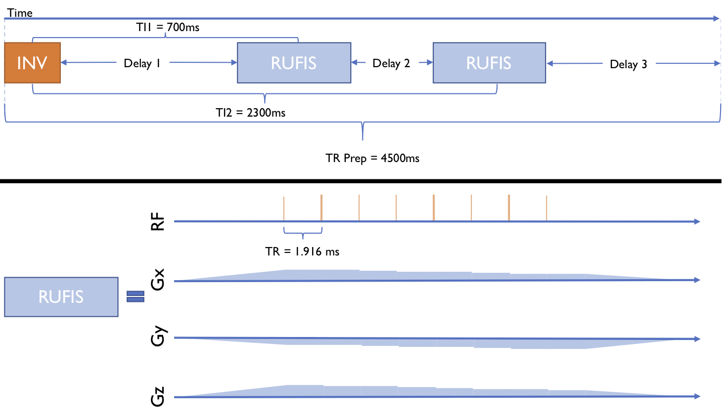
A RUFIS sequence was modified to resemble the MP2RAGE sequence as indicated in figure 1. This consists of an inversion pulse, a delay period, an acquisition segment, a second delay period, a second segment that acquires the same k-space trajectories as the first, and a final recovery delay. Because the centre of k-space is repeatedly sampled during a RUFIS segment the inversion times (TI1/TI2) were defined as the centre of each segment, as the overall k-space weighting will be approximately the average of all weightings during the segment.
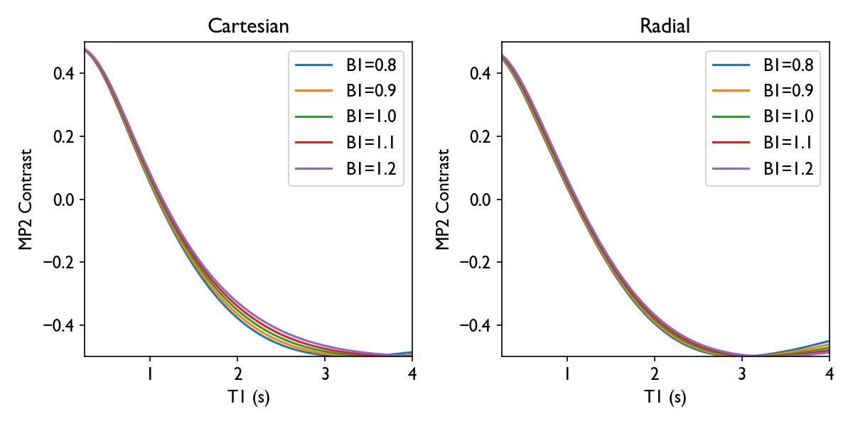
The TR used in a RUFIS sequence is typically much shorter than in a cartesian sequence, which reduces the Ernst angle, as each readout consists of a spoke starting at the k-space centre and not an entire line from one edge to the other. This short TR also means that a greater number of spokes can be acquired in the same time as lines in a cartesian sequence. Hence we simulated the expected MP2 contrast across a range of T1 and B1 and empirically tuned the acquisition segment length, flip-angles and inversion times to produce results similar to that from a common cartesian MP2RAGE protocol. Figure 2 compares the MP2 contrast curve from our optimised protocol and the cartesian protocol. Our finalised protocol was TR/TI1/TI2/TRPrep=2/700/2300/4500ms, FA1/FA2=2/3°, Spokes-Per-Segment 384, receiver bandwidth 20kHz and 0.9mm isotropic resolution, scan-time 10 minutes. A healthy male volunteer was scanned with the sequence on a 3T GE MR750 scanner equipped with a 12-channel head coil. In addition we acquired an Inversion-Recovery cartesian sequence (BRAVO) for comparison of the T1-weighted image only, also at 0.9mm isotropic resolution, with TE/TR/TI=3.2/8.2/450ms and flip-angle 12°, scan-time approx 4m30s (note we did not have a cartesian MP2RAGE sequence available for our platform).
The R2P2 images were reconstructed off-line using a combination of a standard gridding procedure implemented in Matlab and the Berkely Advanced Recon Toolbox5. Coil sensitive were estimated with the BART command "caldir"6 at TI2, and used to create complex-valued images at both TIs. The MP2 T1-weighted image was calculated using the robust method of O'Brien et al7 implemented in the QUIT toolbox8. This image and the cartesian T1-weighted were both used as input to the Freesurfer 6.0.0 "recon-all" pipeline9 to compare performance for cortical and sub-cortical parcellation.
Results
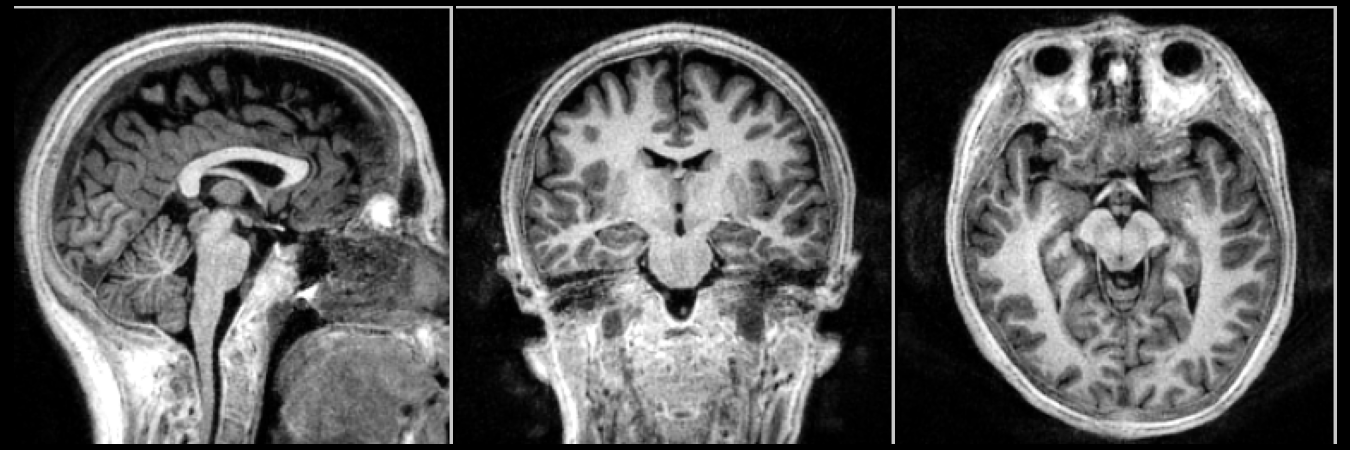
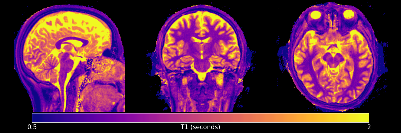
Figure 3 shows the R2P2 T1-weighted image and figure 4 shows the T1-map, giving the expected contrast and plausible T1 values. High contrast similar to cartesian MP2RAGE images in the literature was achieved, with no observable B1 inhomogeneity.
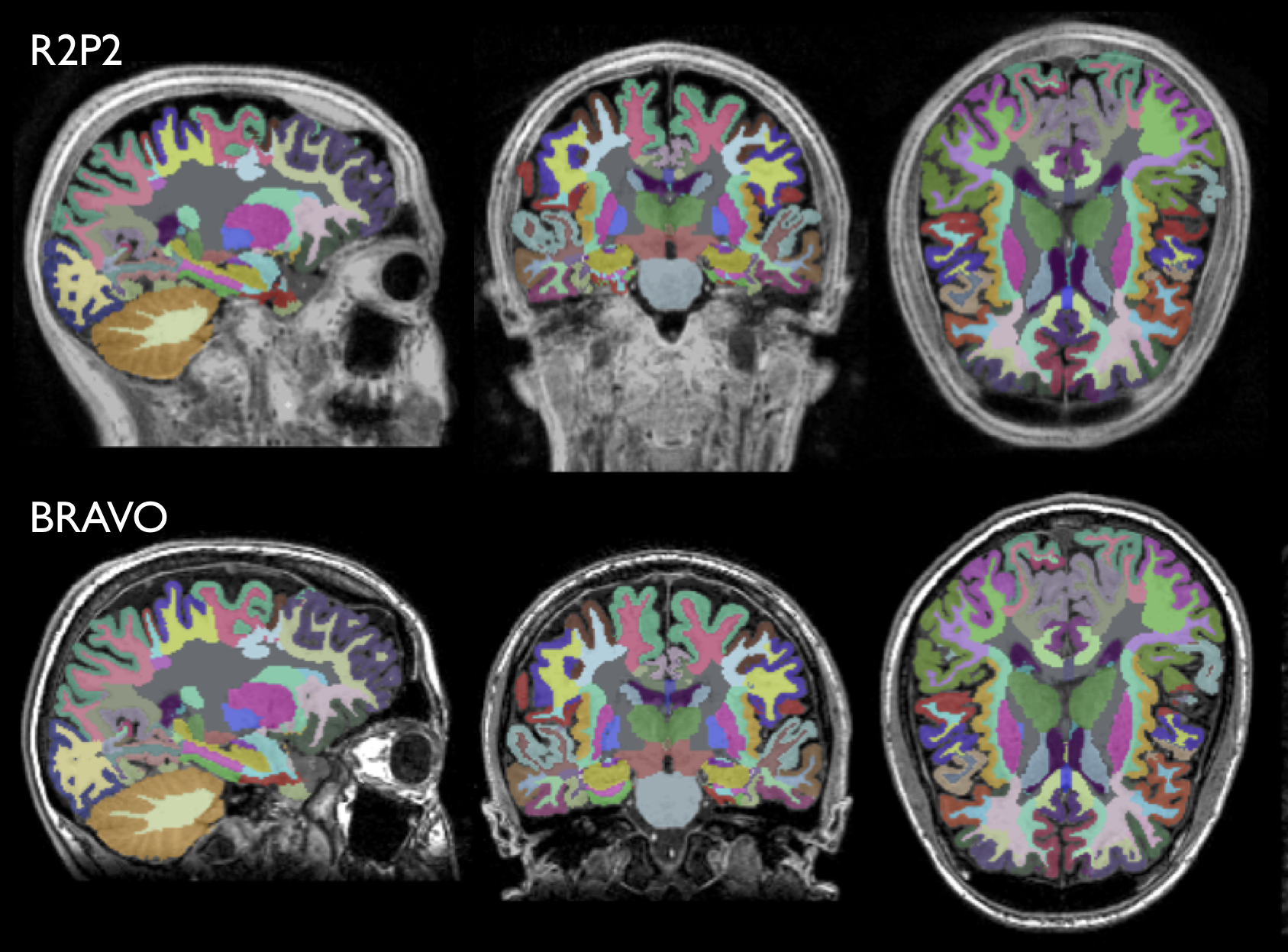
Figure 5 shows the results of the Freesurfer parcellation on both the radial and cartesian data. The radial results match closely to the cartesian sequence, with some minor differences evident in subcortical regions.
Conclusion
The radial MP2-RAGE sequence (R2P2) was demonstrated to give excellent T1-w contrast and T1-maps. This could permit high-resolution structural imaging in patient groups that would otherwise not tolerate an MRI due to the acoustic noise, such as young infants.Acknowledgements
This study represents independent research part funded by the NIHR-Wellcome Trust King's Clinical Research Facility and the National Institute for Health Research (NIHR) Biomedical Research Centre at South London and Maudsley NHS Foundation Trust and King’s College London. The views expressed are those of the author(s) and not necessarily those of the NHS, the NIHR or the Department of Health and Social Care. Funding was also received from General Electric Healthcare.References
[1] David P Madio and Irving J Lowe. Ultra-fast imaging using low flip angles and fids. Magnetic Resonance in Medicine, 34(4):525–529, 1995.
[2] L Sacolick, F Wiesinger, DAC Kelley, MM Khalighim and BK Rutt. 7 Tesla Zero-echo Time Imaging of the Head. Proceedings ESMRMB 2013 #53
[3] Mark Symms, Florian Wiesinger, Mauro Costagli, Doug Kelley, Mirco Cosottini and Michela Tosetti. Silent Corrected Using Second Image (SCUSI) - Application of the MP2RAGE formalism to T1-weighted Zero Time Echo Imaging. Proceedings ISMRM 2018, Paris, #2048
[4] José P Marques, Tobias Kober, Gunnar Krueger, Sietske van der Zwaag, Pierre-François de Moortele, and Rolf Gruetter. MP2RAGE, a self bias-field corrected sequence for improved segmentation and T1-mapping at high field. NeuroImage, 49(2):1271-1281, 2010
[5] Martin Uecker, Frank Ong, Jonathan I Tamir, Dara Bahri, Patrick Virtue, Joseph Y Cheng, Tao Zhang, and Michael Lustig. Berkeley Advanced Reconstruction Toolbox. ISMRM, Toronto 2015, In Proc. Intl. Soc. Mag. Reson. Med. 23:2486
[6] Charles A McKenzie, Ernest N Yeh, Michael A Ohliger, Mark D Price, Daniel K Sodickson. Self-calibrating parallel imaging with automatic coil sensitivity extraction. Magnetic Resonance in Medicine, 47(3):529-538, 2002
[7] Kieran R O'Brien, Tobias Kober, Patric Hagmann, Philippe Maeder, José Marques. Robust T1-Weighted Structural Brain Imaging and Morphometry at 7T Using MP2RAGE. PLOS ONE, 9(6):e99676, 2014
[8] Tobias C Wood. QUIT: Quantitative Imaging Tools. Journal of Open Source Software, 3(26):656, 2018
[9] Bruce Fischl et al. Whole Brain Segmentation: Automated Labelling of Neuroanatomical Structures in the Human Brain. Neuron, 33(3):341-355 2002
Figures