4634
AZTEK: Adaptive Zero TE K-space trajectories1IR4M, CNRS, Orsay, France, 2IR4M, Univ. Paris-Sud, Orsay, France, 3IR4M, Université Paris-Saclay, Orsay, France, 4Applications & Workflow, GE Healthcare, Buc, France, 5Applications & Workflow, GE Healthcare, Menlo Park, CA, United States, 6Applications & Workflow, GE Healthcare, Garching bei München, Germany
Synopsis
Because of short signal lifetimes and respiratory motion, 3D MR lung imaging is still challenging today. Zero TE (ZTE) pulse sequences are promising as they overcome the problem of short T2*. Nevertheless, because of the continuous readout gradients they require, their k-space trajectories are non-optimal for retrospective gating. We propose AZTEK, a 3D radial trajectory featuring several tuning parameters to adapt the acquisition to any moving organ while keeping a smooth transition between consecutive spokes. The increase in image quality was validated with static and moving phantom experiments, and demonstrated with dynamic thoracic imaging performed on a human volunteer.
Introduction
Lung is a challenging organ for proton MRI because of the short T2* values of its parenchyma and because of respiratory motion1,2. 3D radial Ultrashort Echo-Time (UTE)3 and Zero Echo-Time (ZTE)4 pulse sequences, because of the very short TE values they feature, were shown to be particularly adapted to short signal lifetime tissues5–9. Nevertheless, whereas the readout gradient is ramped up and down at each repetition in UTE, with no requirements on the k-space trajectory, this gradient must remain continuous in ZTE, only allowing small angular increments between repetitions10, usually leading to trajectories covering non-uniformly the k-space after retrospective gating. To overcome this problem, we propose AZTEK, an adaptive trajectory suited to ZTE requirements and respiratory resolved lung imaging.Theory
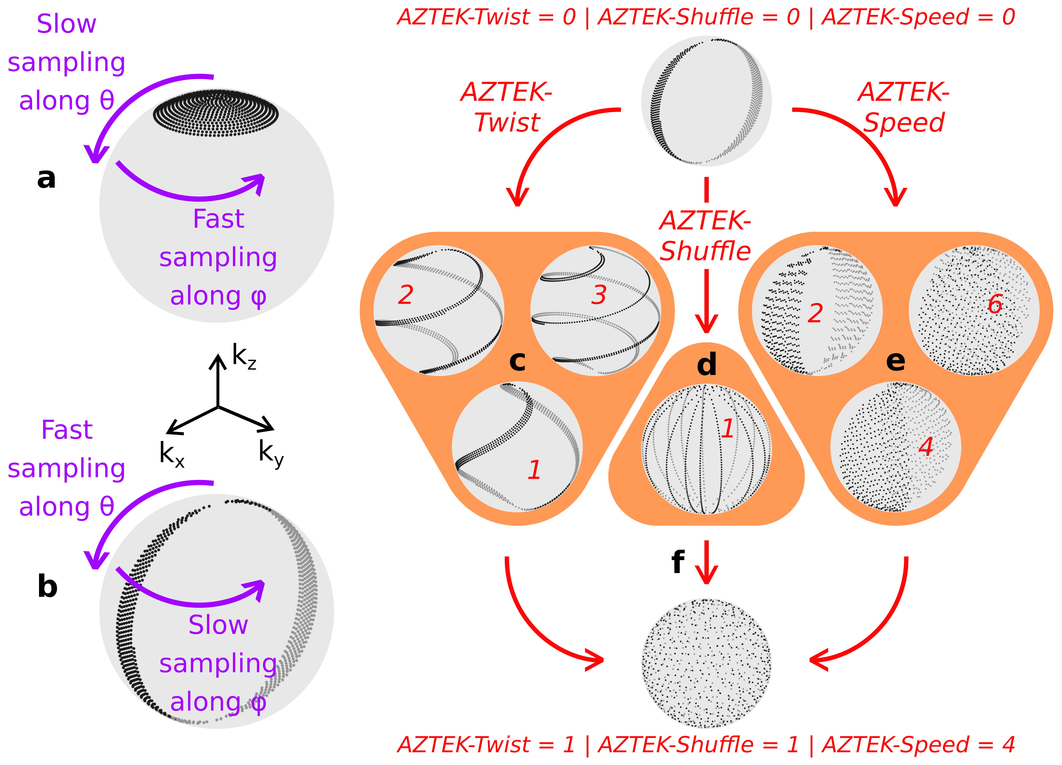
Every spoke of a 3D ‘kooshball’ trajectory is characterized by two angles in spherical coordinates: i) a polar angle, θ, and ii) an azimuthal angle, ϕ. In the standard ZTE sequence, k-space is sampled more rapidly along ϕ than θ (Figure 1a). In AZTEK, we switch the angle orders such that k-space sampling is performed more rapidly along θ than ϕ (Figure 1b). In this case, constant-ϕ half-circle arcs joining at the two poles with θ ranging from 0 to 180° are obtained. To avoid oversampling around the poles, θ steps are modulated accordingly from one spoke to another, and θ values on two adjacent arcs are shifted with a golden ratio basis to ensure sampling uniformity.
AZTEK offers three degrees of freedom to control the spoke sequence and explore the k-space:
- Each arc can be twisted by modulating ϕ as a function of θ. We implemented the parameter AZTEK-Twist, which represents the rate of ϕ shifting as a function of θ (Figure 1c).
- Any increment can be applied for ϕ when a pole is reached. It is done by modifying AZTEK-Shuffle, which is the fraction of the golden angle applied as ϕ shift (Figure 1d).
- Interleaved θ reading can be performed to accelerate undersampled full k-space covering within a normally sampled acquisition. Here, the parameter AZTEK-Speed indicates the number of spokes that are skipped between two successive readouts (Figure 1e).
These three parameters can be jointly tuned to obtain a trajectory, adapted to the targeted dynamics, which uniformly covers the k-space along any retrospective motion gate (Figure 1f).
Methods
ZTE Silenz acquisitions with the standard and adaptive AZTEK trajectories were performed with a GE Signa PET/MR 3.0 T on a water phantom and on a human volunteer freely breathing. The human study was validated by the local ethics committee. Images of the phantom were acquired both in static and dynamic modes. The phantom was put into motion by periodically translating the patient bed during the acquisition with an amplitude of 30 mm and a period of 5 s.
For the phantom, a cubic FOV of 28 cm, an isotropic voxel size of 1.4 mm and a flip angle of 4° were chosen; the body coil was used as the receiver. For the human volunteer, a cubic FOV of 32 cm, an isotropic voxel size of 1.6 mm, a flip angle of 3° and a 30-channel thoracic receive coil were used. Motions were monitored with an abdominal belt. The TR was 1.97 ms, the readout bandwidth was ±31.2 kHz and the scan duration, 1 min 25 s.
Results
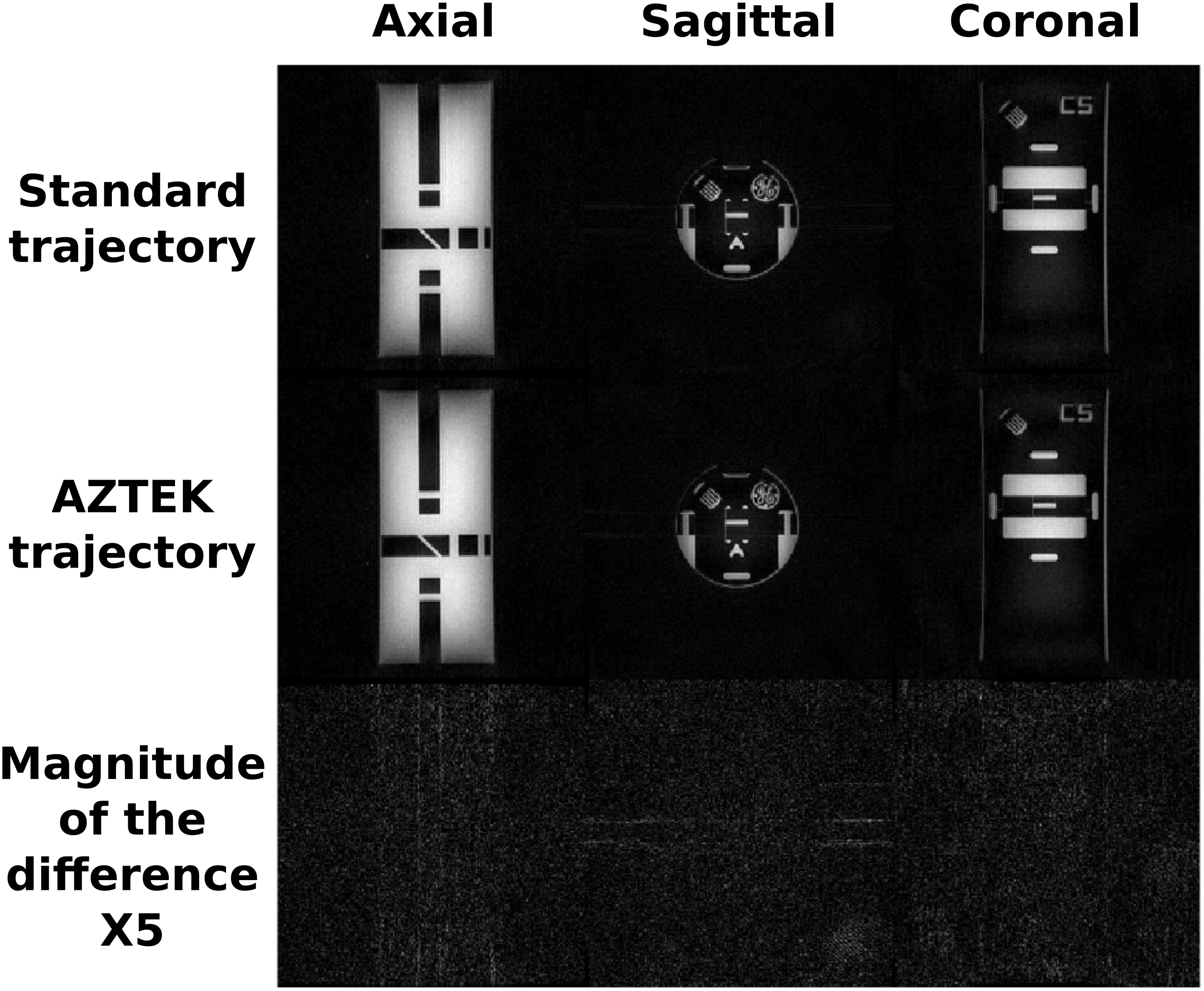
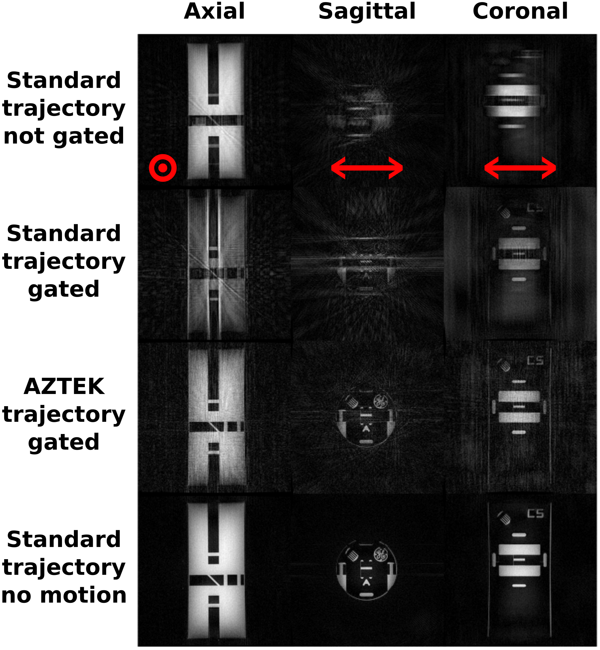
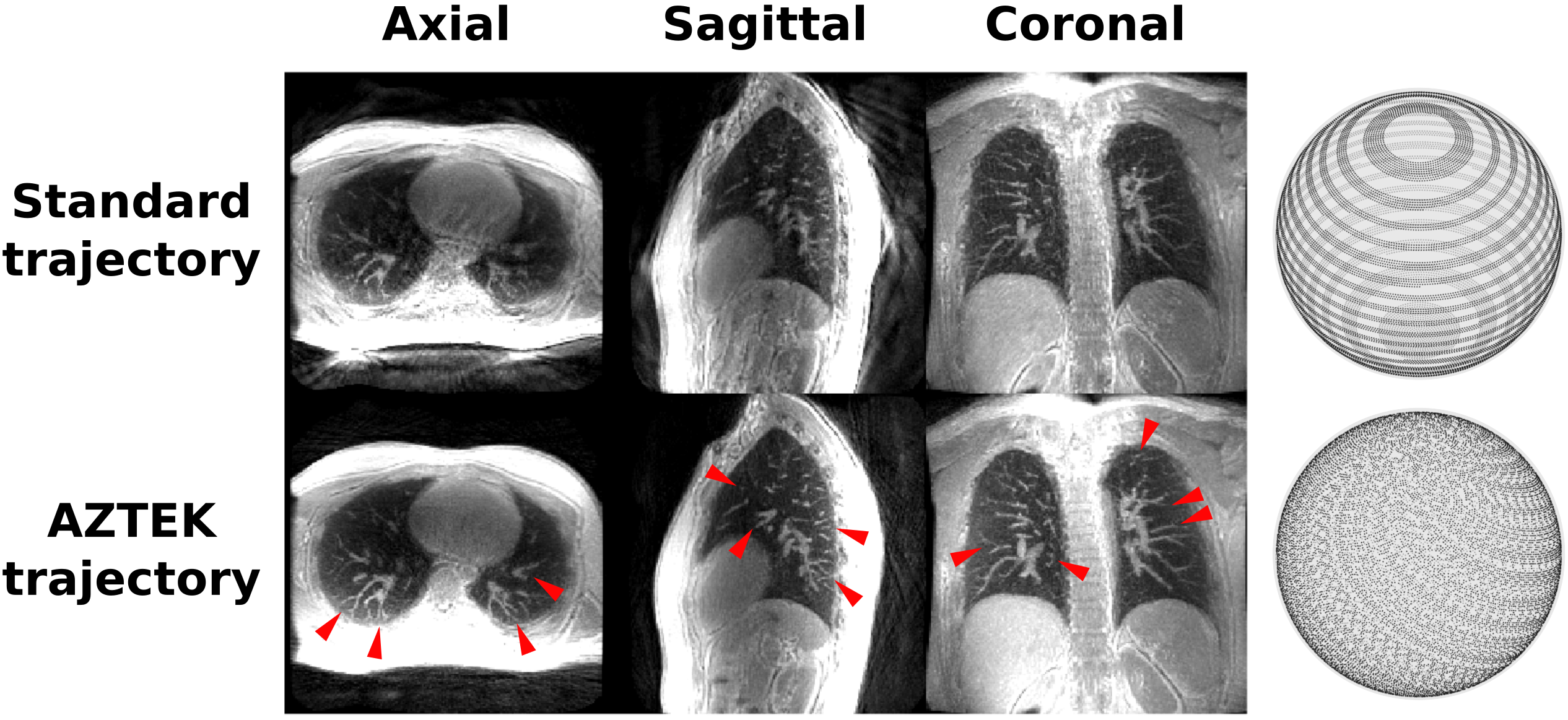
Figure 2 and 3 show respectively the static phantom images and the dynamic phantom gated images obtained with both standard and adaptive trajectories. Figure 4 shows the gated images obtained from the volunteer with the same k-space trajectories.Discussion
We observe comparable static image qualities between the standard and the AZTEK trajectories. With motion, spatially coherent undersampling artifacts observed on gated images with the standard trajectory are not observed anymore with AZTEK. The same result is obtained on human, where some details in the lung become more clearly visible with the use of our trajectory.
AZTEK shows similarities with phyllotaxis trajectories11. However, it offers more flexibility and adaptability on the spoke sequence, while ensuring readout gradient continuity along time and overall uniform angular density in k-space.
Conclusion
This study shows the great potential of AZTEK for 3D dynamic ZTE lung imaging. This trajectory is able to uniformly sample the k-space for any arbitrary retrospective respiratory motion gate, while preserving static image quality, improving dynamic image quality and guarantying smooth readout gradient transitions between spokes, which makes it appropriate to ZTE. Future work will focus on the adaptation of AZTEK to any moving organ, for example, by learning its motion characteristics before the acquisition.Acknowledgements
PET/MR platform affiliated to the France Life Imaging network (grant ANR-11-INBS-0006). We sincerely acknowledge Dr. Timo Schirmer from GE Healthcare for promoting the collaboration.References
1. Kauczor, H.-U. & Kreitner, K.-F. MRI of the pulmonary parenchyma. Eur. Radiol. 9, 1755–1764 (1999).
2. Wild, J. M. et al. MRI of the lung (1/3): methods. Insights Imaging 3, 345–353 (2012).
3. Tyler, D. J., Robson, M. D., Henkelman, R. M., Young, I. R. & Bydder, G. M. Magnetic resonance imaging with ultrashort TE (UTE) PULSE sequences: Technical considerations. J. Magn. Reson. Imaging 25, 279–289 (2007).
4. Madio, D. P. & Lowe, I. J. Ultra-fast imaging using low flip angles and fids. Magn. Reson. Med. 34, 525–529 (1995).
5. Gatehouse, P. D. & Bydder, G. M. Magnetic Resonance Imaging of Short T2 Components in Tissue. Clin. Radiol. 58, 1–19 (2003).
6. Burris, N. S. et al. Detection of Small Pulmonary Nodules with Ultrashort Echo Time Sequences in Oncology Patients by Using a PET/MR System. Radiology 278, 239–246 (2015).
7. Delso, G. et al. Clinical Evaluation of Zero-Echo-Time MR Imaging for the Segmentation of the Skull. J. Nucl. Med. 56, 417–422 (2015).
8. Gibiino, F., Sacolick, L., Menini, A., Landini, L. & Wiesinger, F. Free-breathing, zero-TE MR lung imaging. Magn. Reson. Mater. Phys. Biol. Med. 28, 207–215 (2015).
9. Wiesinger, F. et al. Zero TE MR bone imaging in the head. Magn. Reson. Med. 75, 107–114 (2016).
10. Weiger, M. & Pruessmann, K. P. MRI with Zero Echo Time. in eMagRes (John Wiley & Sons, Ltd, 2007).
11. Piccini, D., Littmann, A., Nielles‐Vallespin, S. & Zenge, M. O. Spiral phyllotaxis: The natural way to construct a 3D radial trajectory in MRI. Magn. Reson. Med. 66, 1049–1056 (2011).
12. Martin Uecker. mrirecon/bart: version 0.4.03. (Zenodo, 2018). doi:10.5281/zenodo.1215477
Figures