4632
Spiral RARE with annular segmentation1Dept. of Radiology, Medical Physics, University Medical Center Freiburg, Freiburg, Germany
Synopsis
We present a new approach to spiral RARE with annular segmentation. Annular segmentation leads to monotonous T2-dependent weighting of signal amplitudes across k-space and thus to very benign artifact behavior. Preliminary results show that single shot images (128x128) of decent quality can be acquired without fat suppression and without field inhomogeneity correction. By cyclic shifting of the spiral segments quantitative T1- and/or T2- images can be acquired in a few seconds. Sequence implementation was performed swiftly and efficiently in MatLab with the vendor independent Pulseq sequence development environment.
INTRODUCTION
Spiral trajectories offer the benefit of very efficient sampling of k-space compared to linear trajectories used in Cartesian or radial sampling and show in addition benign sampling behavior with respect to motion. In most practical applications their sensitivity to off-resonance artifacts requires segmented acquisition. In combination with a RARE(TSE,FSE,…) signal generation module two strategies for segmentation have so far been realized: an annular segmentation scheme1, by which the spiral is segmented into annular portions such that data points are monotonously sampled in time along the spiral. More recently a conventional interleaved ‘inside-out’ segmentation scheme has been demonstrated2 using n identical interleaves rotated by 360/n degrees in the imaging plane. The latter requires normalization of the signal intensities since adjacent points in k-space are acquired with vastly different T2 weighting. The purpose of this abstract is to present an improved method for spiral RARE with annular segmentation.
METHODS
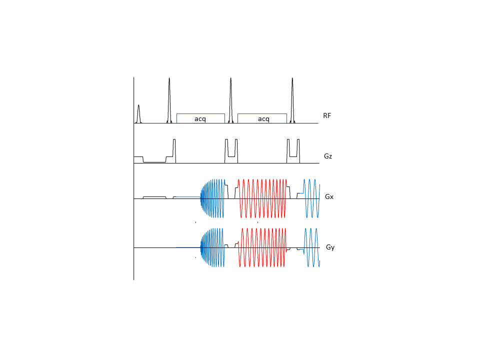
Our trajectories follow the same general scheme as published by Block1 et al. with some notable differences. The pulse sequence is shown in Fig.1. In the previous work the k-space trajectory starts at the center of k-space in each echo interval with a fast traverse accross k-space to the beginning of each annular segment and back to the k-space center before the next refocusing pulse. A disadvantage of this approach is the fact that free induction decays generated by non-perfect 180°-pulses (and any refocusing pathways created thereof) will be encoded with the identical spiral trajectory with – however – T2* weighting and off-resonance signal dephasing building up between excitation and the start of the spiral. The superposition of this ‘gradient echo’ spiral with the proper spin-echo spiral will create artifacts whenever FIDs are not perfectly spoiled. In our approach the k-space point before and after each refocusing pulse lies outside the maximum range of the k-space covered by the acquisition as typically realized in Cartesian RARE implementations (see Fig 1b). The beginning and end of each annular segment is then most efficiently reached in a straight line forming a tangent to each annular segment. Furthermore annular segments are read out alternately as spiral-in and spiral-out trajectories such that points at the juncture between segments show (near) identical T2* and off-resonance dephasing (Fig.2). Images with variable T2-weighting can be acquired by cyclic shift of the annular segments along the echo train rather than not acquiring signals in the echo intervals preceding the desired TEeff as in the previous work.RESULTS
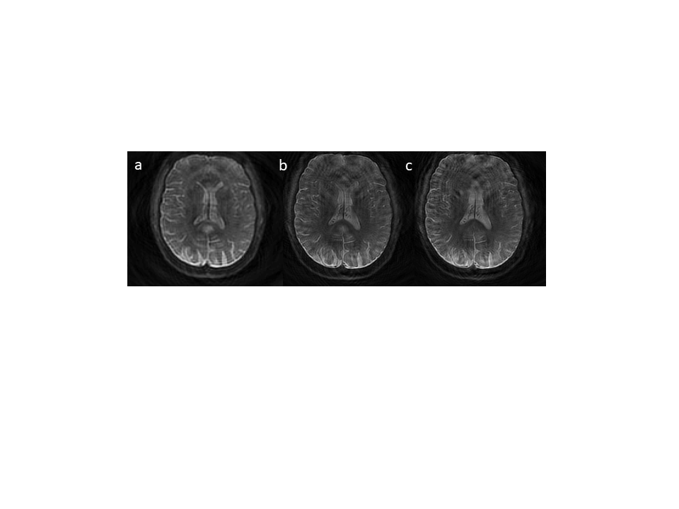
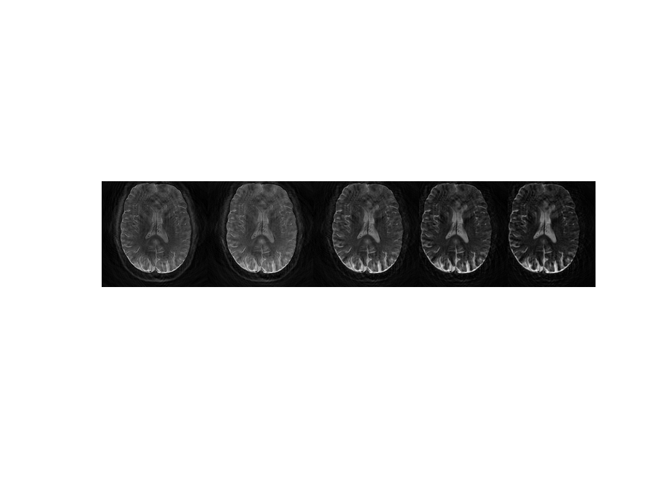
All images were acquired on a 3T scanner (Prisma FIT, Siemens, Erlangen). Spiral trajectories were designed using the variable density spiral toolbox by Brian Hargreaves3 (http://mrsrl.stanford.edu/~brian/vdspiral/). Spirals were designed for a maximum gradient amplitude of 30 mT/m and a maximum slew rate of 150 mT/m/ms. Sequences were realized using the vendor independent Pulseq sequence development platform (https://pulseq.github.io/). Example images (Fig.3) shown are acquired in single shot mode with a fully sampled single density spiral trajectory at variable echo spacing and acquisition times, Fig.4 shows single shot images acquired at different effective echo times TEeff.DISCUSSION
Our results show very encouraging image quality for single shot acquisition. Even without any off-resonance correction image artifacts appear to be tolerable. No fat-saturation was applied, fat in the skull appears therefore blurred due to the known off-resonance behavior of spirals. Off-resonance correction requires a field map but is expected to further improve image quality and to allow non-neuro implementations. With the current gradient system single shot images with an in plane resolution of ~1.5-2 mm can be realized even without acceleration by parallel imaging and/or compressed sensing. Higher nominal resolution for non-accelerated acquisition will lead to blurring due to the extended lengths of the echo train.CONCLUSION
Spiral RARE with annular segmentation appears to be an attractive acquisition mode to generate high-resolution images in short acquisition times. Image contrast and signal behavior are equivalent to Cartesian RARE(TSE, FSE), therefore modifications to extend the length of the echo train by suitable flip angle schemes4,5 can be easily realized as well as contrast modifications like nulling of CSF (FLAIR) by inversion recovery, MT-preparation, fatsat and others. Full quantitative imaging of T1, and T2 can be performed with a few single shot acquisitions in under one minute even without sophisticated acceleration schemes. Apart from this specific sequence the project demonstrates the capabilities of the Pulseq programming environment for rapid prototyping of sequence ideas. The Matlab environment of Pulseq allows to make full use of advanced programming, visualization and debugging capabilities, which are not easy to implement in any vendor specific sequence programming environment.Acknowledgements
This work was supported by Deutsche Forschungsgemeinschaft, Grant/Award Number: EXC 1086, He 1875/26-2 and He 1875/28-1References
1. Block W, Pauly J, Nishimura D. RARE spiral T-2-weighted imaging. Magn.Reson.Med. 1997;37:582–590
2. Li Z, Wang D, Robison RK, et al. Sliding-Slab Three-Dimensional TSE Imaging With a Spiral-In/Out Readout. Magn. Reson. Med. 2016;75:729–738
3. Lee JH, Hargreaves BA, Hu BS, Nishimura DG. Fast 3D imaging using variable-density spiral trajectories with applications to limb perfusion. Magnetic Resonance in Medicine 2003;50:1276–1285
4. Hennig J, Weigel M, Scheffler K. Calculation of flip angles for echo trains with predefined amplitudes with the extended phase graph (EPG)-algorithm: Principles and applications to hyperecho and TRAPS sequences. Magn. Reson. Med. 2004;51:68–80
5. Mugler JP. Optimized Three-Dimensional Fast-Spin-Echo MRI. J. Magn. Reson. Imaging 2014;39:745–767
Figures
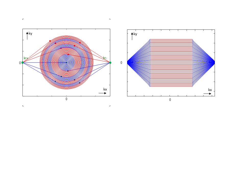
A) k-space trajectory of spiral RARE with annular segmentation. All spiral segments start at point kr+ outside of the acquired k-space and end at the symmetrical point kr- before the next refocusing pulse. Odd numbered segments (blue) are acquired inside-out, even numbered segmented (red) outside-in. Transition from kr+ to each segment resp. to kr- follows a tangential k-space path.
B) shows the equivalent k-space trajectory of a conventional cartesian RARE-sequence.