4613
Simultaneous multislice EPI reconstruction by incorporating split slice-GRAPPA with slice-dependent 2D Nyquist ghost correction (PEC-SP-SG)1Laboratory of Biomedical Imaging and Signal Processing, The University of Hong Kong, Hong Kong, China, 2Department of Electrical and Electronic Engineering, The University of Hong Kong, Hong Kong, China, 3Center for Advanced Imaging, The University of Queensland, Brisbane, Australia
Synopsis
Simultaneous multislice (SMS) EPI reconstruction is challenging due to slice-dependent 2D phase differences between opposite polarities, which is collapsed across slices. Additionally, slice leakage is one major concern in some applications including diffusion and functional MRI. The proposed SMS EPI reconstruction incorporates phase error correction with split slice-GRAPPA (PEC-SP-SG), and was evaluated using simulation, phantom and in vivo experiments. Results show that the proposed approach can offer a robust SMS EPI reconstruction with slice-dependent 2D Nyquist ghost correction, and provide a balance between slice leakage and in-plane artifacts.
Introduction
EPI reconstruction is inherently challenging because of the data inconsistency arising from acquisition imperfections that cause 2D phase difference between opposite readout polarities. It becomes more problematic for simultaneous multislice (SMS) EPI, since such phase difference is not only slice-dependent but also collapsed across slices. Recently, robust SMS EPI reconstruction was implemented in image-space (via PEC-SENSE)1, or in k-space (via PEC-GRAPPA)2, where slice-dependent 2D Nyquist ghost correction is incorporated by taking data from opposite readout polarities as from different virtual channels. Slice leakage is not considered in both approaches. However, slice leakage may become the dominant concern when inconsistent contrast of the calibration scan causes increased slice leakage (e.g. in diffusion MRI where b0 data is used for calibration)3, or with the need of trading in-plane artifacts level for reduced slice leakage (e.g. in functional MRI)4. This has been addressed with the introduction of split slice-GRAPPA (SP-SG)3. The proposed EPI reconstruction incorporates phase error correction with SP-SG (PEC-SP-SG), aiming to offer a robust SMS EPI reconstruction with slice-dependent 2D Nyquist ghost correction, and provide a balance between slice leakage and in-plane artifacts.Method
Proposed Reconstruction
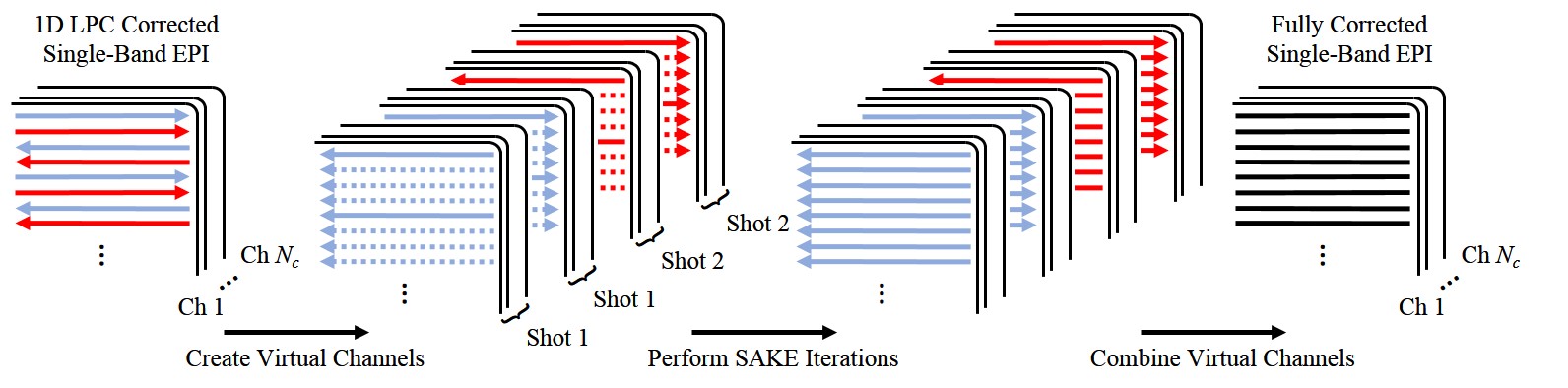
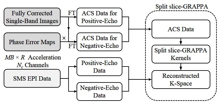
In this study, single-band multi-shot EPI reference scan was acquired and fully corrected with virtual channel simultaneous autocalibrating and k-space estimation (VC-SAKE) (Figure 1), and phase error map was calculated as the phase difference between images separately reconstructed from positive- and negative-echo data in reference scan. Subsequently, autocalibration signals (ACS) data were synthesized from the fully corrected single-band images and phase error maps, by treating data from opposite readout polarities as from different virtual channels. After that, GRAPPA kernels which minimize both slice leakage and in-plane artifacts were trained and utilized for reconstruction (Figure 2).
Data acquisition and image reconstruction
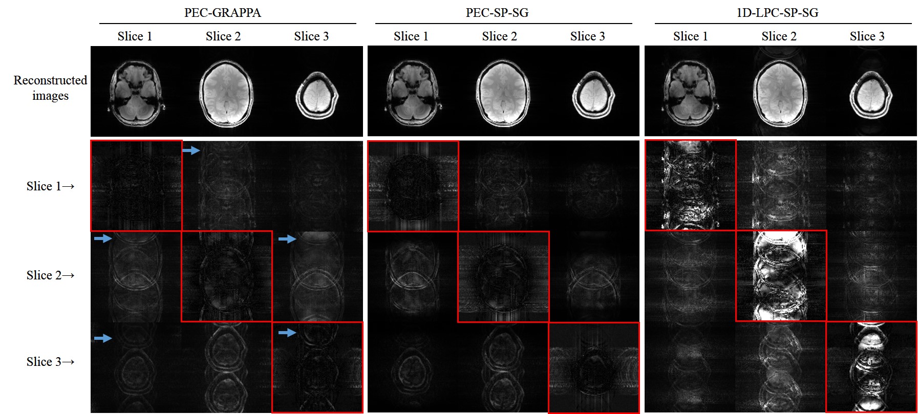
Simulation study: Simulated data were based on GE FLASH data collected using a 7T human scanner (Siemens Healthcare, Erlangen, DE) with a 32-channel coil (Nova Medical, Wilmington, US). The acquisition parameters were TR/TE=1000/4ms, FA=70°, matrix size=128×128, FOV=256×256mm2, slice thickness/gap=2/0mm, slice number=80. Four selected slices were used for synthesizing SMS (R=2, MB=3, and FOV/4 shift) and single-band reference (2-shot) EPI data. Slice-dependent 2D nonlinear phase errors were added to both SMS and single-band EPI data, and inter-shot phase variations were added to single-band EPI data. In-plane artifacts and slice leakage was evaluated using linear system leakage approach (LSLA)3.
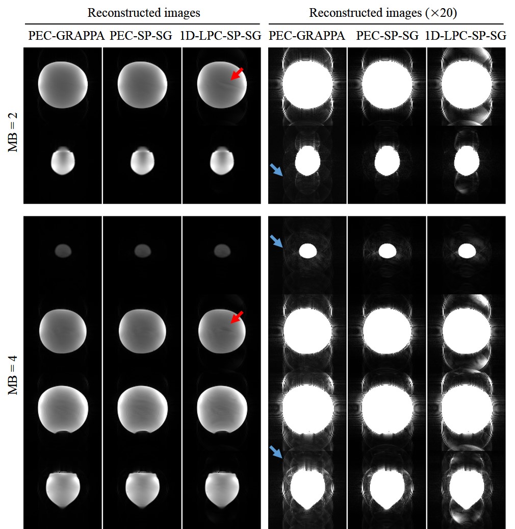
Phantom imaging at 7T: SE-EPI images were acquired on a spherical oil phantom using the same setup at R=2, MB=2, and 4, blipped-CAIPI shift=FOV/4. Other acquisition parameters were TR/TE=2170/30ms, FA=70°, echo-spacing=0.7-0.8ms, bandwidth=1628kHz/pixel, matrix size=128×128, FOV=256×256mm2, slice thickness/gap=2/0mm, slice number=80. Single-band EPI was also performed with matched parameters1,2.
Human brain imaging at 7T: Human brain data were collected using SE-EPI (R=2, MB=4) with TR/TE=539/30ms, FA=50°, slice number=20, number of frames=50, and the same other parameters used in the phantom study1,2. Image reconstruction: Besides PEC-SP-SG, PEC-GRAPPA and 1D-LPC-SP-SG were also implemented for comparison. For 1D-LPC-SP-SG, the slice-dependent phase error was approximated with 1D linear parameters, and calibration data was also corrected with 1D-LPC.
Results
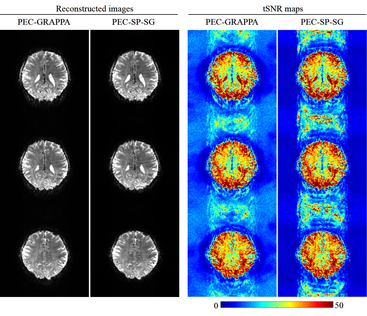
As shown in Figure 3, the PEC-SP-SG has significantly reduce slice leakage compared to PEC-GRAPPA approach. Also note that 1D-LPC-SP-SG has the most distinct residual artifact (both in-plane and inter-slice) due to inaccurate phase error correction. The phantom results (Figure 4) also show that PEC-SP-SG can significantly alleviate slice leakage. In Figure 5, compared to PEC-GRAPPA reconstruction, PEC-SP-SG can provide visually comparable results and similar temporal SNR.Discussion and conclusions
This study demonstrated that the proposed PEC-SP-SG can significantly reduce slice leakage compared to PEC-GRAPPA, while still having similar tSNR performance.
Note that the particular 7T experimental phantom and brain datasets used here do not exhibit strong contrast inconsistency between calibration scan and SMS accelerated scans, thus the slice leakage effect is not prominent. Diffusion MRI data, where contrast inconsistency of calibration scan can induce significant slice leakage, will be used to evaluate the proposed PEC-SP-SG approach3. In functional MRI studies, minimizing slice leakage can significantly reduce false-positive activation4. Future studies will include evaluation with both diffusion and functional MRI studies.
Acknowledgements
This work was supported by the Hong Kong Research Grant Council (C7048-16G and HKU17103015 to E.X.W.).References
1. Lyu, M., et al. Robust SENSE reconstruction of simultaneous multislice EPI with low-rank enhanced coil sensitivity calibration and slice-dependent 2D Nyquist ghost correction. Magn Reson Med 80, 1376-1390 (2018).
2. Liu, Y., et al. PEC-GRAPPA reconstruction of simultaneous multislice EPI with slice-dependent 2D Nyquist ghost correction. Magn Reson Med (2018).
3. Cauley, S.F., Polimeni, J.R., Bhat, H., Wald, L.L. & Setsompop, K. Interslice Leakage Artifact Reduction Technique for Simultaneous Multislice Acquisitions. Magn Reson Med 72, 93-102 (2014).
4. Todd, N., et al. Evaluation of 2D multiband EPI imaging for high-resolution, whole-brain, task-based fMRI studies at 3T: Sensitivity and slice leakage artifacts. NeuroImage 124, 32-42 (2016).
Figures