4606
Clinical application of MAVRIC-SL in reducing metal implant artifacts in anterior cruciate ligament reconstruction1Radiology Department of China Medical University First Hospital, Shenyang, China, 2MR Application China, GE Healthcare, Shen Yang, China, 3GE Healthcare China, Beijing, China
Synopsis
Some anterior cruciate ligament reconstructions have metal implants, and metal scrap may remain in the surgical procedure. Metals in the conventional magnetic resonance sequence, especially in the fat suppression sequences, produce large artifacts that affect the observation of the surrounding structure. This study performed conventional sequences and MAVRIC-SL sequence scan for patients with metal implants after ACLR and analysis the images. Conclusions that the oblique sagittal MAVRIC-SL PDWI FS sequence can be used to assisting in the diagnosis of traditional oblique sagittal T2WI FS and PDWI sequence.
Purpose
Evaluate the feasibility of assisting in the diagnosis of the conventional oblique sagittal T2WI FS sequence and PDWI sequence by o-sag MAVRIC-SL PDWI inversion recovery (IR) sequence.Introduction
Arthroscopic anterior cruciate ligament reconstruction (ACLR) is the main choice for ACL injury. The use of bio-composite interference screw and spiked ligament staple is a common method of ACLR tibial fixation 1. MRI has the advantages that other examination methods can't replace in the ACLR postoperative review. However, as a metal staple, it has large artifacts in conventional sequences, which affects the observation of the fine structure around the implant. 3.0T MRI has been widely used in knee joint examination, but it has bigger metal artifacts than 1.5T 2.Methods
This study analyzed the images of the 26 patients implanted spiked ligament staple, which obtained from conventional sequences and MAVRIC-SL performed in 3.0T MRI. Paired Student t test and Wilcoxon signed rank test were used to compare the quantitative and qualitative data. The consistency of subjective scores was tested by kappa analysis.
image analysis: Quantitative analysis: 1) Selected the maximum artifact slice of each sequence, draw the artifact boundary, and record the region of interest (ROI) area. 2) Record the number of slices which artifact appears. 3) Select the stable signal region of the soleus muscle in the same slice of different sequences, draw a circular ROI, record the average signal intensity(SI)in the ROI;draw the same ROI in the background of the same slice (ie air), record the standard of this ROI’s image signal intensity(SD), namely the noise of the image. Calculate SNR of different sequences using SNR=SI/SD 3.
Qualitative analysis: The three sequences were independently evaluated and compared by two senior radiologists. An artifact degree of the staple and its surrounding structure diagnosis: 1, no artifacts; 2, almost no artifacts; 3, visible artifacts, but does not affect the quality of diagnosis; 4, moderate artifacts, moderate diagnostic quality damage; 5, serious artifacts and cannot be diagnosed. Beside,we evaluation the measurability of distance between the upper edge of the staple and the lower edge of the graft entrance on tibia over the three sequence: 1, completely unmeasurable; 2, measurable but the measured value is not high; 3, measurable, high confidence in measured value.
Results
Quantitative analysis: Artifact area: the area of the MAVRIC-SL is significantly smaller than two conventional sequences (t values are 37.901 and 17.513, p<0.01); the number of artifact slices: MAVRIC-SL is less than that two sequences (t values are 27.608 and 22.797, p<0.01); SNR: MAVRIC-SL is increased (t values are -8.222 and -3.020, p<0.01) (Tab 1).
Qualitative analysis: In terms of artifact degree its surrounding structure diagnosis, the MAVRIC-SL PDWI IR scores were significantly lower (p<0.01) and the distance measurability scores were improved (p<0.01). Kappa values are greater than 0.6(Tab 2).
Discussion
The results of this study clearly show that the MAVRIC-SL PDWI IR can significantly reduce the artifacts caused by the staple, improve the observability of the metal-bone interface, and reduce the deformation of surrounding structures (Fig.1). Due to the position of the staple and screw is adjacent, the magnetic field inhomogeneity and metal artifacts generated in the conventional sequences will cause deformation of the screw and the end of the tibia, which will affect the observation. The metal artifacts will not only exist at the slice of the implant, but also affect the signal uniformity of its adjacent slices and bring errors to the structural observation of adjacent slices. The presence of postoperative complications such as the fixation loosening, Cyclops syndrome 4 or infection poses a greater challenge to the reduction of metal artifacts. But MAVRIC-SL can solve these problems well. In terms of SNR, since MAVRIC-SL uses 3D acquisition, the SNR is improved compared to the conventional sequence of 2D acquisition, and the shortening of the TE time also increase SNR. In addition, in these 26 cases, 23 cases showed metal artifacts other than metal implants. It is believed that these metal artifacts appearing in the joint cavity are residual metal powder during the operation, which is confirmed by CT examination(Fig.2). These artifacts appearing outside the surgical plan may affect the postoperative evaluation indexes such as the continuity of the ligament implant. After the metal artifacts are removed by the MAVRIC-SL, the overall diagnostic confidence of the image is improved.Conclusion
In summary, patients with metal implants in the ACLR can use the oblique sagittal MAVRIC-SL PDWI FS sequence to assist in the diagnosis of the conventional oblique sagittal T2WI FS sequence and PDWI sequence.Acknowledgements
No acknowledgement found.References
1. Lu Q, Wang P, Huang D. A Clinical Investigation for ACL Reconstruction with Semitendinosus and Gracilis Tendon[J]. Chinese Journal of Bone & Joint Injury, 2007.
2. Imai H, Tanaka Y, Nomura N, et al. Three-dimensional quantification of susceptibility artifacts from various metals in magnetic resonance images.[J]. Acta Biomaterialia, 2013, 9(9):8433-8439.
3. Fukuyama A, Imai K, Haba T. Development and Evaluation of a New Method for Measuring of Signal-to-Noise Ratio in Magnetic Resonance Images[J]. 2014.
4. Pinto F G, Thaunat M, Daggett M, et al. Hamstring Contracture After ACL Reconstruction Is Associated With an Increased Risk of Cyclops Syndrome[J]. Orthopaedic Journal of Sports Medicine, 2017,
Figures
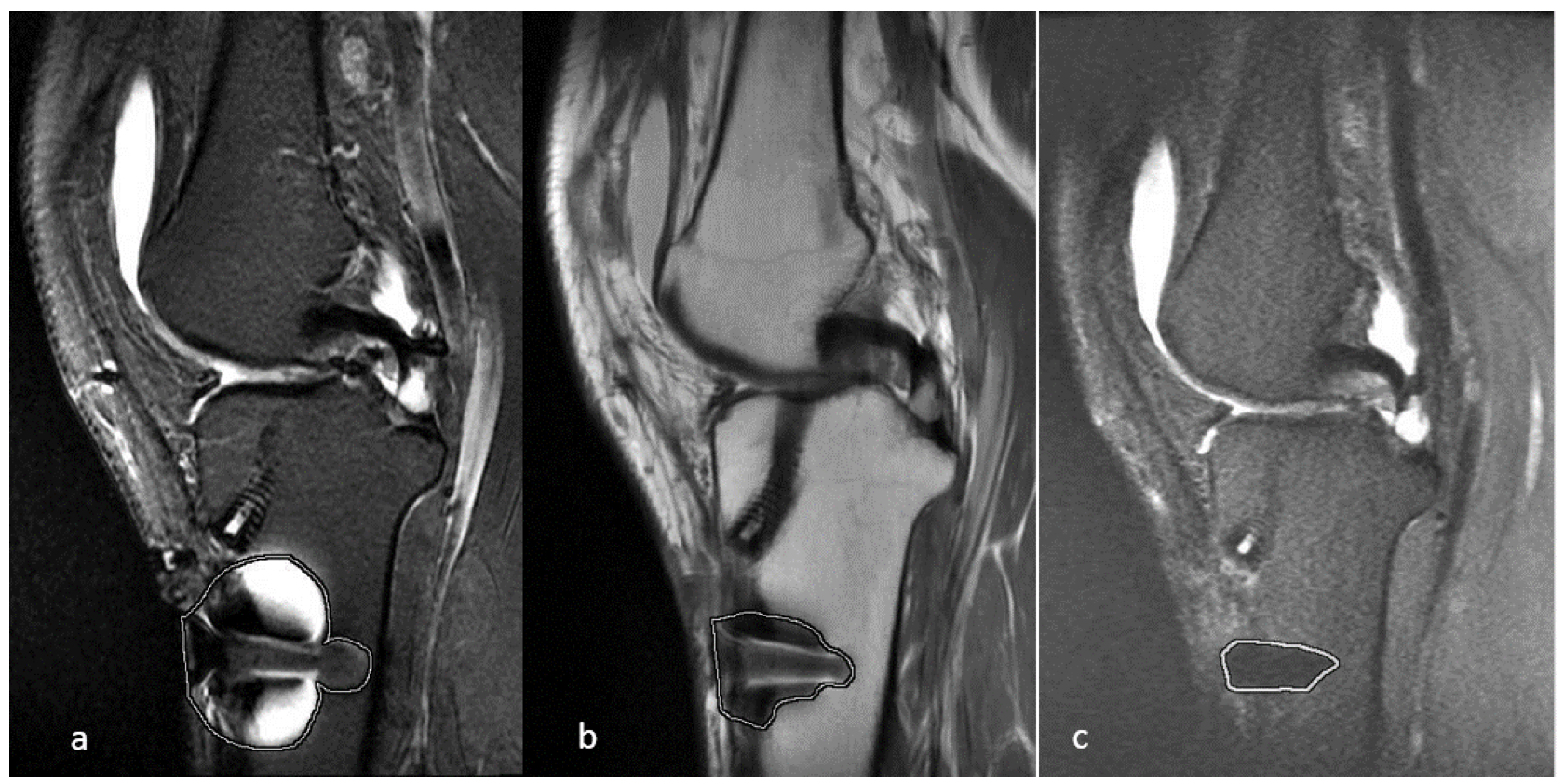
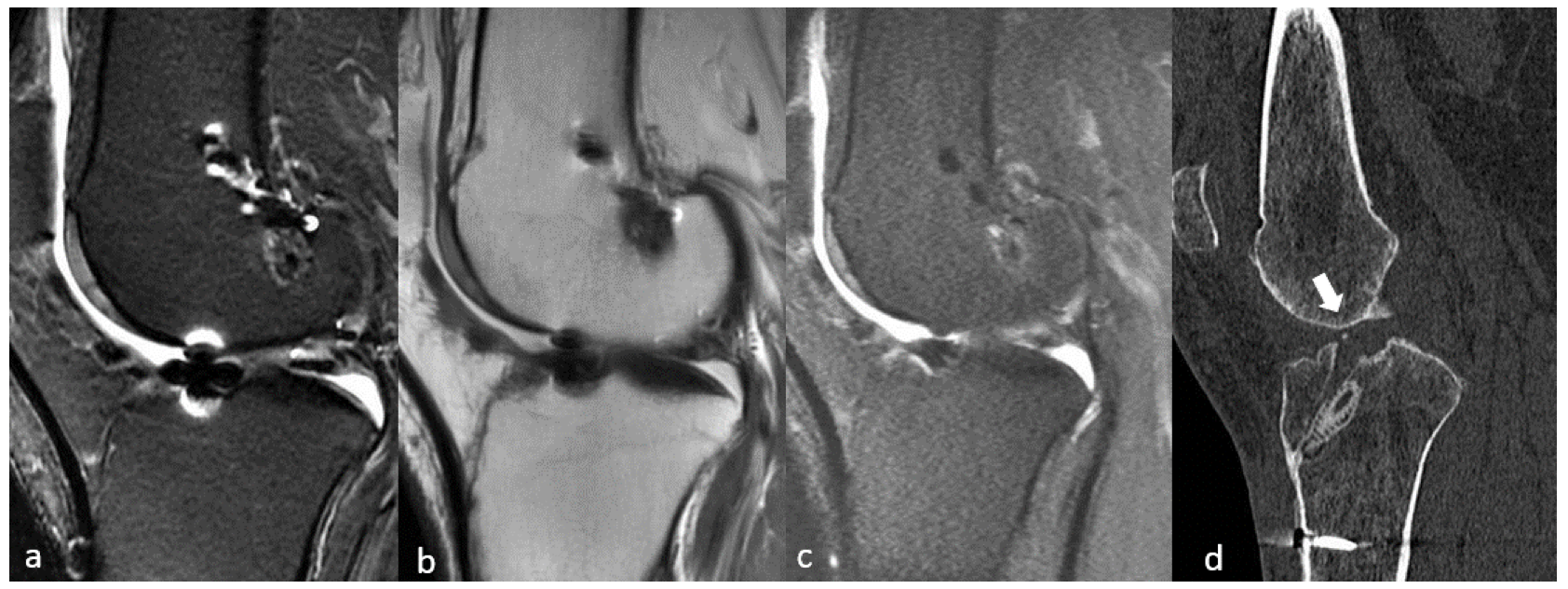
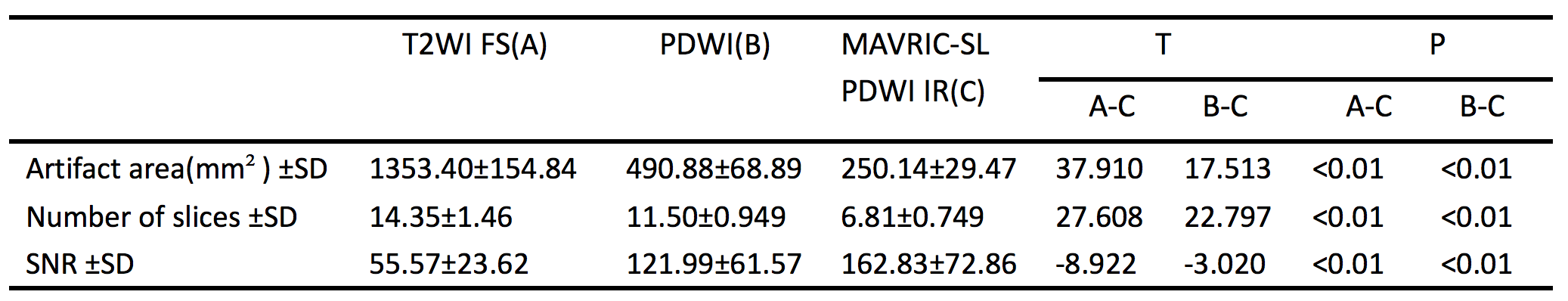
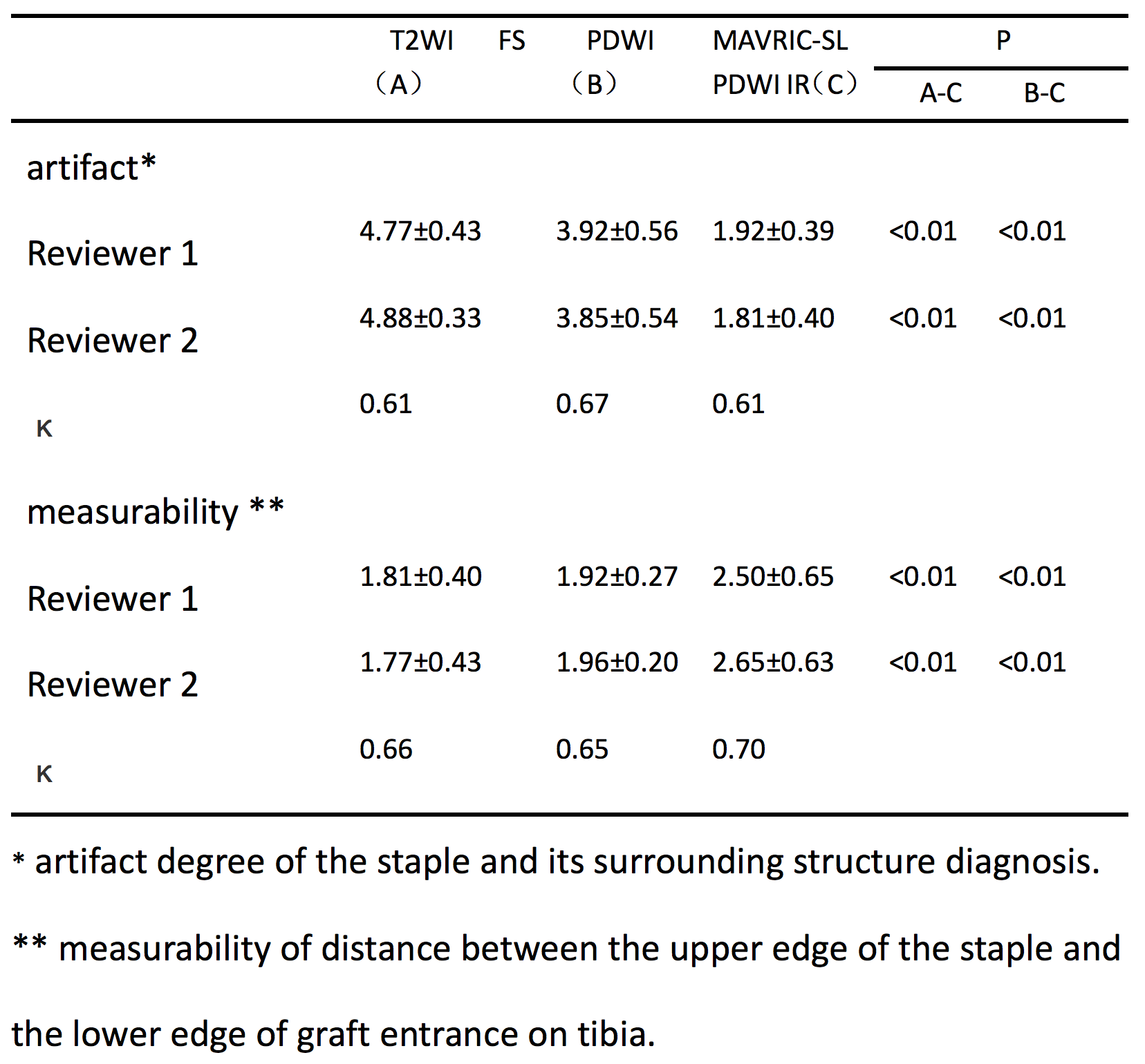
Table 2 The Qualitative analysis of MAVRIC-SL and other three sequences by the two radiologists.