4602
Metal Artifacts Reduction in DWI using Point Spread Function (PSF) Encoding1Department of Biomedical Engineering, Center for Biomedical Imaging Research, Beijing, China, 2Philips Healthcare, Beijing, China, Beijing, China, 3Chao Yang Hospital, Beijing, China, 4Beijing Jishuitan Hospital, Beijing, China, 5Tsinghua University Yu Quan Hospital, Beijing, China
Synopsis
Though metal artifacts have been well-resolved in anatomical imaging by three dimensional multispectral imaging (3D-MSI) methods, diffusion weighted imaging (DWI) near metallic implants still remains a challenge, impeding various clinical applications. Point-Spread-Function encoded EPI (PSF-EPI) combined with Tilted-CAIPI can achieve highly accelerated distortion- and blurring-free high resolution DWI. By using an additional phase encoding, artifacts induced by severe susceptibility inhomogeneity around metal can be reduced even under a high acceleration rate. The reliable performance of PSF-EPI technique in metal artifacts reduction in DWI is demonstrated on phantom, in vitro swine forearm and in vivo patients.
Introduction
The demand for reliable DWI near metallic implants is increasing rapidly in clinic, especially for imaging evaluation of orthopedic surgery 1,2. However, due to severe artifacts induced by metal, current DWI methods are significantly hampered by geometric distortion, bright pile-up and signal loss near implants. To reduce metal artifacts, several 3D multispectral (3D-MSI) imaging approaches, including SEMAC, MAVRIC, and hybrid method, have been developed and shown promise for metal artifacts correction in anatomical imaging 2,3,4,5. Unfortunately, long acquisition time renders these 3D imaging approaches impractical for DWI 6. DWI is mostly conducted by single-shot echo planar imaging (SS-EPI) based sequences. Though fast and insensitive to motion, SS-EPI suffers from susceptibility-induced distortion and T2* blurring. A variety of multi-shot EPI (MS-EPI) methods have been proposed to reduce distortion with practical acquisition time for DWI, such as interleaved EPI (iEPI) 6,7,8. However, the distortion removal in these methods is not complete, especially in the presence of metal. In this study, we adopted a distortion- and blurring- free MS-EPI based technique, Point-Spread-Function encoded EPI (PSF-EPI), to address metal artifacts in DWI 9,10,11.Methods
In the PSF-EPI sequence, an additional spin-warp phase encoding (PE) gradient (termed, PSF-PE) is applied to the PE direction in the conventional 2D EPI sequence, resulting in a 3D kspace dataset with dimensions of $$$kx$$$ (readout), $$$ky$$$ (EPI-PE) and $$$ks$$$ (PSF-PE) 9,10. Since the echo time is constant along PSF-PE at certain $$$ky$$$, distortion- and blurring- free images can be obtained in $$$kx$$$-$$$ks$$$ plane 9. PSF-EPI is also compatible with parallel imaging and partial-Fourier imaging, enabling fast and high resolution DWI 10. For further acceleration, a Tilted-CAIPI strategy was proposed, rendering PSF-EPI DWI practical for clinical purposes 11. Here PSF-PE and EPI-PE were under-sampled by R_PSF and R_PE, respectively.
We first simulated field perturbation induced by metal using COMSOL Multiphysics v5.3 (Comsol, Inc., Burlington, MA, USA). The static field, gradient field and 3D magnetic susceptibility distribution were established according to the real situations. The efficacy of PSF-EPI in metal artifacts reduction in DWI was validated at 3T with phantom, in vitro swine forearm, in vivo spine and brain imaging by comparing it with T2 weighted reference images and an interleaved EPI sequence, iEPI 7. The specific acquisition parameters are illustrated in Results. This study was approved by the Institutional Review Board.
Results
Fig. 1a demonstrates the distribution of frequency band shift when a metallic cylinder object is placed in a static magnetic field with imposed gradient field, indicating distorted excitation profiles. Fig. 1b shows that in-plane frequency changes rapidly on the surface of the cylinder and becomes smooth quickly over a small distance, which enables the recovery of in-plane signal displacements near metal using Tilted-CAIPI.
Fig. 2 illustrates the images from PSF-EPI and iEPI in the phantom experiment, where a deep brain stimulation (DBS) electrode was immobilized to the surface of an ACR phantom. The structure of the grids agrees well with that in the TSE reference image. However, by zooming in, misalignment is observed in the iEPI image while PSF-EPI sequence achieves distortion-free imaging.
In the in vitro study (Fig. 3), proximal screws for humeral fixation were inserted into the swine forearm. Compared with SS-EPI and iEPI, the PSF-EPI technique managed to eliminate signal pile-up near the screws, delineating the screw though through-slice distortion still exists in the reformatted sagittal images. Fig. 3b shows uniform ADC map of PSF-EPI.
Fig. 4 shows the in vivo DWI results of one patient with an anterior cervical plate system in the spine. The acquisition time of the PSF-EPI and iEPI sequences are the same. The PSF-EPI image shows high consistency with the structural image and recovers the entire cervical spinal cord. On the contrary, the iEPI image failed to remove distortions induced by metallic fixations and shows lower SNR.
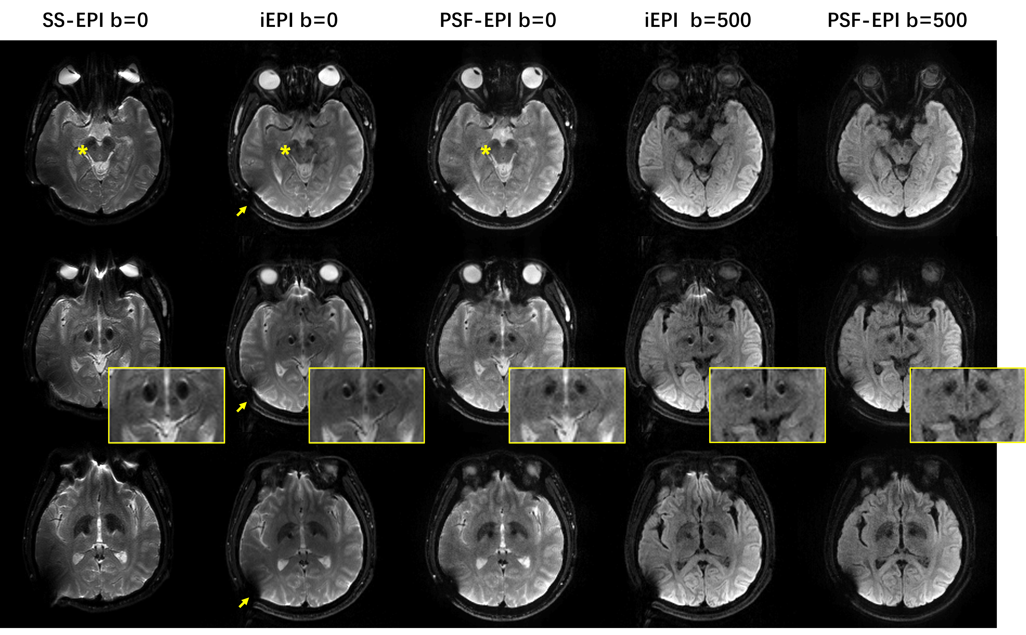
Fig. 5 shows the in vivo DWI results of one patient with a DBS implant in the brain. In comparison, the signal loss around DBS electrodes, which appears as a pair of black holes near brainstem, and implantable extension, is better resolved by PSF-EPI than iEPI. Additionally, signal pile-up remains in the iEPI results.
Discussion and Conclusion
In this study, we demonstrated that PSF-EPI achieved distortion-free imaging through spin-echo formation, thus providing effective metal artifacts suppression in DWI with high resolution and feasible acquisition time. iEPI can also reduce metal-induced distortion but it is still limited by the acquisition bandwidth along the phase-encoding direction. Despite the promising PSF-EPI results, remained signal loss and through-slice distortion need to be solved, which may require enhanced excitation and reconstruction methods.Acknowledgements
We acknowledge the assistance of Zijing Dong and Xiaodong Ma.References
1. Brian Hargreaves, Pauline W. Worters, Kim Butts Pauly, John M. Pauly, Kevin M. Koch, & Garry E. Gold. (2011). Metal induced artifacts in MRI. Ajr Am J Roentgenol, 197(3), 547-555.
2. Koch, K. M., Bhave, S., Gaddipati, A., Hargreaves, B. A., Gui, D., & Peters, R., et al. (2017). Multispectral diffusion-weighted imaging near metal implants. Magnetic Resonance in Medicine, 79(2).
3. Lu, W., Pauly, K. B., Gold, G. E., Pauly, J. M., & Hargreaves, B. A. (2010). SEMAC: slice encoding for metal artifact correction in MRI. Magnetic Resonance in Medicine, 62(1), 66-76.
4. Koch, K. M., Lorbiecki, J. E., Hinks, R. S., & King, K. F. (2010). A multispectral three‐dimensional acquisition technique for imaging near metal implants. Magnetic Resonance in Medicine, 61(2), 381-390.
5. Koch, K. M., Brau, A. C., Chen, W., Gold, G. E., Hargreaves, B. A., & Koff, M., et al. (2011). Imaging near metal with a MAVRIC‐SEMAC hybrid. Magnetic Resonance in Medicine, 65(1), 71-82.
6. Hargreaves, B. A., Taviani, V., Litwiller, D. V., & Yoon, D. (2017). 2D multi-spectral imaging for fast MRI near metal. Magnetic Resonance in Medicine, 79(2), 968-973.
7. Dong, Z., Wang, F., Ma, X., Zhang, Z., Dai, E., & Yuan, C., et al. (2017). Interleaved EPI diffusion imaging using SPIRIT-based reconstruction with virtual coil compression. Magnetic Resonance in Medicine, 79.
8. Holdsworth, S. J., Skare, S., Newbould, R. D., & Bammer, R. (2010). Robust GRAPPA-accelerated diffusion-weighted readout-segmented (RS)-EPI. Magnetic Resonance in Medicine, 62(6), 1629-1640.
9. Zaitsev M, Hennig J, & Speck O. (2004). Point spread function mapping with parallel imaging techniques and high acceleration factors: fast, robust, and flexible method for echo‐planar imaging distortion correction. Magnetic Resonance in Medicine, 52(5): 1156-1166.
10. In, M. H., Posnansky, O., & Speck, O. (2017). High-resolution distortion-free diffusion imaging using hybrid spin-warp and echo-planar PSF-encoding approach. Neuroimage, 148, 20.
11. Dong, Z., Wang, F., Reese, T. G., Manhard, M. K., Bilgic, B., Wald, L. L., ... & Setsompop, K. (2018). Tilted‐CAIPI for highly accelerated distortion‐free EPI with point spread function (PSF) encoding. Magnetic resonance in medicine.
Figures