4601
Streak Artifact Suppression in Radial MRI by Automatic Coil Selection1Biomedical Engineering, Northwestern University, Evanston, IL, United States, 2Radiology, Northwestern University, Chicago, IL, United States
Synopsis
Streak artifact is very common in radial sampled images. One way we can reduce the artifact is to remove individual streaky coils by visually identification. Although it may not be a hard work, it’s time-consuming, especially when it comes to a large number of images. This abstract aims at developing an algorithm that can automatically detect these streaky coils, and suppress streak artifacts in reconstructed images.
Introduction
Radial k-space sampling has many advantages over Cartesian k-space sampling. Compared with Cartesian, radial sampling produces less motion artifacts in general and less aliasing artifacts and higher signal-to-noise ratio when under-sampled. One major drawback of radial k-space sampling is streaking artifacts due to nonlinear gradients at the periphery of the field of view 1. This is a problem because streaking artifacts arising from the periphery could be superimposed on the region of interest. One previous study described a post-processing method to automatically detect streaky coils and evaluated its performance in coronal imaging of the body 2. Our preliminary analysis indicates that this method’s performance for cardiac imaging is suboptimal. In response, we developed another post-processing method tailored for cardiac imaging and evaluated its performance for highly-accelerated cardiac perfusion MRI with radial k-space sampling 3.Methods
Human Subjects & Pulse Sequence: We obtained cardiac perfusion MRI data acquired with 6.4-fold accelerated radial sampling (30 k-space lines per image) from 21 patients (9 males, 12 females, mean age=49.3 ± 14.9 years). Relevant imaging parameters included: FOV=300x300mm2, acquisition matrix=192x192, readout duration = 78 ms.
Image Reconstruction: We performed compressed sensing reconstruction as previously described 3.
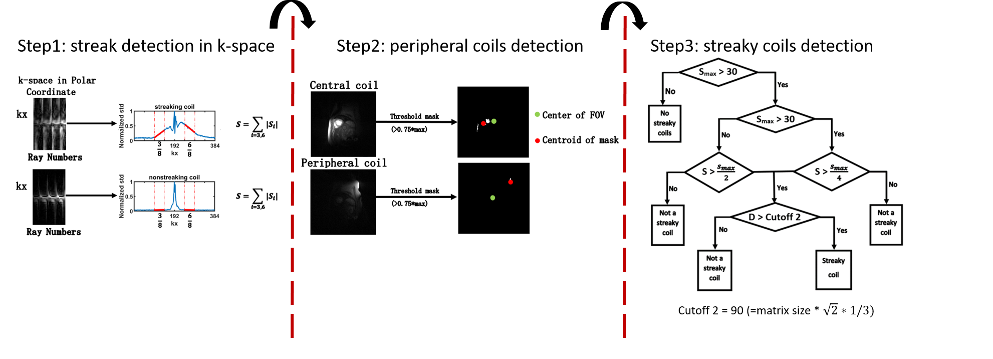
Post-Processing: We developed our method based our observation that radial streaks manifest themselves in k-space and that only peripheral streaky coils should be eliminated for cardiac imaging. Note, streaky coils were identified visually based on zero-filled reconstruction of individual coils, which then served as ground truth. Step 1, we calculated the standard deviation of radial k-space along the ray dimension in polar coordinate space. As shown in Figure 1, streak coils have higher side lobes (see 3/8 and 6/8th k-space segments) compared with non-streaky coils. After dividing the k-space into 8 equal segments, we calculated the slope of the third and sixth segments and summed the absolute value of these two slopes. Step 2, we generated a mask based on 75% of maximum intensity and calculated the Euclidean distance between the centroid of the mask and the center of field of view. Step 3, we combined these two parameters to choose out streaky coils. For details, see Figure 1. The cutoff values of these two parameters were determined empirically based on training data sets from 5 patients.For validation, we applied our algorithm and the method by Xue et al. 2 for cardiac perfusion data from the remaining 16 subjects (2406 images, 3-4 short-axis planes, 0-3 long-axis planes,15-30 coils).
Data Analysis: For validation, we computed the accuracy of streaky coil detection using our method and the method by Xue et al.2. We performed CS reconstruction using all coils, streaky coils removed using our method. We also quantified the mean signal of left ventricular (LV) cavity before contrast arrival and the ratio of mean signal and standard deviation of LV cavity at peak enhancement, compared values of all coils and streaky coils removed using our method using a paired t-test.
Results
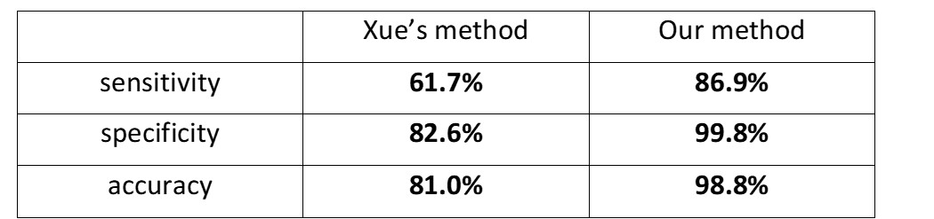
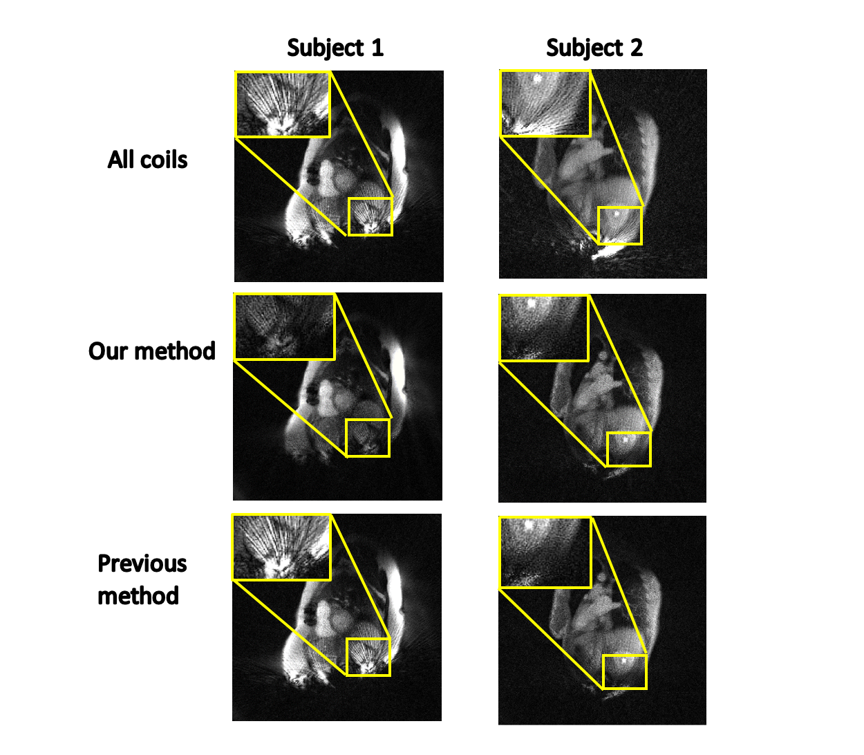
As summarized in Table 1, the sensitivity, specificity, and accuracy were higher for our method than the method by Xue et al.2. Figure 2 shows representative cardiac perfusion images of two different patients. Compared with all coils and streaky coils removed by the previous method, streaky coils removed by our method produced less streaking artifacts. The mean pre-contrast LV cavity signal was significantly (p < 0.001) lower our reconstruction (5.42 ± 2.55 a.u.) than that using all coils (5.56 ± 2.49 a.u.), implying reduced artifact for our reconstruction. The mean ratio of LV cavity signal and standard deviation at peak enhancement by our method was significantly (p < 0.001) higher for our method (19.25 ± 4.48 a.u.) than using all coils (18.23 ± 3.88 a.u.), again implying reduced artifacts for our method.Conclusion
This study demonstrates that the proposed algorithm can accurately detect streaky coils, which then can be removed for optimal reconstruction of cardiac perfusion MR images. Further studies are warranted to evaluate this algorithm for other cardiac and non-cardiac MRI acquisitions.Acknowledgements
This work was supported in part by the following grants:NIH R01HL116895, R01HL138578, R21EB024315, R21AG055954References
1. Du J, Thornton FJ, Fain SB, Korosec FR, Browning F, Grist TM, Mistretta CA. Artifact reduction in undersampled projection reconstruction MRI of the peripheral vessels using selective excitation. Magn Reson Med 2004;51(5):1071-1076.
2. Xue Y, Yu J, Kang HS, Englander S, Rosen MA, Song HK. Automatic coil selection for streak artifact reduction in radial MRI. Magn Reson Med 2012;67(2):470-476.
3. Naresh N, Haji-valizadeh H, Aouad P,
Barrett M, Chow K, Ragin A, Collins J, Carr J, Lee D, Kim D. Accelerated,
first-pass cardiac perfusion pulse sequence with radial k-space sampling,
compressed sensing, and KWIC reconstruction tailored for visual analysis and quantification
of MBF. Magn Reson Med (in press).
Figures