4588
Self-retraced Spiral In-Out 3D Turbo Spin-Echo Imaging1Radiology & Medical Imaging, University of Virginia, Charlottesville, VA, United States, 2Biomedical Engineering, University of Virginia, Charlottesville, VA, United States, 3Application Development, Siemens Healthcare, Erlangen, Germany
Synopsis
The purpose of this work was to perform a preliminary evaluation of a self-retraced spiral in-out trajectory for 3D turbo/fast spin-echo imaging. By sampling k-space locations twice with a single spiral in-out trajectory, off-resonance effects are robustly attenuated and image quality is improved compared to using a standard spiral in-out trajectory.
Introduction & Purpose:
Optimized 3D turbo/fast spin-echo (TSE/FSE) imaging (e.g., SPACE [Siemens], CUBE [GE], VISTA [Philips]) has gained popularity for a variety of clinical applications. While currently-available commercial implementations use conventional Cartesian (rectilinear) sampling of k space, certain applications may benefit from the favorable properties of non-Cartesian k-space trajectories, prompting recent preliminary evaluations of spiral-based implementations1,2. Spin-echo-based pulse sequences such as 3D TSE/FSE are naturally suited to a spiral in-out trajectory2,3, as opposed to the more commonly used spiral-out trajectory. Further, it has been shown that a retraced spiral in-out trajectory, which combines k-space data from two spiral in-out interleaves having waveforms with opposite gradient polarities to sample k-space locations twice, is advantageous for suppressing off-resonance artifacts because the phases accumulated from off resonance have opposing values at corresponding k-space locations for the two interleaves4. For 3D TSE/FSE, however, retraced spiral in-out has the disadvantage that the two spiral in-out interleaves are optimally collected at the same echo position during two different echo trains, making the approach sensitive to any differences between echo trains, such as could be caused by motion. The purpose of this work was to perform a preliminary evaluation of a self-retraced spiral in-out trajectory5 for 3D TSE/FSE, which samples k-space locations twice using a single spiral in-out trajectory.Methods:
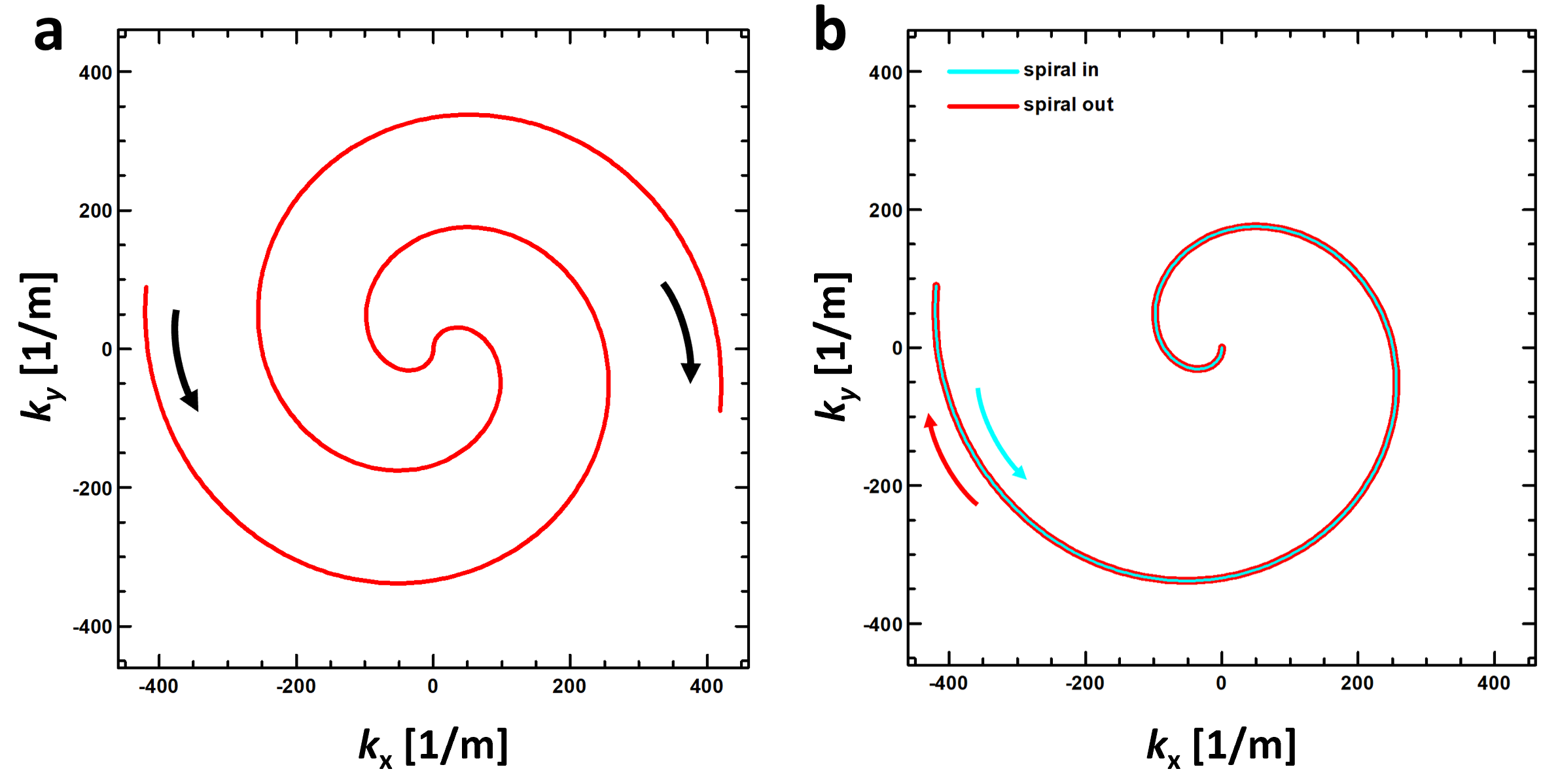
A prototype 3D stack-of-spirals TSE acquisition was implemented based on a commercial version of single-slab, variable-flip-angle, 3D-TSE imaging (SPACE, Siemens Healthcare, Erlangen, Germany) modified to support a stack-of-spirals acquisition. The signal evolution along the spin-echo train was mapped to the through-plane (3D) Cartesian phase-encoding direction to obtain the desired image-contrast properties1. The pulse sequence supported spiral out, either with the waveform centered within an echo spacing for maximum efficiency or positioned such that the center of k space is sampled at the spin echo2, standard (i.e., single trajectory, not retraced) spiral in-out3, and self-retraced spiral in-out5. Figure 1 illustrates the standard and self-retraced spiral in-out trajectories.
The basic properties of the 3D-TSE pulse sequence using a self-retraced spiral in-out trajectory were evaluated using measurements in a spatial-resolution phantom on 1.5T (MAGNETOM Aera, Siemens Healthcare, Erlangen, Germany) and 3T (MAGNETOM Prisma, Siemens Healthcare) commercial MR scanners. Trajectory durations of 3.6 ms and 7.2 ms were used for spiral out and spiral in-out trajectories, respectively.
Results & Discussion:
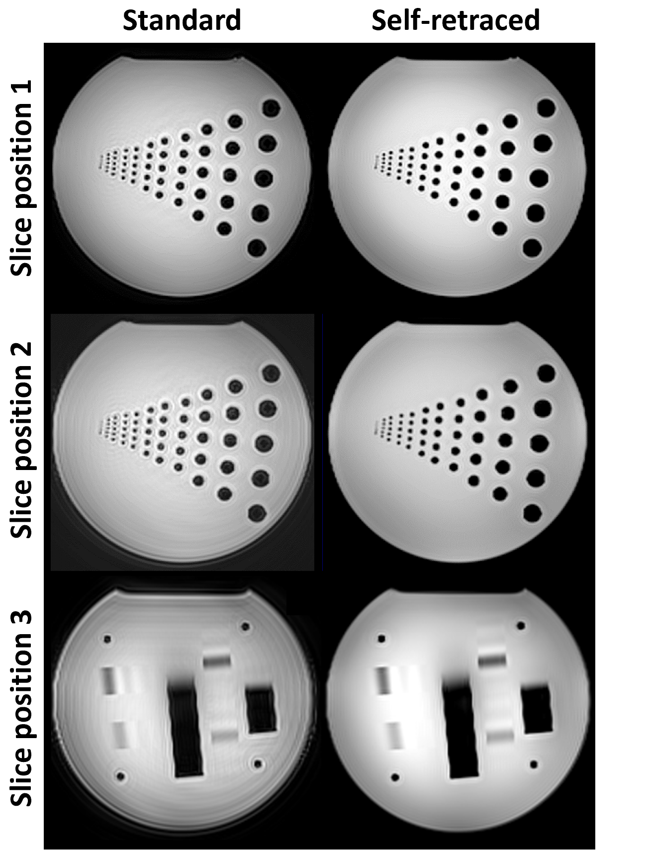
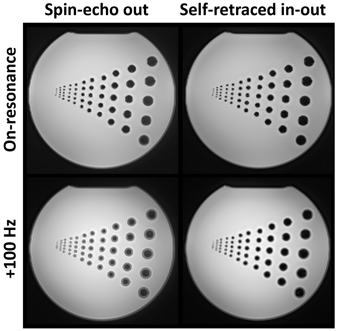
Figure 2 compares the image quality for self-retraced spiral in-out to that for standard spiral in-out. The images for self-retraced spiral in-out showed good quality throughout the phantom, while those for standard spiral in-out were visibly degraded in comparison, particularly for slice positions toward either end of the phantom (Figure 2, middle and bottom rows). The image-quality degradation for standard spiral in-out may have been due to the differential effects of eddy currents, concomitant gradients, and/or off resonance on data sampled at conjugate k-space positions; these effects are inherently compensated with the self-retraced spiral in-out trajectory. Figure 3 compares the image quality for self-retraced spiral in-out to that for standard spiral out (with the center of k space sampled at the spin echo) for acquisitions on resonance and at 100 Hz off resonance (36° of phase accumulation per millisecond). On resonance, the spiral out and self-retraced spiral in-out images appeared essentially identical. However, at 100 Hz off resonance, the spiral out image clearly showed off-resonance blurring (particularly visible around the resolution elements), whereas the off-resonance effects were much less pronounced for the self-retraced spiral in-out image, consistent with behavior previously described for retraced spiral in-out trajectories4.Conclusions & Future Work:
This preliminary study has demonstrated the feasibility of using a self-retraced spiral in-out trajectory for 3D-TSE imaging to obtain robustness to off-resonance effects and improved image quality compared to that for a standard spiral in-out trajectory. Given the favorable performance of the self-retraced spiral in-out trajectory, future work will focus on in-vivo testing of applications for which spiral-based 3D-TSE imaging may provide an advantage compared to conventional Cartesian-based 3D-TSE imaging in terms of acquisition speed or sensitivity to motion.Acknowledgements
No acknowledgement found.References
1. Fielden S et al. Variable‐flip angle 3D‐turbo spin echo imaging utilizing spiral acquisitions. Proc ISMRM 19 (2011); 2820.
2. Li Z et al. Sliding-slab three-dimensional TSE imaging with a spiral-in/out readout. Magn Reson Med 2016; 75:729-738.
3. Meyer CH et al. Fast spiral T2-weighted imaging. Proc SMR 2 (1994); 467.
4. Fielden SW, Meyer CH. A simple acquisition strategy to avoid off-resonance blurring in spiral imaging with redundant spiral-in/out k-Space trajectories. Magn Reson Med 2015; 73:704-710.
5. Glover GH, Law CS. Spiral-in/out BOLD fMRI for increased SNR and reduced susceptibility artifacts. Magn Reson Med 2001; 46:515-522.
Figures